


Article Figures & Data
Figures
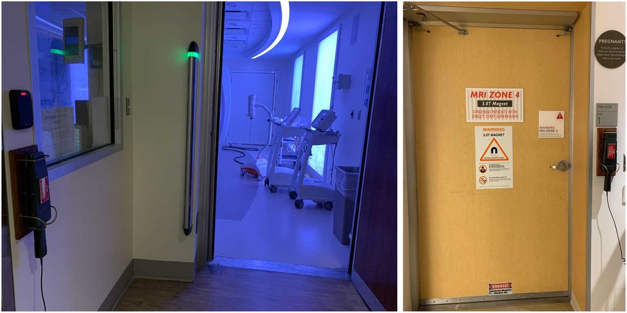
- FIGURE 1.
On the left is secure MRI doorway with ferromagnetic detector and 5-G line demarcated by floor color change and roof lighting. On the right is example of secure PET/MRI door with zone 4 signage, wall-mounted ferromagnetic detector, and pregnancy sign. (Courtesy of New York–Presbyterian Cornell Weill Medical Center and Memorial Sloan Kettering Cancer Center.)
- FIGURE 2.
Typical PET/MRI footprint with gauss lines for 200 G (blue); 100 G (solid red); 50, 10, and 5 G (all dashed red); and 3 and 1 G (dashed green).
- FIGURE 3.
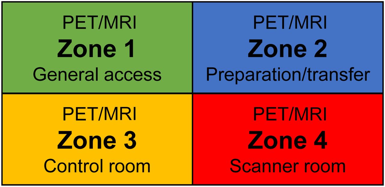
Example of signage used to demarcate MRI zones. Color coding can be used on floor plans and emergency exit plans for first responders, and color-coded floor tape and door trim can be used to help identify zones.
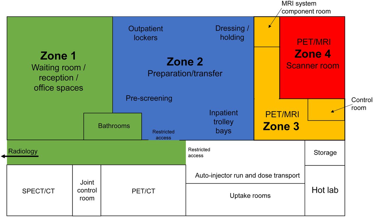
- FIGURE 4.
Schematic of typical department design incorporating PET/MRI highlighting color-coded zones. Noncolored zones are specific to nuclear medicine and will have radiation-based restrictions.


Jump to section
Related Articles
Cited By...
- PET-MRI for evaluation of response to radiochemotherapy in patients with locally advanced cervical cancer
- PET-MRI for evaluation of response to radiochemotherapy in patients with locally advanced cervical cancer
- PET/MRI, Part 4: Clinical Applications
- PET/MRI, Part 2: Technologic Principles
- Virtually Celebrating the Advances of Nuclear Medicine and Molecular Imaging: 2021