


Article Figures & Data
Figures
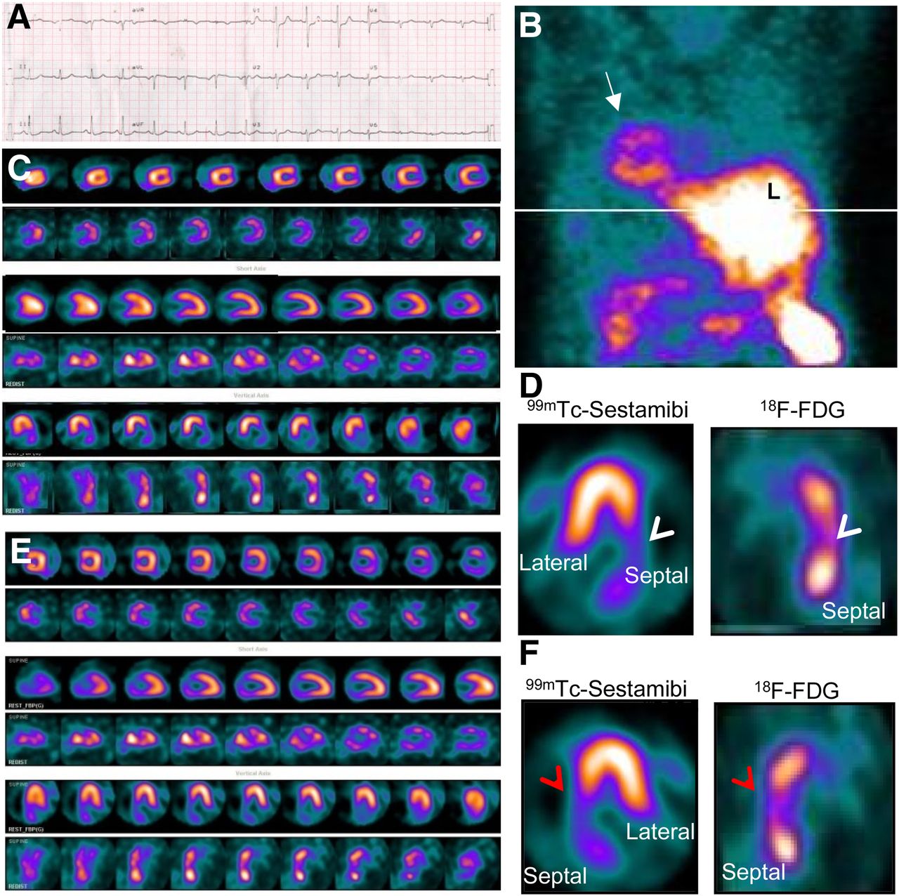
- FIGURE 1.
A 66-y-old man who had dextrocardia with situs inversus presented with non–ST-segment elevation myocardial infarction. (A) Poor R-wave progression was seen on V3–V6 chest leads on electrocardiography, which was obtained with chest leads arranged on right chest wall. (B) Maximum-intensity-projection rest perfusion SPECT image showed heart in right chest (arrow) and liver on left side. (C and D) Processing of raw data in feet-first supine position showed interchanged lateral wall and septum in reconstructed horizontal long-axis image (arrowheads). (E and F) Orientation of septum and lateral wall was corrected in repeat processing, which was done after entering dataset for feet-first prone position instead of feet-first supine to match conventional nomenclature of display image (arrowheads). Perfusion was absent from mid-anteroseptal, basal anteroseptal, mid-inferoseptal, and basal inferoseptal segments whereas 18F-FDG uptake was present in same segments, thus suggesting viable myocardium (matched perfusion and metabolism).
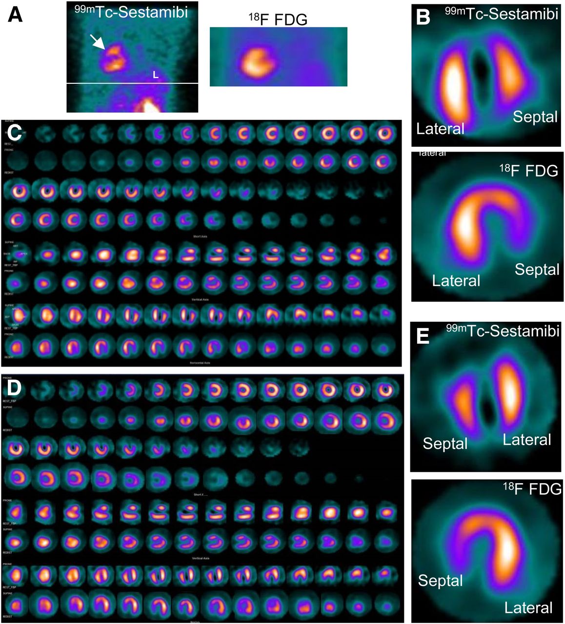
- FIGURE 2.
A 61-y-old woman had dextrocardia with situs inversus. (A) On maximum-intensity projection, heart was seen in right chest and liver on left side (arrow). (B and C) Processing of raw data in feet-first supine position showed interchanged lateral wall and septum in reconstructed 3-axes display image (C) and horizontal long-axis image (B). (D and E) Orientation of septum and lateral wall was corrected in repeat processing (D), which was done after entering dataset for feet-first prone position instead of feet-first supine to match conventional nomenclature of display image (E). Perfusion was absent from apex, apical septum, mid- and basal anterior walls, and mid- and basal anteroseptal segments whereas 18F-FDG uptake was present in same segments, suggesting viable myocardium (matched perfusion and metabolism).
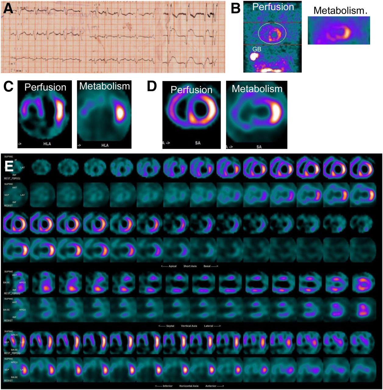
- FIGURE 3.
A 42-y-old man with mesocardia presented with breathlessness. Electrocardiography (A) showed ST-segment elevation in V4 and V5, Q waves in V1 and V4–V6, and right bundle branch block. (B) Maximum-intensity projections showed heart in midline of chest and gallbladder and liver on right side. (C–E) Processing of raw data in feet-first supine position showed lateral wall and septum positioned normally on reconstructed horizontal long-axis image (C) and short-axis image (D) and on display images (E). Rest perfusion slices showed absence of perfusion in apex, apical anterior, apical septal, apical inferior, apical lateral, basal anteroseptal, and mid-inferior segments. Perfusion was reduced in entire anterior wall and septum. 18F-FDG PET images did not show uptake in corresponding myocardial segments, suggesting absence of viable myocardium in infarcted segments (matched perfusion and metabolism).
Tables
Variable Patient 1 Patient 2 Patient 3 Age (y) 66 61 42 Sex M F M Presentation NSTEMI Breathlessness Chest pain Type of dextrocardia Complete Complete Incomplete (mesocardia) Situs inversus? Yes Yes No Patient positioning SPECT Feet-first supine Feet-first supine Feet-first supine 18F-FDG PET Head-first supine Head-first supine Head-first supine Imaging arc during acquisition SPECT 45° LAO to 135° RPO clockwise 45° LAO to 135° RPO clockwise 180° right lateral to left lateral 18F-FDG PET 360° 360° 360° Routine processing* SPECT Feet-first supine Feet-first supine Feet-first supine 18F-FDG PET Head-first supine Head-first supine Head-first supine Cardiac wall orientation Interchanged septum and lateral wall Interchanged septum and lateral wall Not affected Change in processing Required Required Not required SPECT Feet-first prone Feet-first prone — 18F-FDG PET Head-first prone Head-first prone — Cardiac wall orientation Corrected Corrected — ↵* Using Emory Cardiac Toolbox.
NSTEMI = non–ST-segment elevation myocardial infarction.


