
Main menu

  • Home
  • Content
    • Current
      • JNMT Supplement
    • Ahead of print
    • Past Issues
    • Continuing Education
    • JNMT Podcast
    • SNMMI Annual Meeting Abstracts
  • Subscriptions
    • Subscribers
    • Rates
    • Journal Claims
    • Institutional and Non-member
  • Authors
    • Submit to JNMT
    • Information for Authors
    • Assignment of Copyright
    • AQARA Requirements
  • Info
    • Reviewers
    • Permissions
    • Advertisers
    • Corporate & Special Sales
  • About
    • About Us
    • Editorial Board
    • Contact Information
  • More
    • Alerts
    • Feedback
    • Help
    • SNMMI Journals
  • SNMMI
    • JNMT
    • JNM
    • SNMMI Journals
    • SNMMI

User menu

  • Subscribe
  • My alerts
  • Log in
  • My Cart

Search

  • Advanced search
Journal of Nuclear Medicine Technology
  • SNMMI
    • JNMT
    • JNM
    • SNMMI Journals
    • SNMMI
  • Subscribe
  • My alerts
  • Log in
  • My Cart
Journal of Nuclear Medicine Technology

Advanced Search

  • Home
  • Content
    • Current
    • Ahead of print
    • Past Issues
    • Continuing Education
    • JNMT Podcast
    • SNMMI Annual Meeting Abstracts
  • Subscriptions
    • Subscribers
    • Rates
    • Journal Claims
    • Institutional and Non-member
  • Authors
    • Submit to JNMT
    • Information for Authors
    • Assignment of Copyright
    • AQARA Requirements
  • Info
    • Reviewers
    • Permissions
    • Advertisers
    • Corporate & Special Sales
  • About
    • About Us
    • Editorial Board
    • Contact Information
  • More
    • Alerts
    • Feedback
    • Help
    • SNMMI Journals
  • Watch or Listen to JNMT Podcast
  • Visit SNMMI on Facebook
  • Join SNMMI on LinkedIn
  • Follow SNMMI on Twitter
  • Subscribe to JNMT RSS feeds
Review ArticleTeaching Case Studies

Waxing and Waning Presentation of Isolated Cardiac Sarcoidosis on Sequential 18F-FDG PET Examinations

Wesam Ostwani, Carolyn Hanna, Aaron E. Brice and David C. Wymer
Journal of Nuclear Medicine Technology December 2020, 48 (4) 378-380; DOI: https://doi.org/10.2967/jnmt.120.246322
Wesam Ostwani
Department of Cardiac Imaging, Malcom Randall VA Medical Center, College of Medicine, University of Florida, Gainesville, Florida
  • Find this author on Google Scholar
  • Find this author on PubMed
  • Search for this author on this site
Carolyn Hanna
Department of Cardiac Imaging, Malcom Randall VA Medical Center, College of Medicine, University of Florida, Gainesville, Florida
  • Find this author on Google Scholar
  • Find this author on PubMed
  • Search for this author on this site
Aaron E. Brice
Department of Cardiac Imaging, Malcom Randall VA Medical Center, College of Medicine, University of Florida, Gainesville, Florida
  • Find this author on Google Scholar
  • Find this author on PubMed
  • Search for this author on this site
David C. Wymer
Department of Cardiac Imaging, Malcom Randall VA Medical Center, College of Medicine, University of Florida, Gainesville, Florida
  • Find this author on Google Scholar
  • Find this author on PubMed
  • Search for this author on this site
  • Article
  • Figures & Data
  • Info & Metrics
  • PDF
Loading

Abstract

We describe the case of a patient who had suspected myocardial ischemia, showed normal findings on multiple perfusion scans, and showed isolated cardiac sarcoidosis on 18F-FDG-PET. Also discussed are the diagnosis and the monitoring of disease response using imaging follow-up.

  • isolated cardiac sarcoidosis
  • cardiac sarcoidosis
  • 18F-FDG PET

Sarcoidosis is a multisystem, granulomatous disease of unknown etiology, and noncaseating granulomas are the histopathologic hallmark (1). Cardiac sarcoidosis can lead to advanced heart block, arrhythmias, cardiomyopathy, and sudden cardiac death. Reported rates of isolated cardiac sarcoidosis among patients with cardiac sarcoidosis vary widely, ranging from 27% to 54%. Diagnosis is challenging and not well defined in the literature (2).

CASE REPORT

A 69-y-old African American man with a history of atrial fibrillation, high-grade atrioventricular block with a dual-chamber pacemaker, nonsustained ventricular tachycardia, multiple prior negative perfusion studies (Fig. 1), and end-stage renal disease requiring hemodialysis presented with chest pain and exertional dyspnea.

FIGURE 1.
  • Download figure
  • Open in new tab
  • Download powerpoint
FIGURE 1.

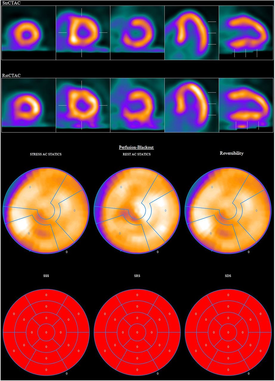
Myocardial perfusion scan with normal findings. AC = attenuation-corrected; SDS = summed difference score; SRS = summed rest score; SSS = summed stress score.

The findings on physical examination were unremarkable. Laboratory values demonstrated mildly elevated but flat troponin T, serially 0.12 ng/mL. Electrocardiography showed a ventricle-paced rhythm. Echocardiography showed normal biventricular systolic function.

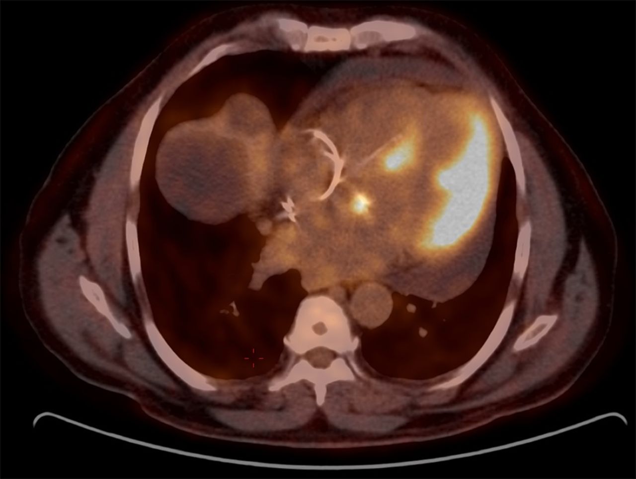
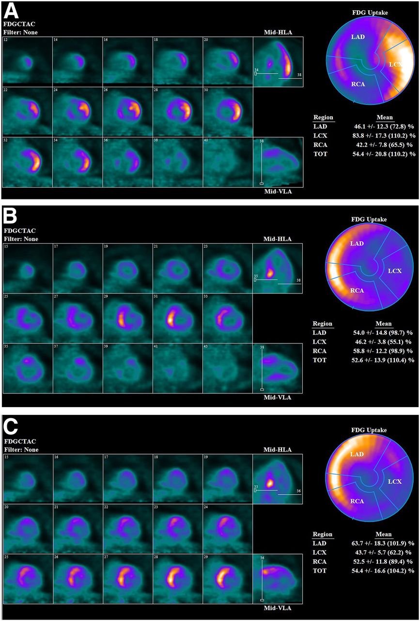
Given the patient’s race (African Americans are more prone to sarcoidosis), unexplained symptoms, heart block, and nonsustained ventricular tachycardia, 18F-FDG PET was performed. The initial 18F-FDG PET study demonstrated lateral left ventricular wall uptake (Figs. 2, 3A, and 4A) suggestive of cardiac sarcoidosis. No extracardiac sarcoidosis was found on workup. Left ventricular endomyocardial biopsy was not performed because it was likely to have a low yield and present an elevated risk. Contrast-enhanced MRI was also contraindicated because of the patient’s end-stage renal disease. The patient began taking 40 mg of prednisone daily for 5 mo, and his pacemaker was upgraded to a defibrillator. After 3 mo of treatment, a repeat 18F-FDG PET examination showed a partial response, with a significant reduction in uptake in the lateral left ventricular wall but new uptake in the septal wall (Figs. 3B and 4B).

FIGURE 2.
  • Download figure
  • Open in new tab
  • Download powerpoint
FIGURE 2.

Axial 18F-FDG PET/CT image showing intense uptake in left ventricular lateral wall and lesser septal uptake.

FIGURE 3.
  • Download figure
  • Open in new tab
  • Download powerpoint
FIGURE 3.

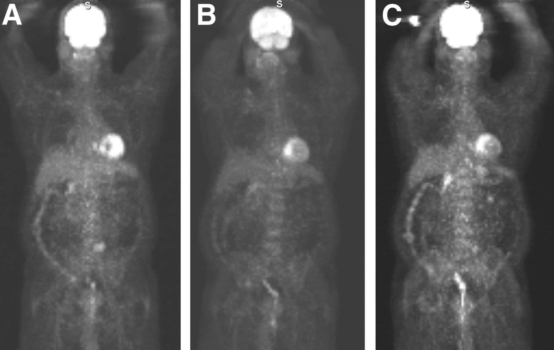
Whole-body 18F-FDG PET images obtained at 3-mo intervals, showing no extracardiac uptake. (A) Initial examination, with intense lateral wall uptake. (B) Improved findings in lateral wall but new basal–to–mid-septal uptake while patients was taking 40 mg of prednisone daily. (C) Persistent uptake in septal wall and new uptake in basal–to–mid-anterior wall while patient was taking 20 mg of prednisone daily.

FIGURE 4.
  • Download figure
  • Open in new tab
  • Download powerpoint
FIGURE 4.

Cardiac 18F-FDG PET metabolism at 3-mo intervals, including axial, horizontal long-axis, vertical long-axis, and bull’s-eye views that again show uptake pattern seen in Figure 3. FDGCTAC = CT attenuation-corrected 18F-FDG images; HLA = horizontal long axis; LAD = left anterior descending coronary artery; LCX = left circumflex coronary artery; RCA = right coronary artery; TOT = total; VLA = vertical long axis.

The patient began taking mycophenolate mofetil, but this was stopped after 1 mo because of oral sores related to herpes virus. His prednisone dose was decreased to 20 mg per day. Repeat 18F-FDG PET after 3 mo revealed a continued response to treatment in the lateral wall but persistent uptake in the septal wall and new uptake in the basal–to–mid-anterior wall (Figs. 3C and 4C).

Currently, the patient is stable, with no cardiovascular symptoms or evidence of worsening cardiomyopathy or ventricular tachycardia. He has continued taking 20 mg of prednisone per day, with plans to restart mycophenolate at a lower dose and titrate upward as tolerated.

DISCUSSION

On the basis of available data, the approximate rate of isolated cardiac sarcoidosis among patients with cardiac sarcoidosis is 25%. There is limited sensitivity for the diagnosis of cardiac sarcoidosis, with current criteria requiring evidence of extracardiac disease or positive results on endomyocardial biopsy, which itself has limited sensitivity (20%–40%) because of the patchy nature of the disease. Diagnostic sensitivity for isolated cardiac sarcoidosis is therefore even more limited. Advanced cardiac imaging techniques using a combined approach of 18F-FDG PET and cardiac MRI could improve early detection of isolated cardiac sarcoidosis and improve the accuracy of diagnosis by providing data on both inflammation and scarring or fibrosis. Whole-body 18F-FDG PET is also useful for screening for extracardiac involvement (2). Cardiac MRI was not performed for our patient because of his history of kidney failure and the risk for nephrogenic systemic fibrosis. Endomyocardial biopsy was not performed because of its low sensitivity, pacemaker dependence, and increased risk.

Most experts agree that corticosteroids with or without immunosuppressive therapy should be used in the treatment of cardiac sarcoidosis. 18F-FDG PET reassessment of response to treatment is recommended at 3-mo intervals (1). Our patient showed a mixed response to treatment at both the 3-mo and the 6-mo intervals (Figs. 3 and 4). The data are insufficient to guide management for patients with a mixed response.

CONCLUSION

Diagnosis of isolated cardiac sarcoidosis is currently not well defined, and symptoms can overlap those of myocardial ischemia. The presence of ventricular tachycardia and a high-grade atrioventricular block should raise suspicion that cardiac sarcoidosis may be present. Given the variable response to current therapy, frequent 3-mo follow-up with cardiac imaging and monitoring of clinical response is recommended to guide treatment.

DISCLOSURE

No potential conflict of interest relevant to this article was reported.

Footnotes

  • Published online Jun. 9, 2020.

REFERENCES

  1. 1.↵
    1. Birnie DH,
    2. Nery PB,
    3. Ha AC,
    4. Beanlands RS
    . Cardiac sarcoidosis. J Am Coll Cardiol. 2016;68:411–421.
    OpenUrlFREE Full Text
  2. 2.↵
    1. Okada DR,
    2. Bravo PE,
    3. Vita T,
    4. et al
    . Isolated cardiac sarcoidosis: a focused review of an under-recognized entity. J Nucl Cardiol. 2018;25:1136–1146.
    OpenUrl
  • Received for publication April 3, 2020.
  • Accepted for publication May 5, 2020.
PreviousNext
Back to top

In this issue

Journal of Nuclear Medicine Technology: 48 (4)
Journal of Nuclear Medicine Technology
Vol. 48, Issue 4
December 1, 2020
  • Table of Contents
  • About the Cover
  • Index by author
Print
Download PDF
Article Alerts
Sign In to Email Alerts with your Email Address
Email Article

Thank you for your interest in spreading the word on Journal of Nuclear Medicine Technology.

NOTE: We only request your email address so that the person you are recommending the page to knows that you wanted them to see it, and that it is not junk mail. We do not capture any email address.

Enter multiple addresses on separate lines or separate them with commas.
Waxing and Waning Presentation of Isolated Cardiac Sarcoidosis on Sequential 18F-FDG PET Examinations
(Your Name) has sent you a message from Journal of Nuclear Medicine Technology
(Your Name) thought you would like to see the Journal of Nuclear Medicine Technology web site.
Citation Tools
Waxing and Waning Presentation of Isolated Cardiac Sarcoidosis on Sequential 18F-FDG PET Examinations
Wesam Ostwani, Carolyn Hanna, Aaron E. Brice, David C. Wymer
Journal of Nuclear Medicine Technology Dec 2020, 48 (4) 378-380; DOI: 10.2967/jnmt.120.246322

Citation Manager Formats

  • BibTeX
  • Bookends
  • EasyBib
  • EndNote (tagged)
  • EndNote 8 (xml)
  • Medlars
  • Mendeley
  • Papers
  • RefWorks Tagged
  • Ref Manager
  • RIS
  • Zotero
Share
Waxing and Waning Presentation of Isolated Cardiac Sarcoidosis on Sequential 18F-FDG PET Examinations
Wesam Ostwani, Carolyn Hanna, Aaron E. Brice, David C. Wymer
Journal of Nuclear Medicine Technology Dec 2020, 48 (4) 378-380; DOI: 10.2967/jnmt.120.246322
Twitter logo Facebook logo LinkedIn logo Mendeley logo
  • Tweet Widget
  • Facebook Like
  • Google Plus One
Bookmark this article

Jump to section

  • Article
    • Abstract
    • CASE REPORT
    • DISCUSSION
    • CONCLUSION
    • DISCLOSURE
    • Footnotes
    • REFERENCES
  • Figures & Data
  • Info & Metrics
  • PDF

Related Articles

  • No related articles found.
  • PubMed
  • Google Scholar

Cited By...

  • No citing articles found.
  • Google Scholar

More in this TOC Section

  • High-Sensitivity Troponin Elevation in a Young Woman with Typical Chest Pain: The Heart of the Matter
  • Pulmonary Adenocarcinoma Revealed by Parathyroid Scintigraphy: An Incidental Case to Remember
  • Prominent Right Ventricular Tracer Uptake: A Harbinger of Multivessel Coronary Artery Disease
Show more Teaching Case Studies

Similar Articles

Keywords

  • Isolated Cardiac Sarcoidosis
  • cardiac sarcoidosis
  • 18F-FDG PET
SNMMI

© 2025 SNMMI

Powered by HighWire