


Article Figures & Data
Figures
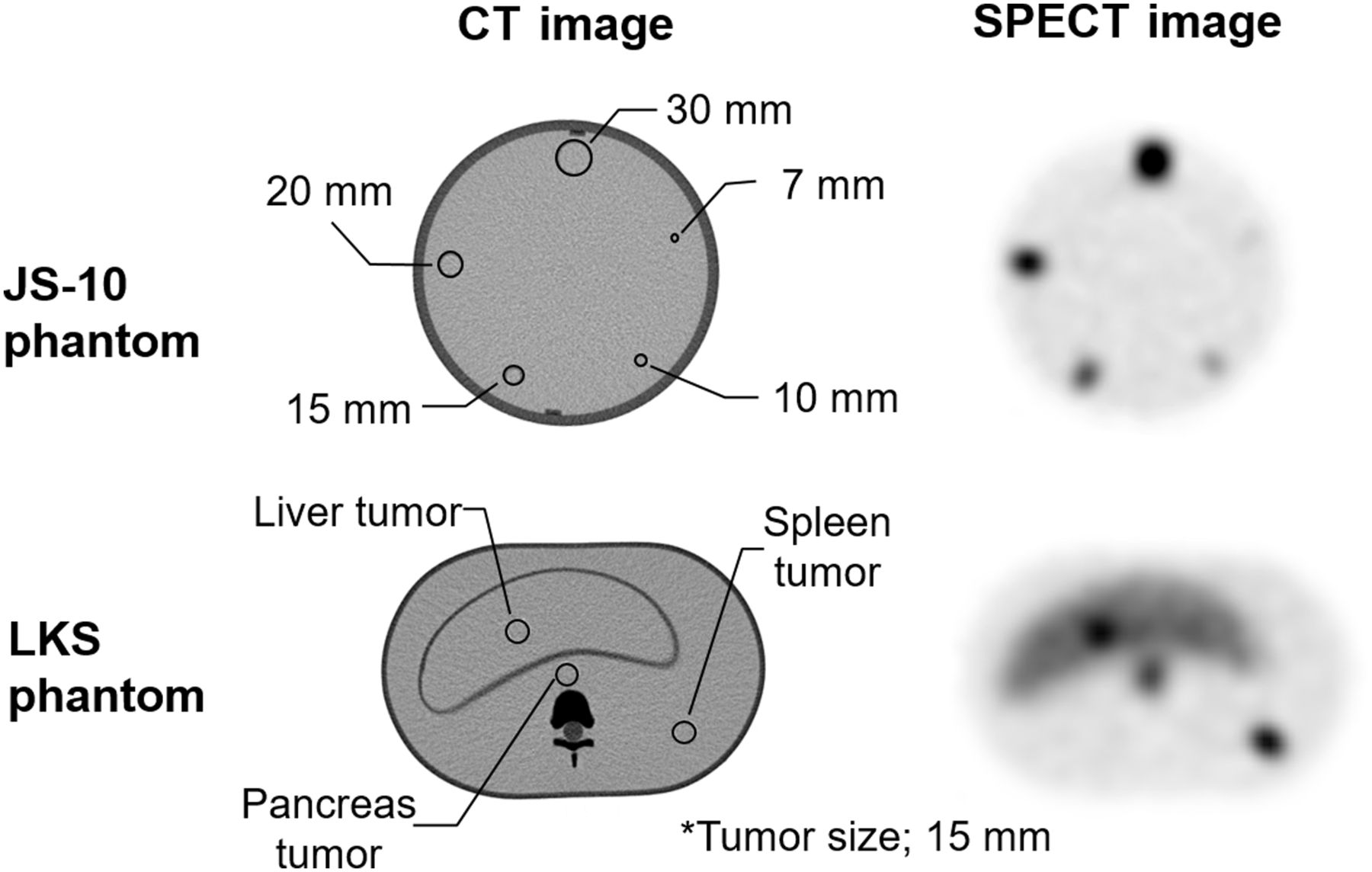
- FIGURE 1.
Comparison of SPECT QA (JS-10) phantom images for EWW, image reconstruction conditions, and CT images and of anthropomorphic abdominal (LKS) phantom for image collection conditions.
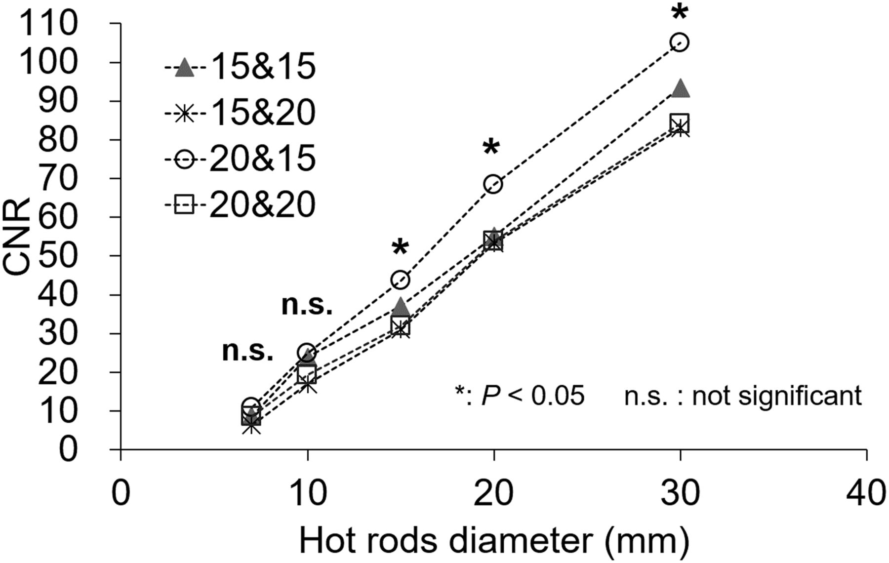
- FIGURE 2.
CNR of hot rods for optimum EWW in phantom study. EWW setting of 171 keV ± 10% and 245 keV ± 7.5% was significantly better than other settings (P < 0.05).
- FIGURE 3.
Optimization of OSEM technique between CNR and numbers of iterations in phantom study. OSEM reconstruction conditions of 8 subsets and 6 iterations gave significantly highest CNR (P < 0.05).
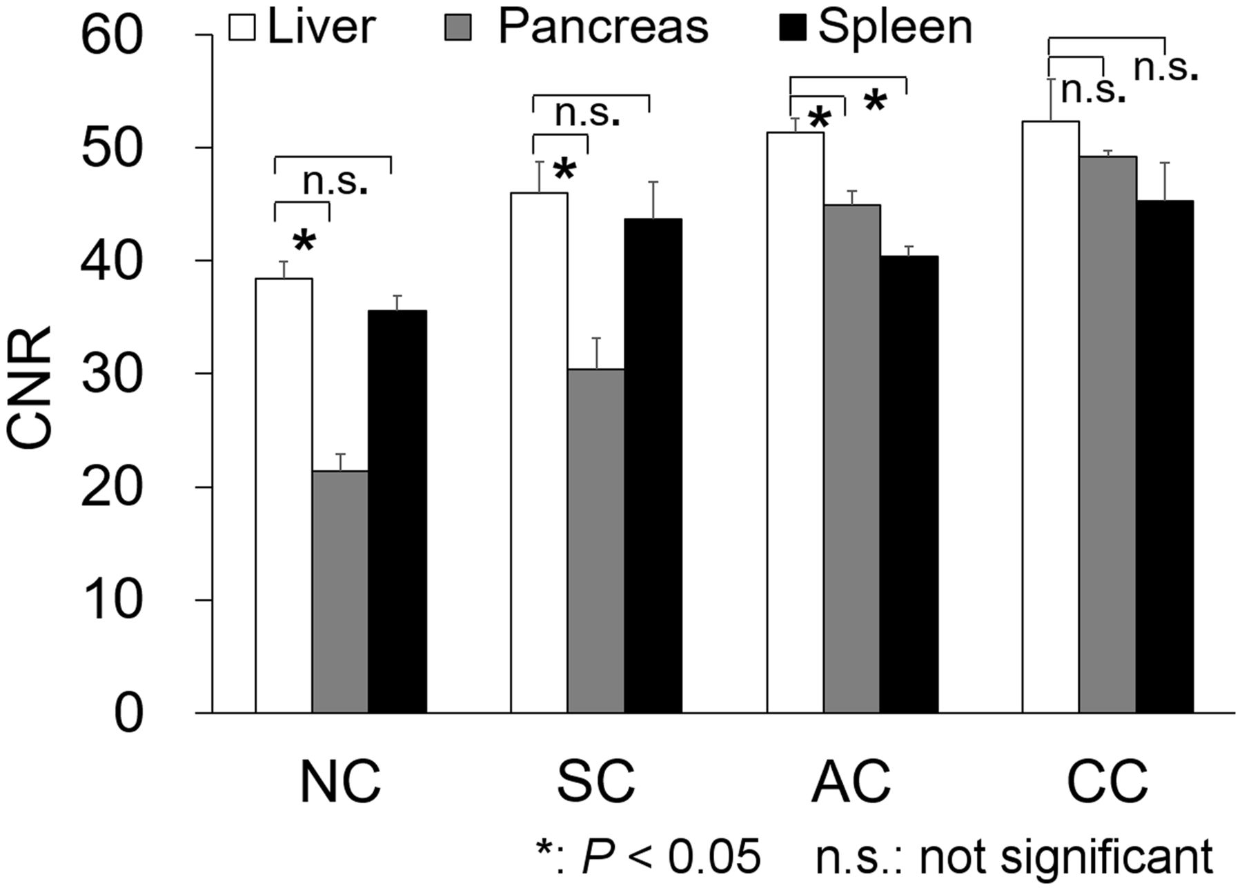
- FIGURE 4.
Comparison of 4 techniques of SPECT image correction (NC, SC, AC, and CC) in phantom study. Although underestimation occurred in pancreas when NC and SC were used, CC showed no significant difference (P = 0.83). In particular, CC gave significantly better CNR for pancreas than did NC or SC (P < 0.05).
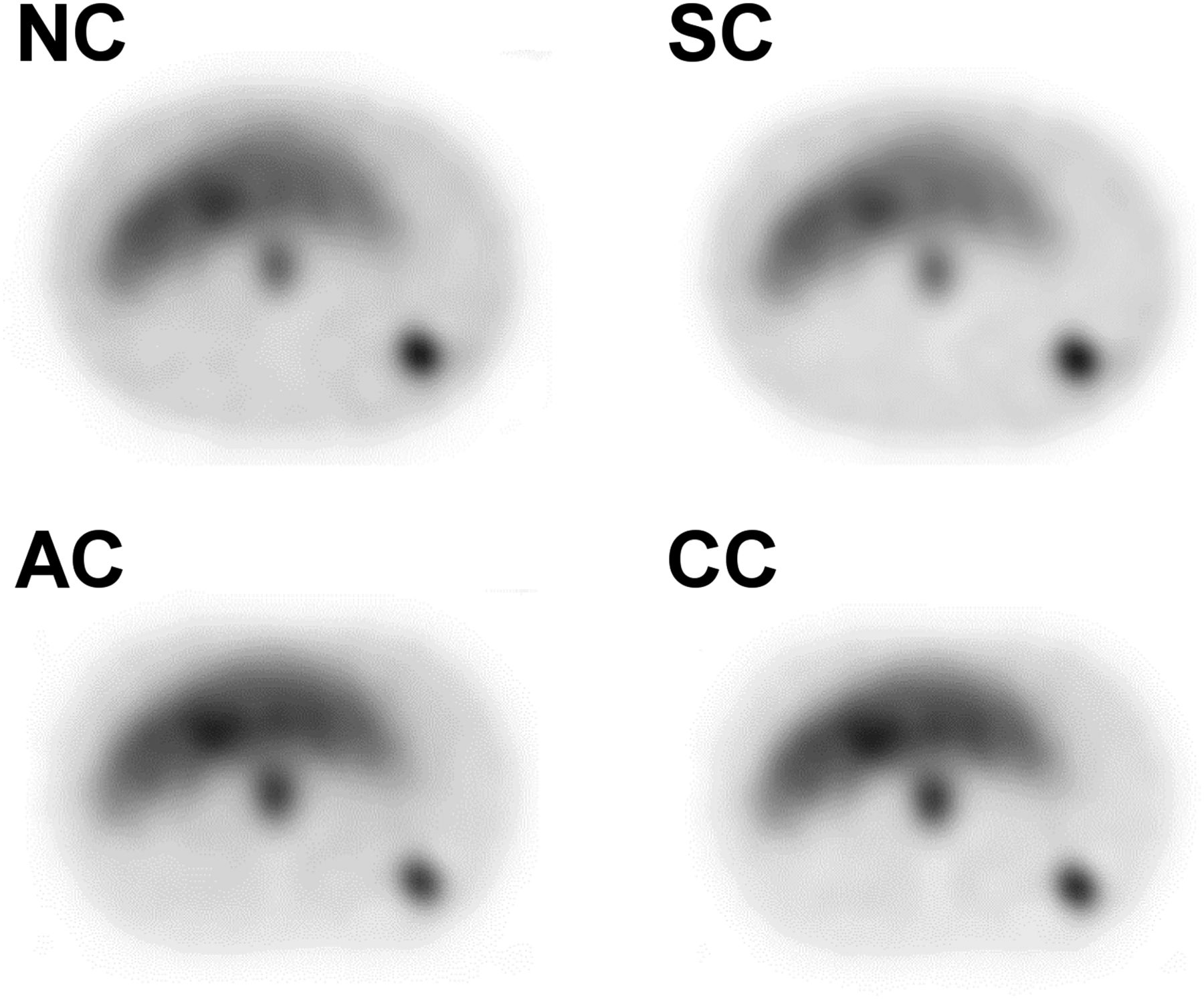
- FIGURE 5.
Axial phantom SPECT images corrected by 4 methods (NC, SC, AC, and CC).
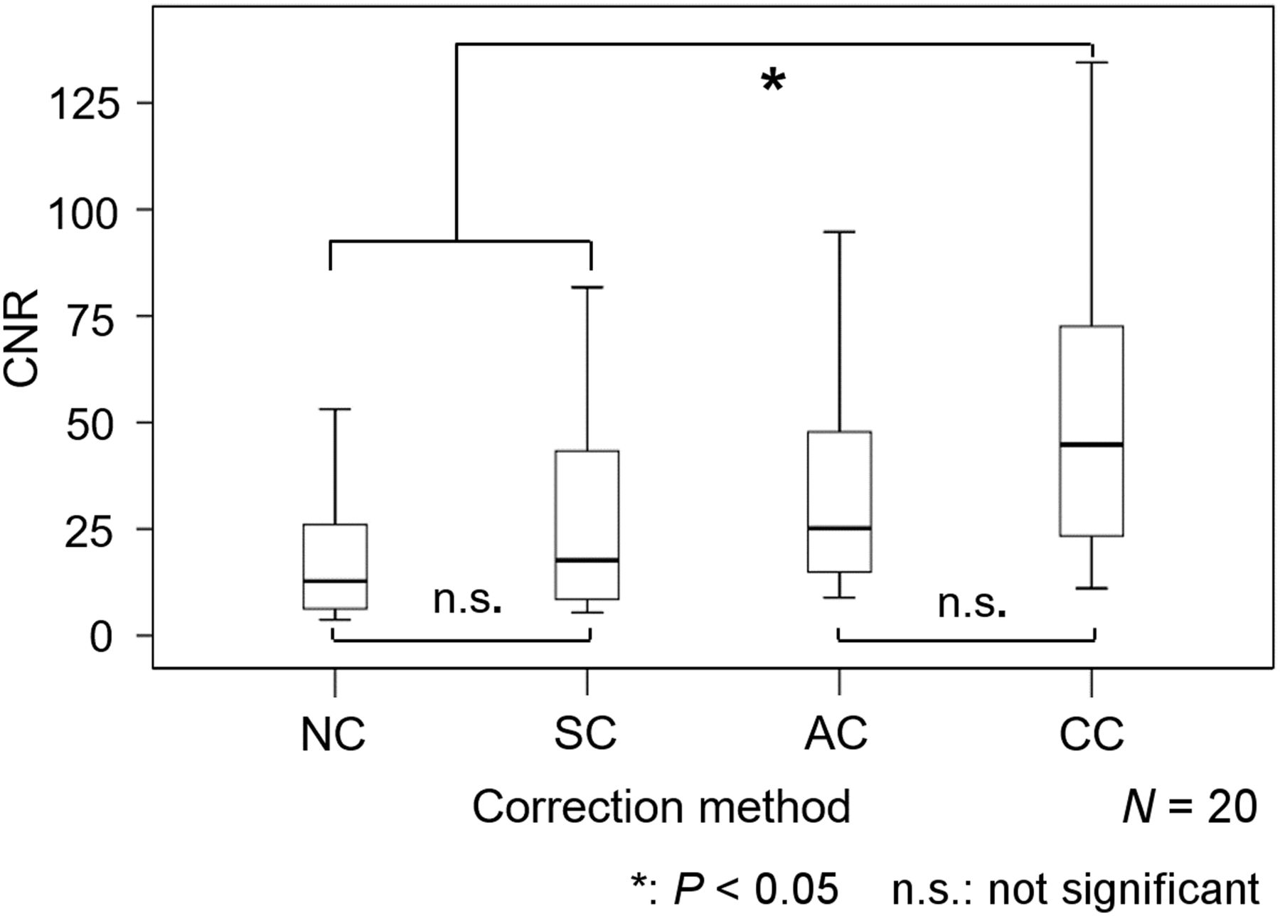
- FIGURE 6.
Box plot of CNR evaluated by 4 methods (NC, SC, AC, and CC) in 20 GEP NET patients. NC and SC showed no significant difference (P = 0.86), whereas CC showed significant differences from NC and SC.
- FIGURE 7.
Case of G1 NET of pancreatic head without metastasis. (A) Highly enhanced lesion (arrow) in pancreas is noted in early dynamic MR image. (B) 111In-pentetreotide SPECT images with NC, SC, AC, and CC depict abnormal uptake in corresponding upper abdominal area (arrows). Although images with SC and AC show comparable visualization of lesion with NC, CC delineates lesion most clearly.


Tables
Characteristic Data Age (y) 66.0 ± 15.6 (37–81) Sex Male 13 Female 7 Final diagnosis Pancreatic NET 1 Stage G1 7 Stage G2 2 Stage unclear 6 Duodenal NET 2 Rectal NET 2 Lymph node metastases of NET 1 Qualitative data are numbers; continuous data are mean ± SD followed by range (n = 20 patients).


Jump to section
Related Articles
Cited By...
- No citing articles found.