


Article Figures & Data
Figures
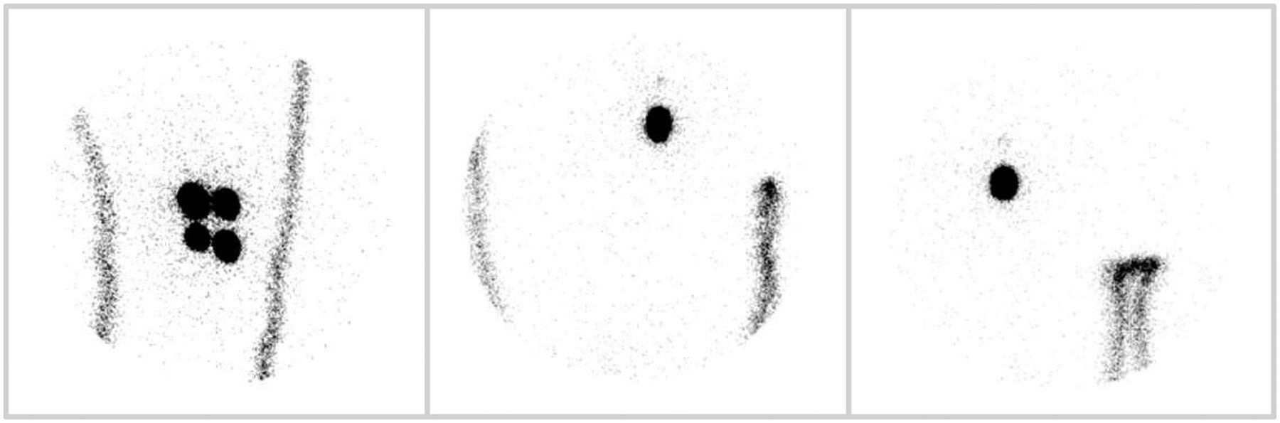
- FIGURE 1.
Leg melanoma with visualized node in groin. This image shows localization of SLN in groin area when injection is performed at thigh. Image provides idea of generalized regions in which lymph nodes are frequently found.
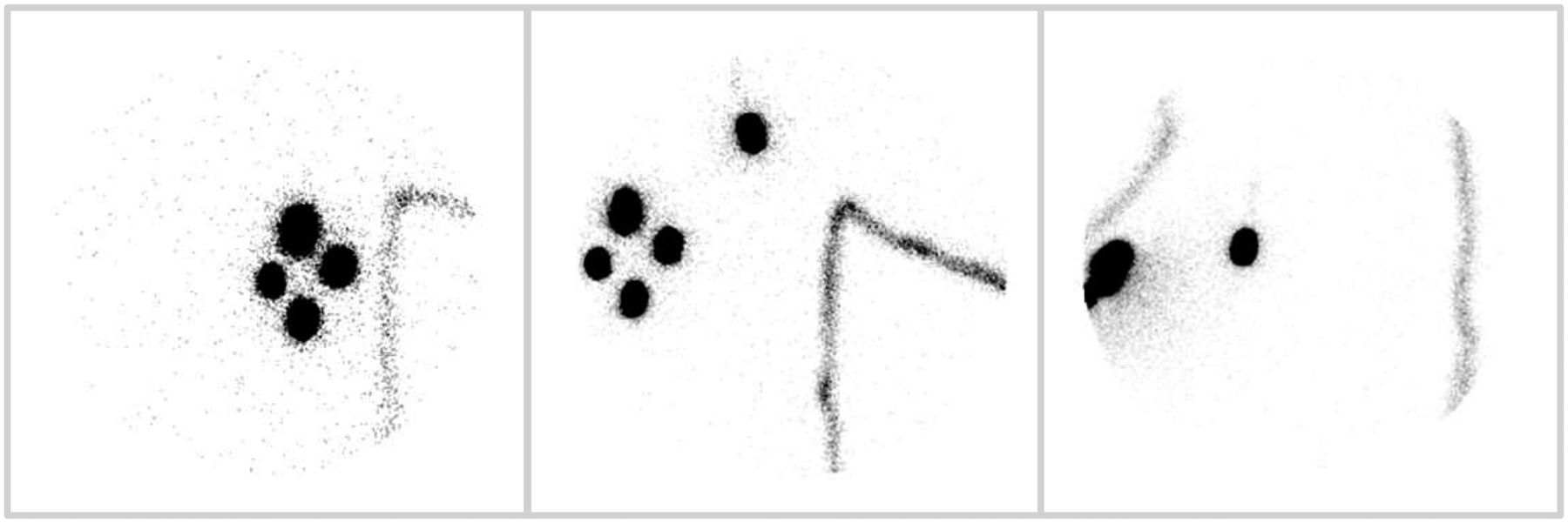
- FIGURE 2.
Breast lesion with visualized lymph node in axilla. Compare with Figure 3 to see benefits of using transmission source when imaging SLNs.
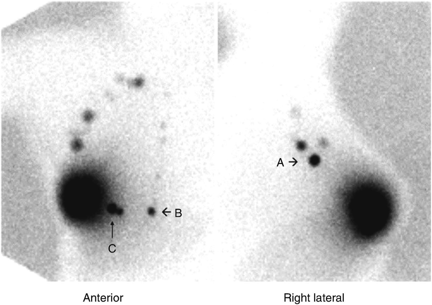
- FIGURE 3.
Breast lesion lymphoscintigraphy with 57Co transmission source (7). This image shows how transmission source can be effective tool when trying to relate internal node to external site on body. By showing body outline, image makes it easier to mark node for surgery. A = SLN from lateral view; B and C = other significant nodes, from anterior view, in ROI. These nodes may also be removed for biopsy.


Tables
- TABLE 1
Differences in Melanoma Subtypes, Their Impact on the Body, and Who Is Most at Risk
Subtype Incidence Location Growth pattern Metastasis potential Sex most at risk Superficial spreading 70% Non–weight-bearing surfaces of foot Slow and radial Low Women Nodular 15%–30% Non–weight-bearing surfaces of foot Rapid and vertical High Men Lentigo maligna 4%–10% Anterior lower leg in women; head and trunk in men Slow and radial Low Women Acral lentiginous 2%–8% Palms, soles, nail beds Radial at first, then vertical High Women


