


Article Figures & Data
Figures
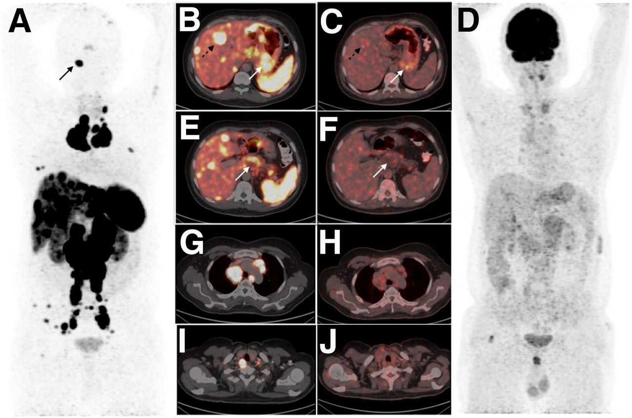
- FIGURE 1.
(A and D) Early and delayed static images of 99mTc-sestamibi scintigraphy performed for hyperparathyroidism showing 2 foci of increased tracer uptake with retention near upper poles of both lobes of thyroid gland (solid arrows). (G and B) Early SPECT maximum-intensity projection image and coronal fused SPECT/CT image localizing increased tracer uptake in 2 lesions in posterosuperior location of both lobes of thyroid gland (solid arrows) suggestive of parathyroid adenomas. (A, B, D, E, G, and H) 99mTc-sestamibi planar and SPECT/CT images showing tracer avidity in the enlarged mediastinal and cervical lymph nodes (dashed arrows). (C, F, I, and J) Regional neck 18F-fluorocholine PET maximum-intensity projection image and cross-sectional fused PET/CT images showing tracer-avid lesions near upper poles of thyroid lobes, suggestive of bilateral superior parathyroid adenomas (solid arrows) along with intense tracer-avid enlarged mediastinal and cervical lymph nodes (dashed arrows).
- FIGURE 2.
(A) 68Ga-DOTANOC PET maximum-intensity projection image showing SSTR-expressing extensive metastatic disease of neuroendocrine origin. (B, E, G, and I) Transaxial fused 68Ga-DOTANOC PET/CT images showing intensely tracer-avid lesions in stomach and liver (B; solid and dashed arrows); cystic lesion in pancreas (E; arrow); and multiple enlarged lymph nodes in mediastinum (G) and cervical regions (I). (C, D, F, H, and J) 18F-FDG PET maximum-intensity projection image (D) and corresponding transaxial fused 18F-FDG PET/CT images (C, F, H, and J) showing low-grade 18F-FDG avidity in the above-mentioned lesions suggestive of well differentiated nature of the disease.
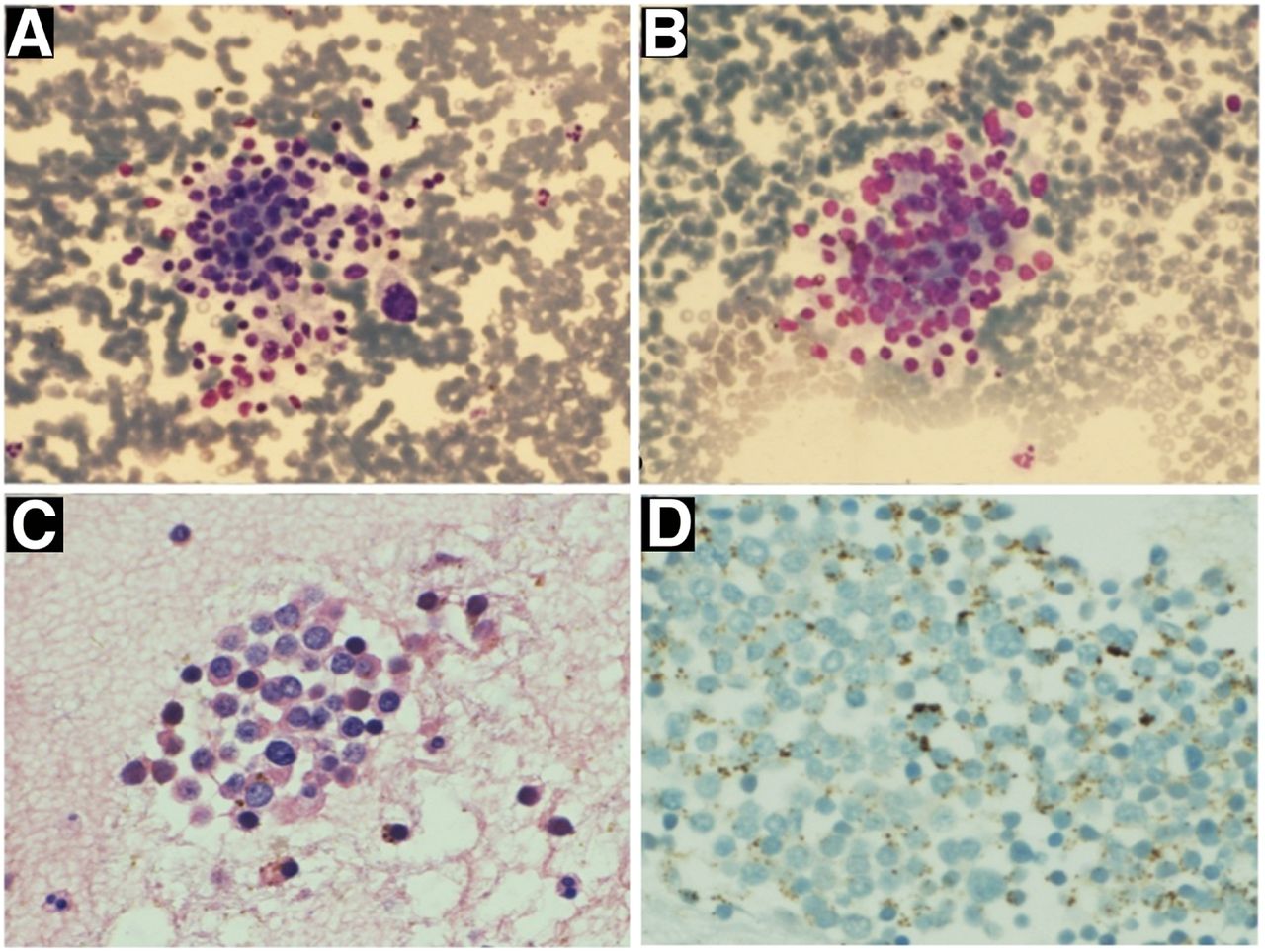
- FIGURE 3.
(A and B) Ultrasonography-guided fine-needle aspiration cytology from enlarged cervical lymph node revealing clusters of tumor cells with mildly pleomorphic nuclei, showing sudden anisonucleosis (May–Grünwald staining, ×20). (C) Additional cluster of tumor cells with stippled chromatin and moderate amount of cytoplasm (hematoxylin and eosin staining, ×20) suggestive of metastatic NET. (D) Immunocytochemistry for Ki-67 proliferation index was found to be less than 2%.
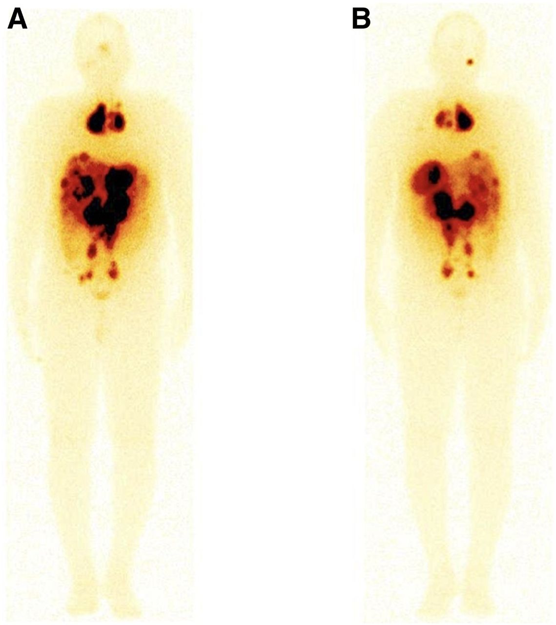
- FIGURE 4.
Whole-body anterior (A) and posterior (B) posttherapy images (177Lu-DOTATATE therapy; first cycle) showing tracer avidity in the lesions of metastatic neuroendocrine tumor.


Jump to section
Related Articles
Cited By...
- No citing articles found.