
Main menu

  • Home
  • Content
    • Current
      • JNMT Supplement
    • Ahead of print
    • Past Issues
    • Continuing Education
    • JNMT Podcast
    • SNMMI Annual Meeting Abstracts
  • Subscriptions
    • Subscribers
    • Rates
    • Journal Claims
    • Institutional and Non-member
  • Authors
    • Submit to JNMT
    • Information for Authors
    • Assignment of Copyright
    • AQARA Requirements
  • Info
    • Reviewers
    • Permissions
    • Advertisers
    • Corporate & Special Sales
  • About
    • About Us
    • Editorial Board
    • Contact Information
  • More
    • Alerts
    • Feedback
    • Help
    • SNMMI Journals
  • SNMMI
    • JNMT
    • JNM
    • SNMMI Journals
    • SNMMI

User menu

  • Subscribe
  • My alerts
  • Log in
  • Log out
  • My Cart

Search

  • Advanced search
Journal of Nuclear Medicine Technology
  • SNMMI
    • JNMT
    • JNM
    • SNMMI Journals
    • SNMMI
  • Subscribe
  • My alerts
  • Log in
  • Log out
  • My Cart
Journal of Nuclear Medicine Technology

Advanced Search

  • Home
  • Content
    • Current
    • Ahead of print
    • Past Issues
    • Continuing Education
    • JNMT Podcast
    • SNMMI Annual Meeting Abstracts
  • Subscriptions
    • Subscribers
    • Rates
    • Journal Claims
    • Institutional and Non-member
  • Authors
    • Submit to JNMT
    • Information for Authors
    • Assignment of Copyright
    • AQARA Requirements
  • Info
    • Reviewers
    • Permissions
    • Advertisers
    • Corporate & Special Sales
  • About
    • About Us
    • Editorial Board
    • Contact Information
  • More
    • Alerts
    • Feedback
    • Help
    • SNMMI Journals
  • Watch or Listen to JNMT Podcast
  • Visit SNMMI on Facebook
  • Join SNMMI on LinkedIn
  • Follow SNMMI on Twitter
  • Subscribe to JNMT RSS feeds
Review ArticleTeaching Case Studies

Sister Mary Joseph Nodule in Advanced Pancreatic Adenocarcinoma Identified on 18F-FDG PET/MRI

Ming Yang, Alix C. Hopp, Tanios S. Bekaii-Saab and Joseph M. Collins
Journal of Nuclear Medicine Technology December 2019, 47 (4) 341-342; DOI: https://doi.org/10.2967/jnmt.119.225961
Ming Yang
1Department of Radiology, Mayo Clinic, Scottsdale, Arizona; and
  • Find this author on Google Scholar
  • Find this author on PubMed
  • Search for this author on this site
Alix C. Hopp
1Department of Radiology, Mayo Clinic, Scottsdale, Arizona; and
  • Find this author on Google Scholar
  • Find this author on PubMed
  • Search for this author on this site
Tanios S. Bekaii-Saab
2Department of Hematology/Oncology, Mayo Clinic, Scottsdale, Arizona
  • Find this author on Google Scholar
  • Find this author on PubMed
  • Search for this author on this site
Joseph M. Collins
1Department of Radiology, Mayo Clinic, Scottsdale, Arizona; and
  • Find this author on Google Scholar
  • Find this author on PubMed
  • Search for this author on this site
  • Article
  • Figures & Data
  • Info & Metrics
  • PDF
Loading

Abstract

Sister Mary Joseph nodule is an umbilical metastatic lesion typically originating from gastrointestinal or gynecologic malignancies. In pancreatic cancer, Sister Mary Joseph nodule is a sign of advanced disease with an associated poor prognosis. 18F-FDG PET/MRI provides an imaging tool in the identification of Sister Mary Joseph nodule and helps to improve the staging of pancreatic cancer.

  • molecular imaging
  • oncology: pancreas
  • PET/MRI
  • pancreatic cancer
  • Sister Mary Joseph nodule

Sister Mary Joseph nodule refers to a metastatic lesion of the umbilicus, with the primary malignancy originating in the gastrointestinal or genitourinary system. Pancreatic cancer accounts for 7%–9% of the reported cases of Sister Mary Joseph nodule, with nearly 90% of tumors occurring at the pancreatic body and tail (1–2).

18F-FDG PET/CT has been used to detect Sister Mary Joseph nodule, given its excellent staging ability (3). Here, we describe a Sister Mary Joseph nodule in advanced pancreatic cancer discovered on 18F-FDG PET/MRI. Combining PET molecular imaging and diagnostic abdominal MRI information, PET/MRI is a powerful imaging tool in the diagnosis and staging of pancreatic cancer.

CASE REPORT

A 75-y-old man with biopsy-proven pancreatic adenocarcinoma had a rising serum level of the tumor biomarker carbohydrate antigen 19-9, at 41,154 U/mL, after 1 cycle of modified FOLFIRINOX chemotherapy (folinic acid, fluorouracil, irinotecan, and oxaliplatin). Diagnostic whole-body CT performed at an outside institution revealed a new liver metastasis in addition to the known primary pancreatic body tumor. Restaging hybrid 18F-FDG PET/MRI, including a torso PET/MRI survey scan and focused 1-bed-position contrast-enhanced diagnostic pancreatic PET/MRI, was performed to reassess the tumor burden. On both the torso PET/MRI survey and the focused diagnostic pancreatic PET/MR images, a 2.3-cm hypermetabolic lesion with an MRI signal pattern of a solid, enhancing mass was identified at the retroumbilical region (Figs. 1 and 2). In retrospect, this mass lesion was present on the abdominal CT image but was diagnosed as umbilical fungal infection. The patient had been on 6 wk of antifungal therapy for suspected periumbilical fungal infection, which had improved since the start of chemotherapy. An ultrasound-guided core-needle biopsy was performed and confirmed metastatic pancreatic adenocarcinoma of the umbilicus, also known as a Sister Mary Joseph nodule. On the basis of this proof of disseminated pancreatic cancer, FOLFIRINOX therapy was resumed.

FIGURE 1.
  • Download figure
  • Open in new tab
  • Download powerpoint
FIGURE 1.

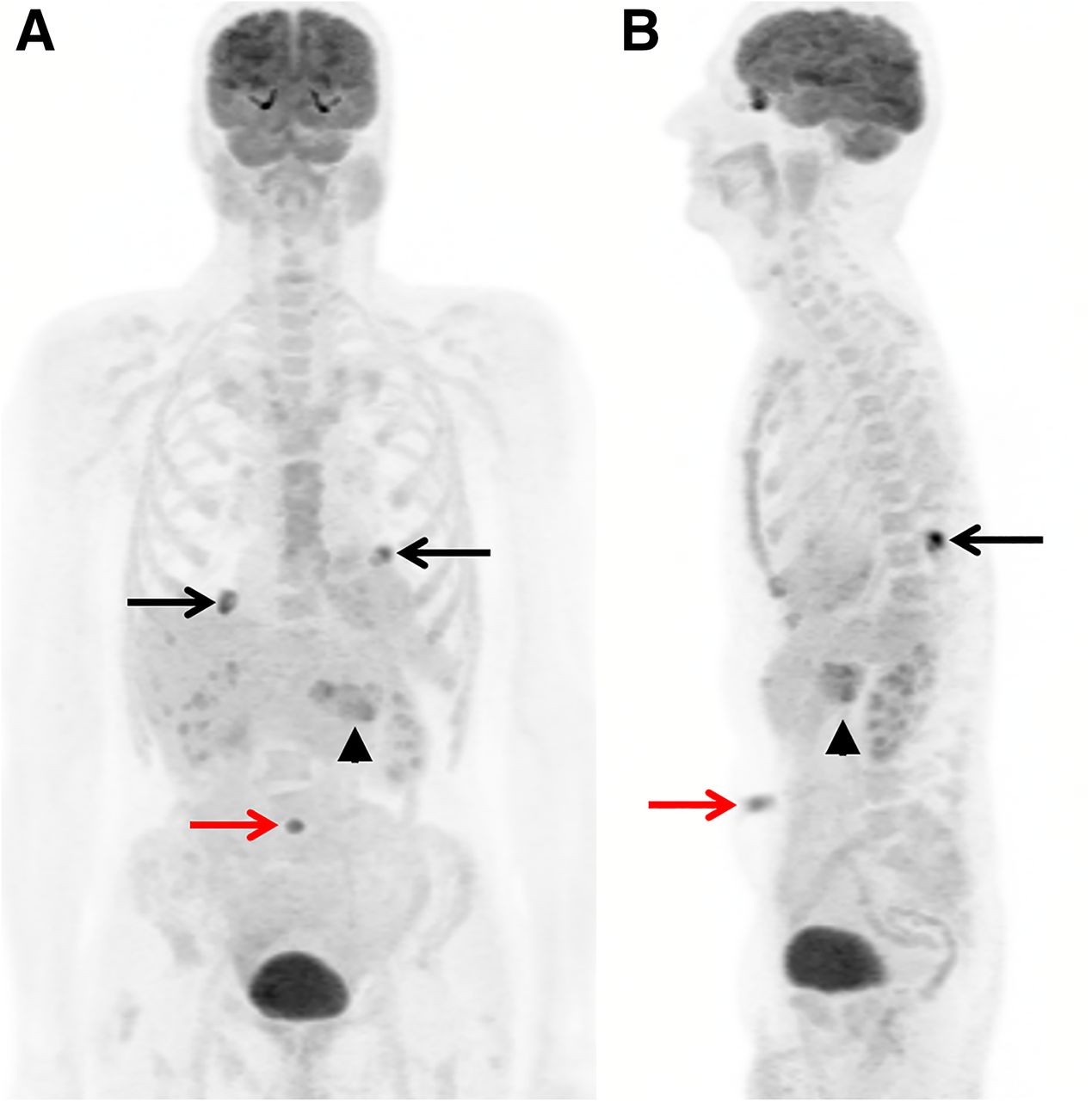
On torso PET/MRI survey, maximum-intensity-projection anterior (A) and lateral (B) views show tracer-avid lesion at umbilical region (red arrows) with SUVmax of 7.1 and primary pancreatic tumor (arrowheads) with SUVmax of 6.7. Two additional benign inflammatory lesions were noted bilaterally in lung bases (black arrows). Liver lesion with low 18F-FDG uptake was not discernable on maximum-intensity projections.

FIGURE 2.
  • Download figure
  • Open in new tab
  • Download powerpoint
FIGURE 2.

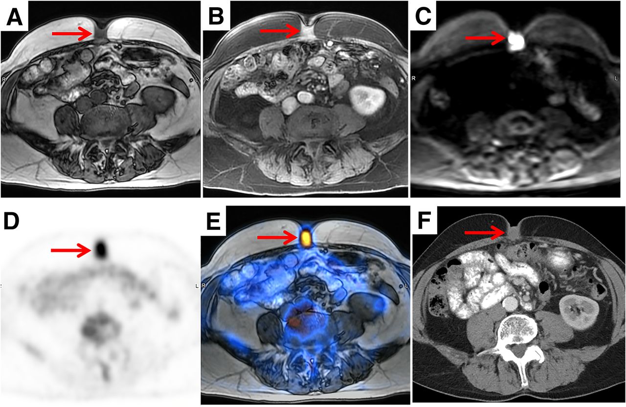
(A–C) Focused abdominal PET/MRI demonstrates 2.3-cm lesion (arrows) with low T1 signal (A), intense enhancement on postcontrast image (B), and restricted diffusion on diffusion-weighted image (C). (D and E) Lesion exhibited intense tracer uptake on PET (D) and PET/MRI (E) images. (F) This lesion was initially diagnosed clinically as umbilical fungal infection and was overlooked on contrast-enhanced abdominal CT study obtained at another institution.

DISCUSSION

The Sister Mary Joseph nodule represents an umbilical metastatic lesion that typically originates from gastrointestinal and gynecologic malignancies. The most common primary sites are stomach (21%), ovary (18%), colon (12%), and pancreas (9%). Other sites of rarer primaries include uterus, biliary tree, breast, and lung (1–2). The condition was named for Sister Mary Joseph, the surgical assistant to Dr. William Mayo of Mayo Clinic. She first noticed the association between an umbilical nodule and advanced intraabdominal malignancies while preparing the patient’s skin before surgery.

The presence of the Sister Mary Joseph nodule is a sign of widespread abdominal or pelvic malignancy and usually indicates a poor prognosis (2). The tumor markers, including carbohydrate antigen 19-9, carcinoembryonic antigen, and carbohydrate antigen 125, should be examined. The mechanism of tumor spread to the umbilicus has been postulated as direct invasion from the peritoneum through hematogenous or lymphatic pathways or along the embryonic remnants of the ligamentum teres or umbilical ligaments (3–4). In addition to metastasis, the differential diagnosis of a neoplastic umbilical nodule includes primary carcinoma, endometriosis, hernia, and granuloma, among others (5).

To our knowledge, this is the first report of identification of a Sister Mary Joseph nodule using PET/MRI. 18F-FDG PET/MRI is superior to PET/CT in providing excellent soft-tissue delineation of the nodule on the conjunctive abdominal MRI.

CONCLUSION

18F-FDG PET/MRI is a novel imaging tool in the diagnosis and staging of pancreatic adenocarcinoma. It provides an easier target for biopsy to establish the diagnosis of widespread pancreatic cancer with umbilical metastasis.

DISCLOSURE

No potential conflict of interest relevant to this article was reported.

Footnotes

  • Published online Apr. 24, 2019.

REFERENCES

  1. 1.↵
    1. Yendluri V,
    2. Centeno B,
    3. Springett GM
    . Pancreatic cancer presenting as a Sister Mary Joseph’s nodule: case report and update of the literature. Pancreas. 2007;34:161–164.
    OpenUrlCrossRefPubMed
  2. 2.↵
    1. Chiang CH,
    2. Lin MH
    . Sister Mary Joseph nodule associated with pancreatic adenocarcinoma. J Formos Med Assoc. 2015;114:92–93.
    OpenUrl
  3. 3.↵
    1. Métivier D Jr.,
    2. Bonardel G,
    3. Rouquie D,
    4. et al
    . 18F-FDG PET/CT imaging of Sister Mary Joseph’s nodule. Clin Nucl Med. 2012;37:486–488.
    OpenUrlPubMed
  4. 4.↵
    1. Inanir S,
    2. Oksuzoglu K
    . FDG PET/CT imaging of calcified Sister Mary Joseph nodule. Clin Nucl Med. 2016;41:e458–e459.
    OpenUrl
  5. 5.↵
    1. Coll DM,
    2. Meyer JM,
    3. Mader M,
    4. et al
    . Imaging appearances of Sister Mary Joseph nodule. Br J Radiol. 1999;72:1230–1233.
    OpenUrlAbstract
PreviousNext
Back to top

In this issue

Journal of Nuclear Medicine Technology: 47 (4)
Journal of Nuclear Medicine Technology
Vol. 47, Issue 4
December 1, 2019
  • Table of Contents
  • About the Cover
  • Index by author
Print
Download PDF
Article Alerts
Sign In to Email Alerts with your Email Address
Email Article

Thank you for your interest in spreading the word on Journal of Nuclear Medicine Technology.

NOTE: We only request your email address so that the person you are recommending the page to knows that you wanted them to see it, and that it is not junk mail. We do not capture any email address.

Enter multiple addresses on separate lines or separate them with commas.
Sister Mary Joseph Nodule in Advanced Pancreatic Adenocarcinoma Identified on 18F-FDG PET/MRI
(Your Name) has sent you a message from Journal of Nuclear Medicine Technology
(Your Name) thought you would like to see the Journal of Nuclear Medicine Technology web site.
Citation Tools
Sister Mary Joseph Nodule in Advanced Pancreatic Adenocarcinoma Identified on 18F-FDG PET/MRI
Ming Yang, Alix C. Hopp, Tanios S. Bekaii-Saab, Joseph M. Collins
Journal of Nuclear Medicine Technology Dec 2019, 47 (4) 341-342; DOI: 10.2967/jnmt.119.225961

Citation Manager Formats

  • BibTeX
  • Bookends
  • EasyBib
  • EndNote (tagged)
  • EndNote 8 (xml)
  • Medlars
  • Mendeley
  • Papers
  • RefWorks Tagged
  • Ref Manager
  • RIS
  • Zotero
Share
Sister Mary Joseph Nodule in Advanced Pancreatic Adenocarcinoma Identified on 18F-FDG PET/MRI
Ming Yang, Alix C. Hopp, Tanios S. Bekaii-Saab, Joseph M. Collins
Journal of Nuclear Medicine Technology Dec 2019, 47 (4) 341-342; DOI: 10.2967/jnmt.119.225961
Twitter logo Facebook logo LinkedIn logo Mendeley logo
  • Tweet Widget
  • Facebook Like
  • Google Plus One
Bookmark this article

Jump to section

  • Article
    • Abstract
    • CASE REPORT
    • DISCUSSION
    • CONCLUSION
    • DISCLOSURE
    • Footnotes
    • REFERENCES
  • Figures & Data
  • Info & Metrics
  • PDF

Related Articles

  • No related articles found.
  • PubMed
  • Google Scholar

Cited By...

  • No citing articles found.
  • Google Scholar

More in this TOC Section

  • High-Sensitivity Troponin Elevation in a Young Woman with Typical Chest Pain: The Heart of the Matter
  • Pulmonary Adenocarcinoma Revealed by Parathyroid Scintigraphy: An Incidental Case to Remember
  • Prominent Right Ventricular Tracer Uptake: A Harbinger of Multivessel Coronary Artery Disease
Show more Teaching Case Studies

Similar Articles

Keywords

  • molecular imaging
  • Oncology: Pancreas
  • PET/MRI
  • Pancreatic cancer
  • Sister Mary Joseph nodule
SNMMI

© 2025 SNMMI

Powered by HighWire