


Article Figures & Data
Figures
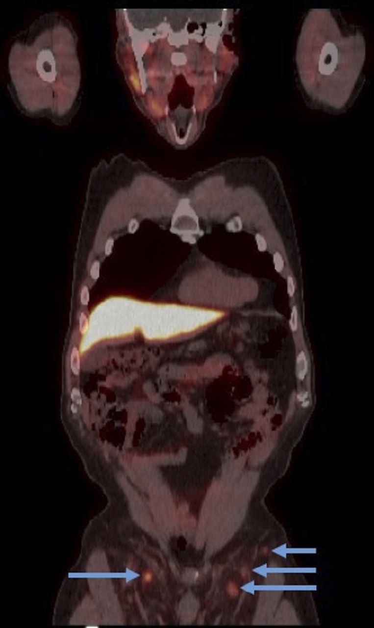
- FIGURE 1.
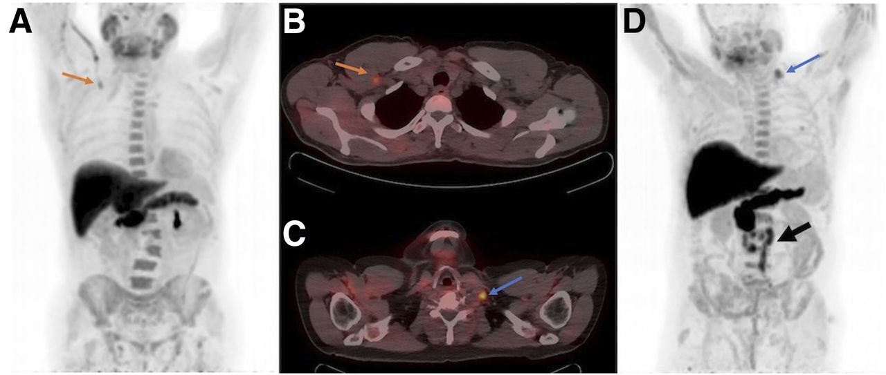
(A) 18F-fluciclovine intravenous injection via right antecubital vein demonstrates increased uptake in right axillary vein on maximum-intensity projection (arrow). (B) Focal uptake in subclavian space may mimic or mask metastatic lymph node uptake on PET/CT transaxial image (arrow). (C and D) In patient injected via right antecubital vein, focus of increased 18F-fluciclovine uptake in left supraclavicular space correlates with enlarged suggestive lymph node on PET/CT transaxial image (C) and maximum-intensity projection (D) (blue arrows). Additional diffuse retroperitoneal metastatic lymph nodes are noted (black arrow).
- FIGURE 2.
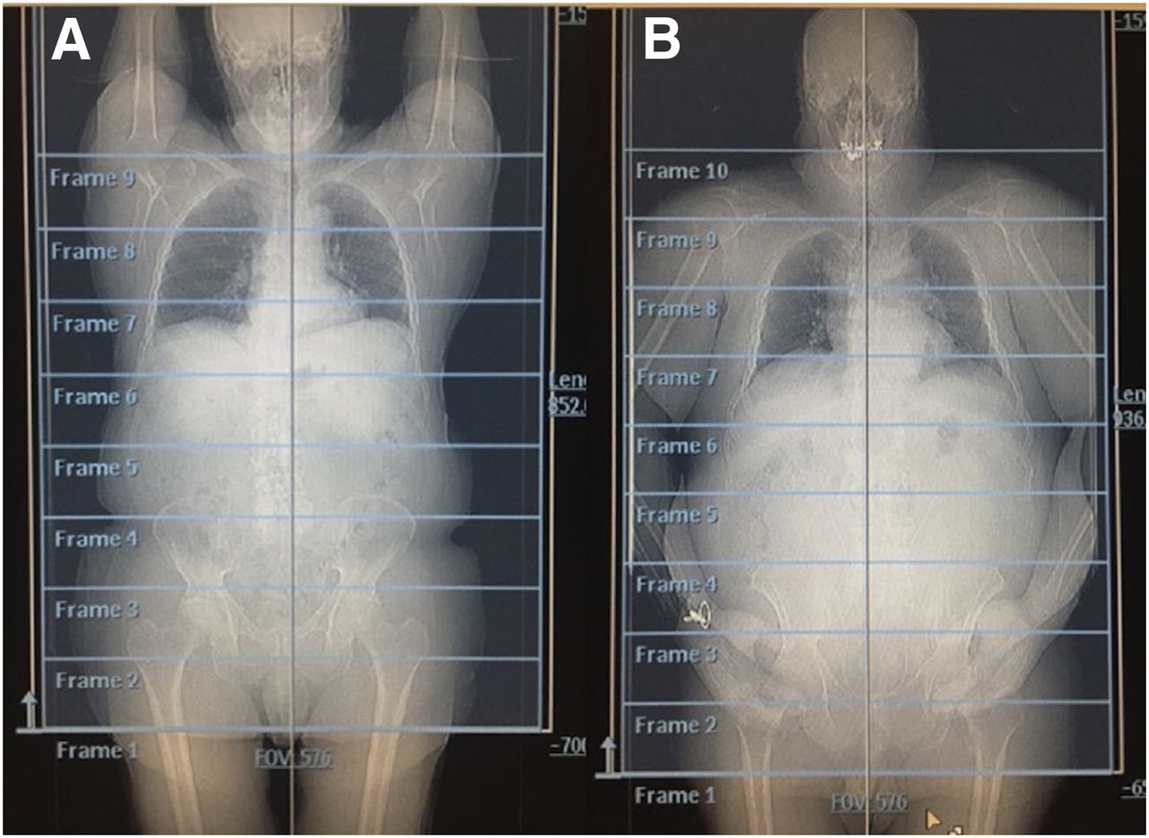
CT scout images demonstrating recommended positioning of patient with arms up (A) and alternative, less preferred, position with arms down (B).
- FIGURE 3.
Respiration artifact from rapid breathing pattern on 18F-fluciclovine PET maximum-intensity projection appears as artificially decreased tracer uptake (arrow) by liver dome.
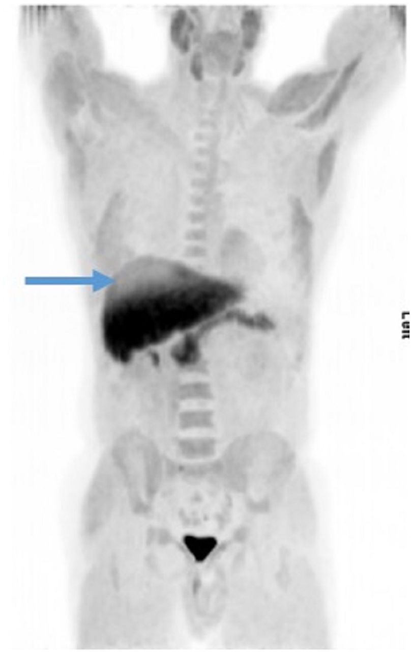
- FIGURE 4.
Normal biodistribution of 18F-fluciclovine on PET maximum-intensity projection shows highest uptake within liver and pancreas.
- FIGURE 5.
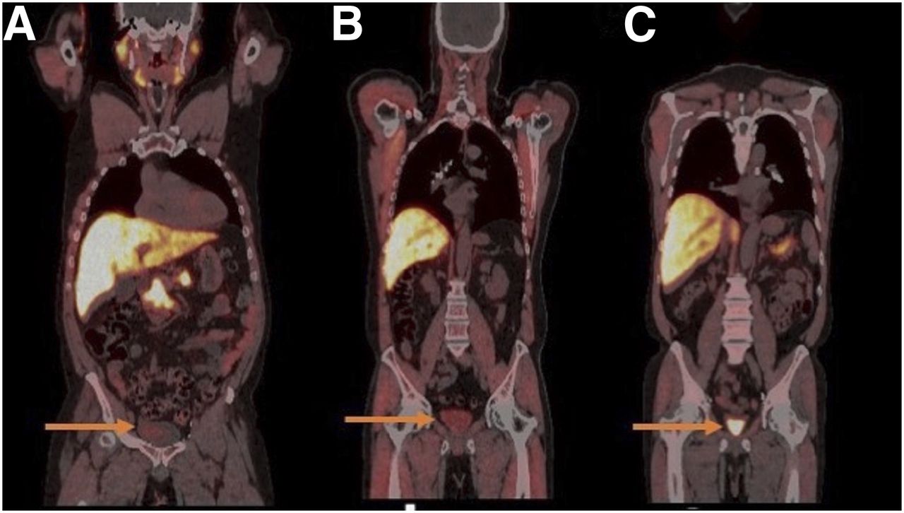
18F-fluciclovine PET/CT image demonstrating moderate uptake bilaterally in reactive inguinal lymph nodes (arrows).
- FIGURE 6.
18F-fluciclovine PET/CT coronal images demonstrating mild (SUVmean > blood pool) (A) and moderate (SUVmean > marrow < liver) (B) urine radioactivity in patients who did not void before injection of 18F-fluciclovine, compared with intense urine radioactivity (SUVmean > liver) (C) in patient who voided.


Tables
- TABLE 1
Differences Between Imaging Protocols for 18F-Fluciclovine PET/CT and 18F-FDG PET/CT
Parameter 18F-fluciclovine PET/CT 18F-FDG PET/CT Patient preparation Ask patients to fast for at least 4 h, including water restriction Ask patients to fast for at least 4 h, with no water restriction Ask patients not to void for 1 h before 18F-fluciclovine injection and imaging Ask patients to void immediately before imaging starts Injection site Right arm Right or left arm (if applicable, arm contralateral to cancer side) Image acquisition Start PET imaging 4 min after injection Start PET imaging 30–90 min after injection Image caudocranially, from mid thighs to skull base Image craniocaudally (field of image is cancer-specific)


Jump to section
- Article
- Abstract
- PATIENT SCHEDULING
- PATIENT PREPARATION
- RADIOPHARMACEUTICAL INJECTION
- PATIENT POSITIONING
- CT ACQUISITION
- PET ACQUISITION
- QUALITY CONTROL
- NORMAL BIODISTRIBUTION OF 18F-FLUCICLOVINE
- IMAGE INTERPRETATION
- 18F-FLUCICLOVINE PITFALLS
- CONCLUSION
- DISCLOSURE
- Acknowledgments
- APPENDIX A: BEST PRACTICES FOR 18F-FLUCICLOVINE PET/CT
- Footnotes
- REFERENCES
- Figures & Data
- Info & Metrics