
Main menu

  • Home
  • Content
    • Current
      • JNMT Supplement
    • Ahead of print
    • Past Issues
    • Continuing Education
    • JNMT Podcast
    • SNMMI Annual Meeting Abstracts
  • Subscriptions
    • Subscribers
    • Rates
    • Journal Claims
    • Institutional and Non-member
  • Authors
    • Submit to JNMT
    • Information for Authors
    • Assignment of Copyright
    • AQARA Requirements
  • Info
    • Reviewers
    • Permissions
    • Advertisers
    • Corporate & Special Sales
  • About
    • About Us
    • Editorial Board
    • Contact Information
  • More
    • Alerts
    • Feedback
    • Help
    • SNMMI Journals
  • SNMMI
    • JNMT
    • JNM
    • SNMMI Journals
    • SNMMI

User menu

  • Subscribe
  • My alerts
  • Log in
  • My Cart

Search

  • Advanced search
Journal of Nuclear Medicine Technology
  • SNMMI
    • JNMT
    • JNM
    • SNMMI Journals
    • SNMMI
  • Subscribe
  • My alerts
  • Log in
  • My Cart
Journal of Nuclear Medicine Technology

Advanced Search

  • Home
  • Content
    • Current
    • Ahead of print
    • Past Issues
    • Continuing Education
    • JNMT Podcast
    • SNMMI Annual Meeting Abstracts
  • Subscriptions
    • Subscribers
    • Rates
    • Journal Claims
    • Institutional and Non-member
  • Authors
    • Submit to JNMT
    • Information for Authors
    • Assignment of Copyright
    • AQARA Requirements
  • Info
    • Reviewers
    • Permissions
    • Advertisers
    • Corporate & Special Sales
  • About
    • About Us
    • Editorial Board
    • Contact Information
  • More
    • Alerts
    • Feedback
    • Help
    • SNMMI Journals
  • Watch or Listen to JNMT Podcast
  • Visit SNMMI on Facebook
  • Join SNMMI on LinkedIn
  • Follow SNMMI on Twitter
  • Subscribe to JNMT RSS feeds
Review ArticleContinuing Education

Intelligent Imaging: Anatomy of Machine Learning and Deep Learning

Geoff Currie
Journal of Nuclear Medicine Technology December 2019, 47 (4) 273-281; DOI: https://doi.org/10.2967/jnmt.119.232470
Geoff Currie
School of Dentistry and Health Sciences, Charles Sturt University, Wagga Wagga, Australia
  • Find this author on Google Scholar
  • Find this author on PubMed
  • Search for this author on this site
  • Article
  • Figures & Data
  • Supplemental
  • Info & Metrics
  • PDF
Loading

Article Figures & Data

Figures

  • Additional Files
  • FIGURE 1.
    • Download figure
    • Open in new tab
    • Download powerpoint
    FIGURE 1.

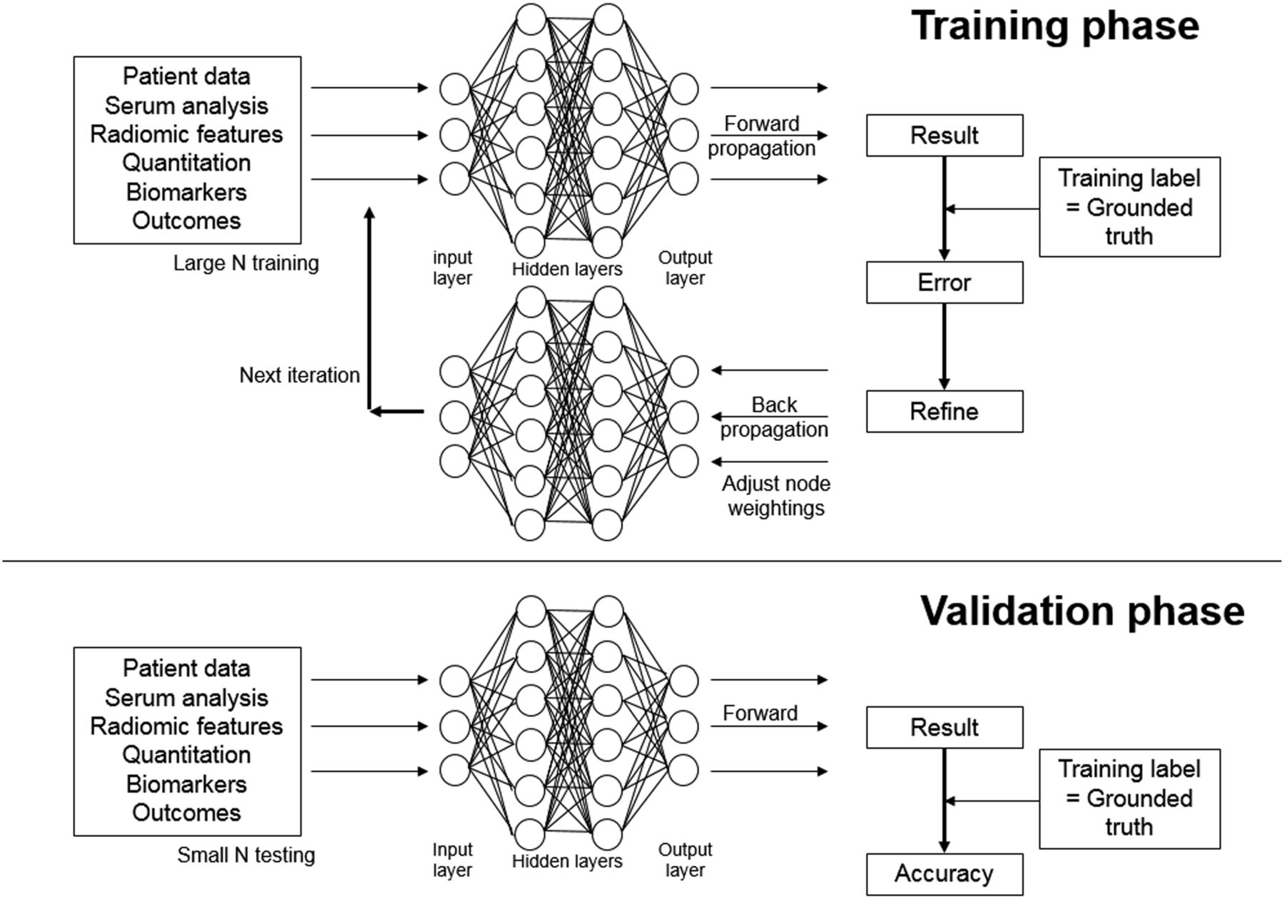
    The training phase of a neural network using extracted features as inputs. The grounded truth defines the ANN as a supervised ANN. This ANN structure is also what might be used as an analysis tool in parallel with traditional statistical analysis—importing data from a spreadsheet, for example. This example, which depicts all nodes as being connected to all others in adjacent layers, represents “fully connected layer,” which is more typical of CNNs. The validation phase evaluates trained ANN against a new database of known cases to determine accuracy. (Adapted from (1).)

  • FIGURE 2.
    • Download figure
    • Open in new tab
    • Download powerpoint
    FIGURE 2.

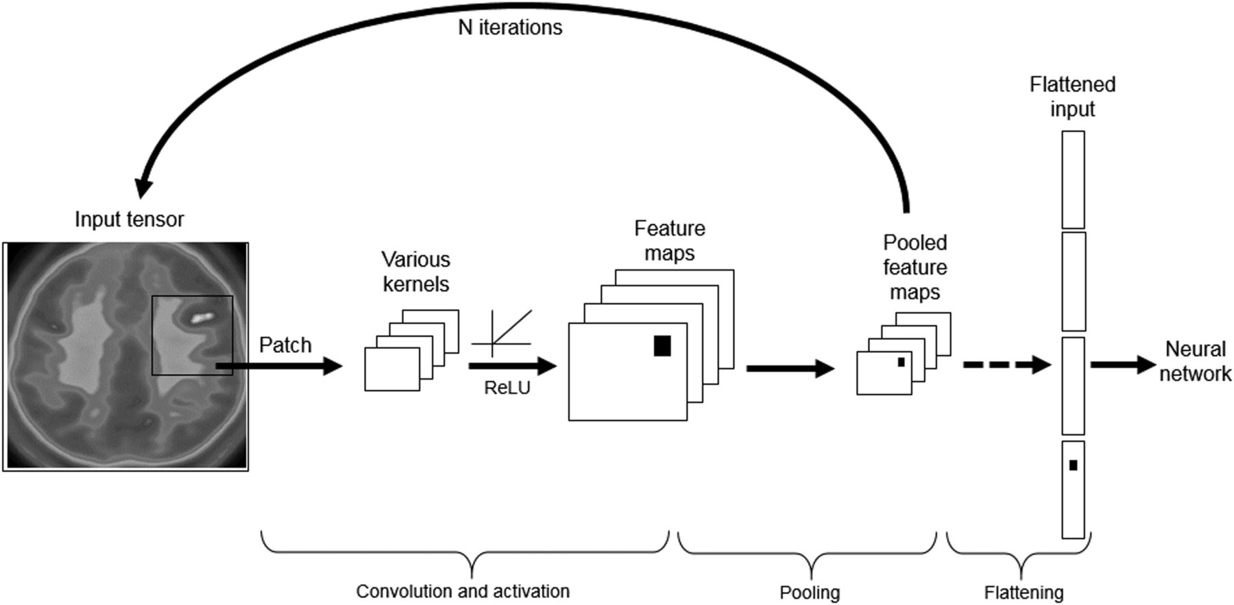
    Basic structure of a CNN, with the network extracting radiomic features, producing convolution function, pooling data through rectified linear unit (ReLU) kernel, and flattening pooled feature map for input into fully connected hidden layers of the neural network. (Reprinted from (1).)

  • FIGURE 3.
    • Download figure
    • Open in new tab
    • Download powerpoint
    FIGURE 3.

    Anatomy of an ANN. Single node (C) can have multiple inputs (X) with different weighting factors (W) and bias (B) but single output (Y) via activation function (A). Multiple lines exiting each node are same output being delivered to multiple next-layer nodes.

  • FIGURE 4.
    • Download figure
    • Open in new tab
    • Download powerpoint
    FIGURE 4.

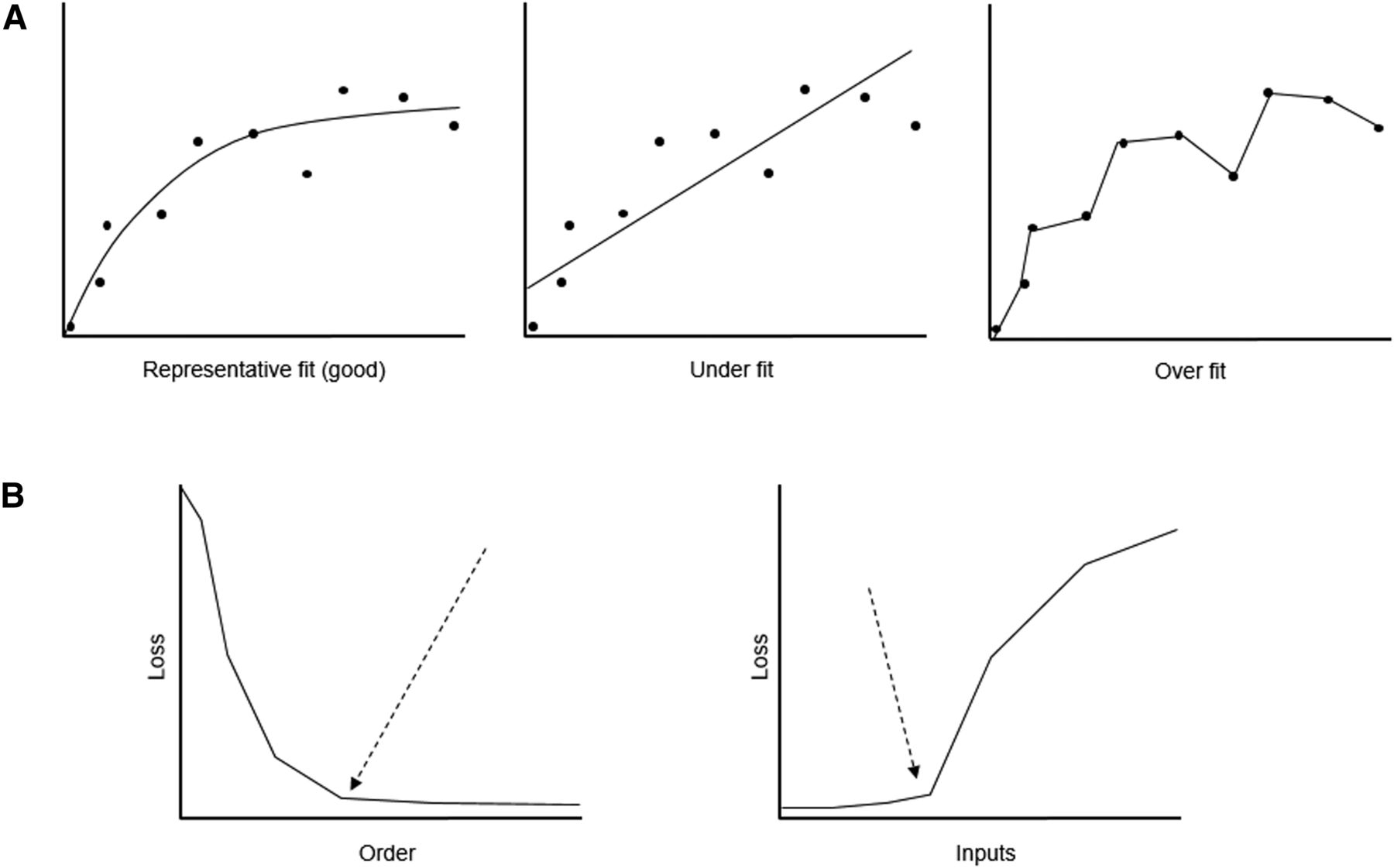
    (A) Schematic representation of good fit vs. under- and overfitting associated with selection loss. (B, left) Optimization of selection loss to determine ANN complexity and node number (order) using decremental order algorithm, with arrow indicating reasonable cutoff for total node number. (B, right) Optimization of selection loss to determine inputs (features) to be included using growing-input algorithm, with arrow indicating reasonable cutoff for inputs.
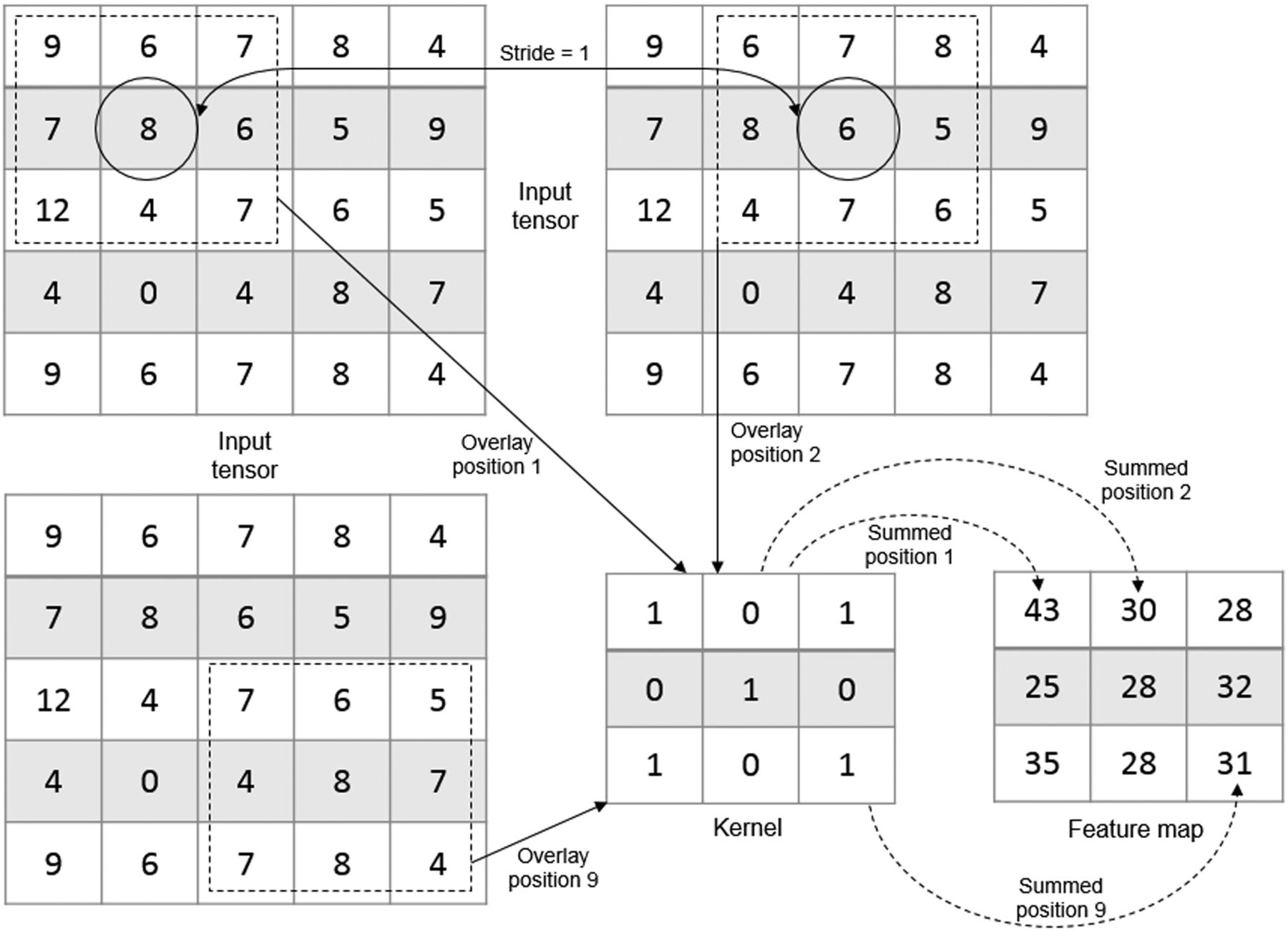
  • FIGURE 5.
    • Download figure
    • Open in new tab
    • Download powerpoint
    FIGURE 5.

    Convolution uses 3 × 3 kernel to run sequential (in this case, successive, to provide stride of 1) 3 × 3 array of elements. Weighted sum of kernel for 3 × 3 input tensor creates single representative value in feature map. Multiple feature maps are produced by different kernels.

  • FIGURE 6.
    • Download figure
    • Open in new tab
    • Download powerpoint
    FIGURE 6.

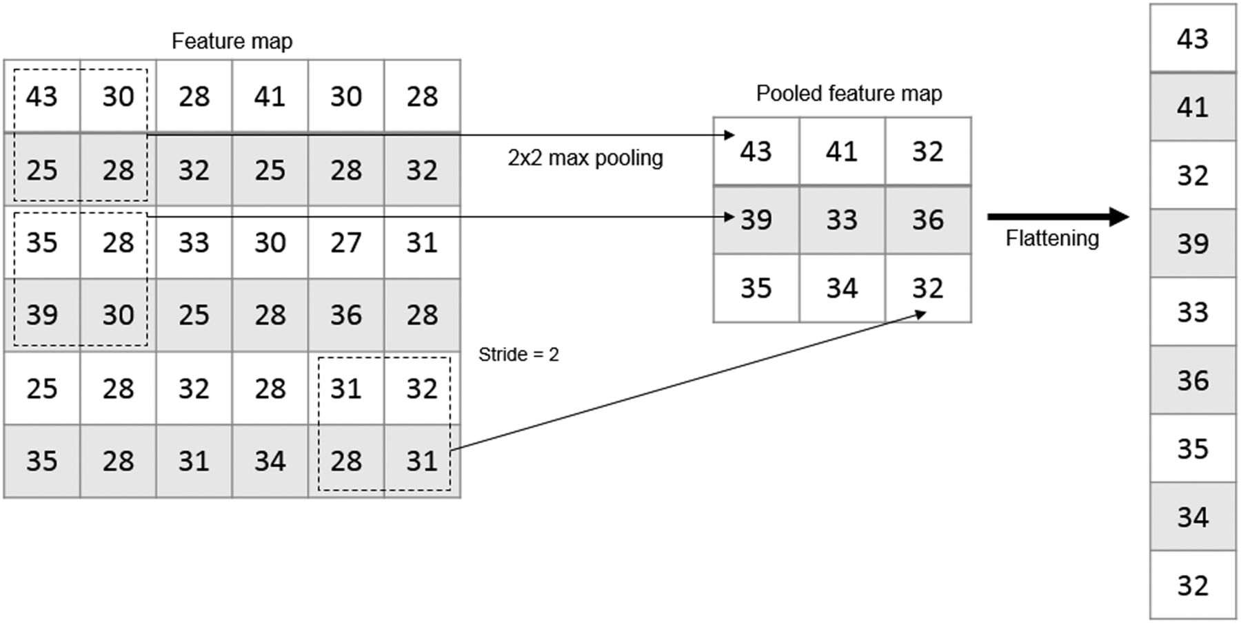
    Pooling using max pooling method and 2 × 2 array produces pooling of maximum count among 4 connected elements (patch) to represent those data in pooled feature map. Use of consecutive blocks of 2 × 2 elements means stride of 2. Final pooled feature map immediately before input into neural network can then be flattened from 2-dimensional data into single dimension; this approach avoids need for global pooling.
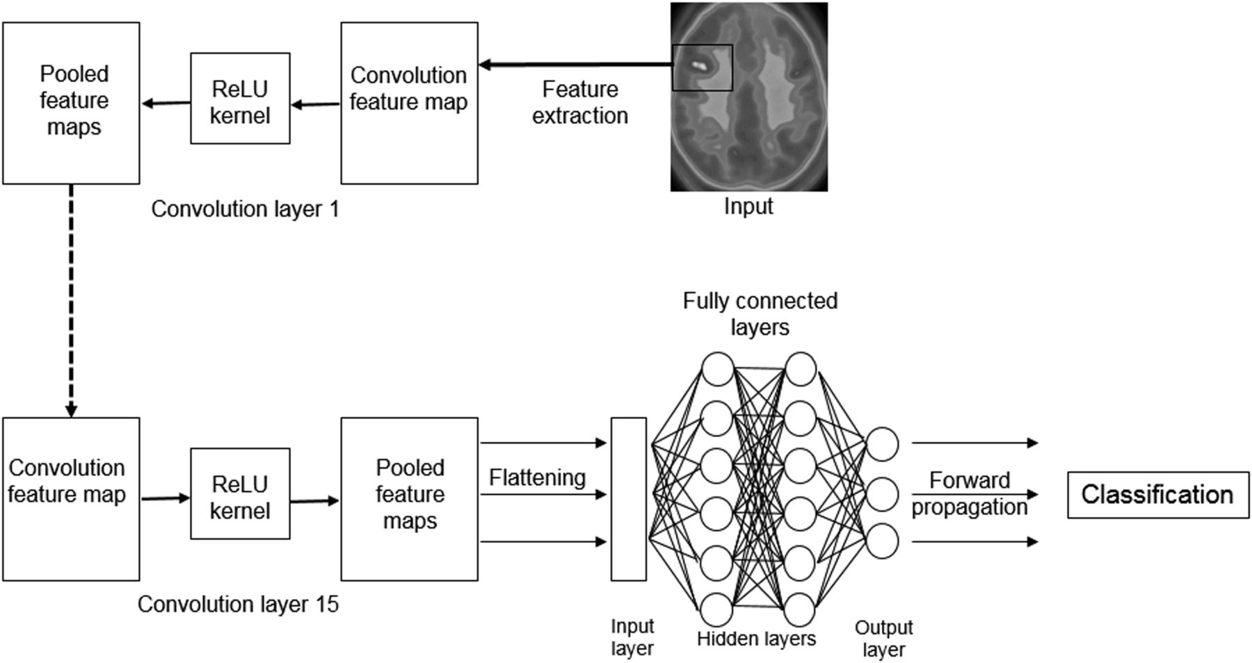
  • FIGURE 7.
    • Download figure
    • Open in new tab
    • Download powerpoint
    FIGURE 7.

    CNN will have several convolution and pooling layers before flattening and input to neural network. Several kernels can be used on same input tensor to produce layers of feature maps via rectified linear units (ReLU) for pooling and eventually flattening.

Additional Files

  • Figures
  • Supplemental Data

    Files in this Data Supplement:

    • Supplemental Data
PreviousNext
Back to top

In this issue

Journal of Nuclear Medicine Technology: 47 (4)
Journal of Nuclear Medicine Technology
Vol. 47, Issue 4
December 1, 2019
  • Table of Contents
  • About the Cover
  • Index by author
Print
Download PDF
Article Alerts
Sign In to Email Alerts with your Email Address
Email Article

Thank you for your interest in spreading the word on Journal of Nuclear Medicine Technology.

NOTE: We only request your email address so that the person you are recommending the page to knows that you wanted them to see it, and that it is not junk mail. We do not capture any email address.

Enter multiple addresses on separate lines or separate them with commas.
Intelligent Imaging: Anatomy of Machine Learning and Deep Learning
(Your Name) has sent you a message from Journal of Nuclear Medicine Technology
(Your Name) thought you would like to see the Journal of Nuclear Medicine Technology web site.
Citation Tools
Intelligent Imaging: Anatomy of Machine Learning and Deep Learning
Geoff Currie
Journal of Nuclear Medicine Technology Dec 2019, 47 (4) 273-281; DOI: 10.2967/jnmt.119.232470

Citation Manager Formats

  • BibTeX
  • Bookends
  • EasyBib
  • EndNote (tagged)
  • EndNote 8 (xml)
  • Medlars
  • Mendeley
  • Papers
  • RefWorks Tagged
  • Ref Manager
  • RIS
  • Zotero
Share
Intelligent Imaging: Anatomy of Machine Learning and Deep Learning
Geoff Currie
Journal of Nuclear Medicine Technology Dec 2019, 47 (4) 273-281; DOI: 10.2967/jnmt.119.232470
Twitter logo Facebook logo LinkedIn logo Mendeley logo
  • Tweet Widget
  • Facebook Like
  • Google Plus One
Bookmark this article

Jump to section

  • Article
    • Abstract
    • ANATOMY OF ML
    • ANATOMY OF A CNN
    • DISCUSSION
    • CONCLUSION
    • DISCLOSURE
    • Footnotes
    • REFERENCES
  • Figures & Data
  • Supplemental
  • Info & Metrics
  • PDF

Related Articles

  • PubMed
  • Google Scholar

Cited By...

  • Validation of Convolutional Neural Networks for Fast Determination of Whole-Body Metabolic Tumor Burden in Pediatric Lymphoma
  • Remodeling 99mTc-Pertechnetate Thyroid Uptake: Statistical, Machine Learning, and Deep Learning Approaches
  • Intelligent Imaging: Developing a Machine Learning Project
  • Topical Sensor for the Assessment of PET Dose Administration: Metric Performance with an Autoinjector
  • 2019: A Year of Reflection
  • 2019: A Year of Reflection
  • Google Scholar

More in this TOC Section

  • Illuminating the Hidden: Standardizing Cardiac MIBG Imaging for Sympathetic Dysfunction
  • PET/CT Case Series: Unmasking the Mystery of Cardiac Sarcoidosis
  • Delivery Methods of Radiopharmaceuticals: Exploring Global Strategies to Minimize Occupational Radiation Exposure
Show more Continuing Education

Similar Articles

Keywords

  • nuclear medicine
  • artificial neural network
  • deep learning
  • CONVOLUTIONAL NEURAL NETWORK
  • artificial intelligence
SNMMI

© 2025 SNMMI

Powered by HighWire