


Article Figures & Data
Figures
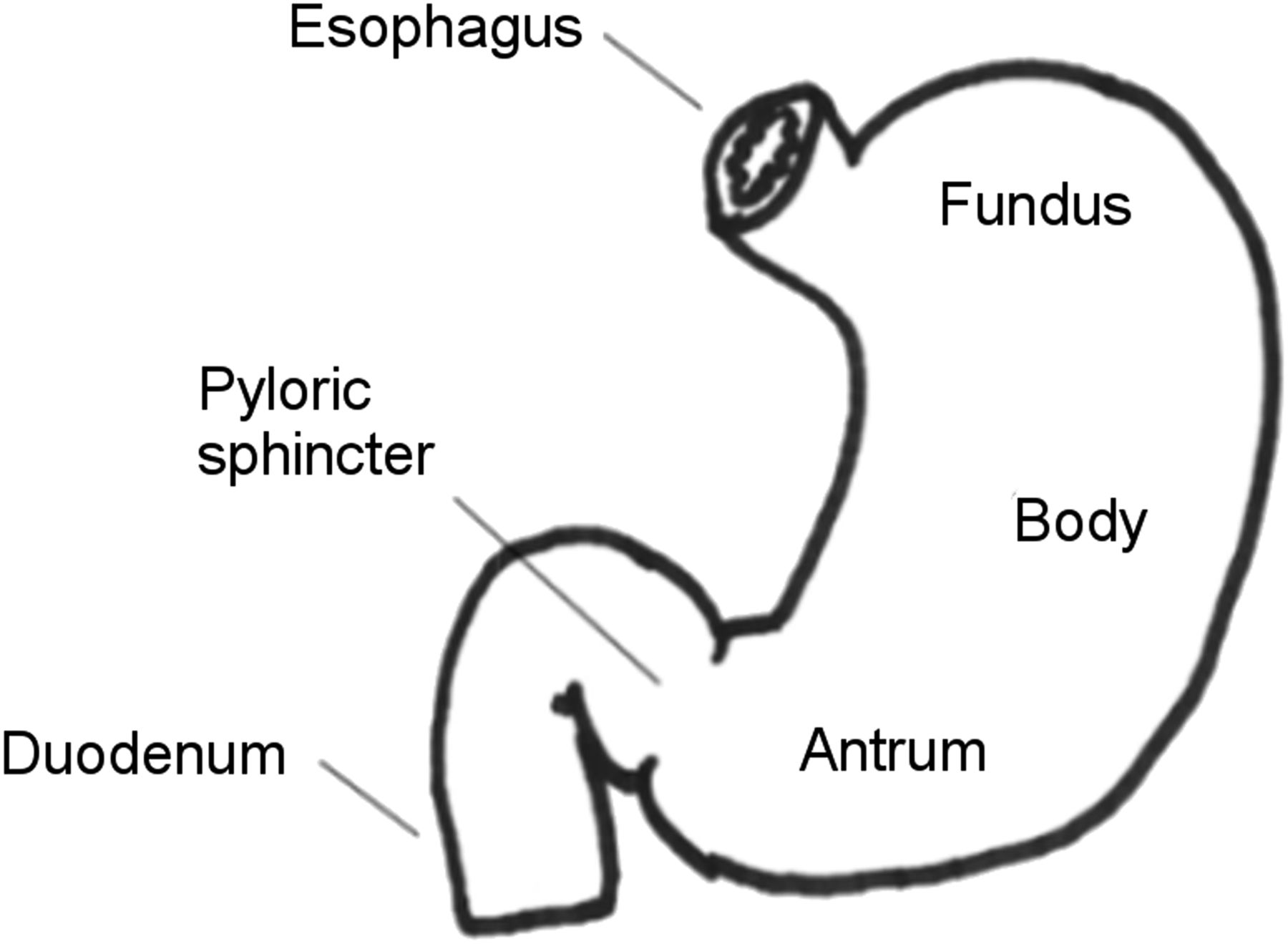
- FIGURE 1.
Stomach regions. The proximal fundus functions as a reservoir (accommodation) for food while the distal antrum grinds and mixes food.
- FIGURE 2.
Normal gastric emptying study demonstrating correct regions of interest in both the anterior and posterior projections on initial, 1-hour, 2-hour, and 4-hour images. This image was originally published in JNMT. Vijayakumar V. Assessment of the Practical Role of a Radionuclide Low-Fat-Meal Solid Gastric Emptying Study. J Nucl Med Technol. 2006; 34:82–85. © SNMMI.
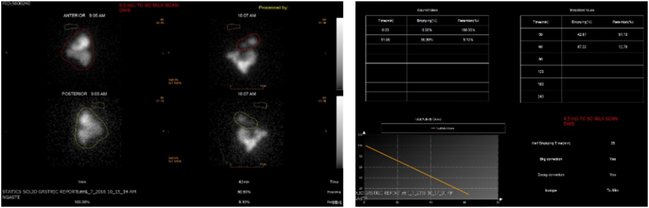
- FIGURE 3.
Normal liquid gastric emptying study using 0.5 99mTc-SC added to milk. Anterior and posterior images (right) at 0 and 60 minutes. Half-time emptying curve (left). Images courtesy of Leonie L. Gordon, MD, FACNM Medical University of South Carolina, Charleston, SC.
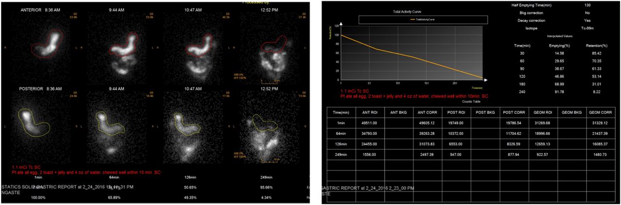
- FIGURE 4.
Normal solid gastric emptying study. (Top) Anterior and posterior images at 0 and approximately 1, 2 and 4 hours. (Bottom) Region counts from the anterior and posterior images and geometric mean. The percent retention at 4 hours is 8.2%. Images courtesy of Leonie L. Gordon, MD, FACNM Medical University of South Carolina, Charleston, SC.
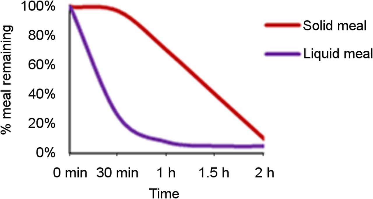
- FIGURE 5.
Normal gastric emptying curves. For solid meal (red), there is an initial 20-30 m lag period as the antrum reduces meal particle size and mixes with gastric acid. After the lag period, the solid material empties from the stomach in a linear fashion. The liquid meal (purple) immediately begins to leave the stomach and empties in an exponential pattern.
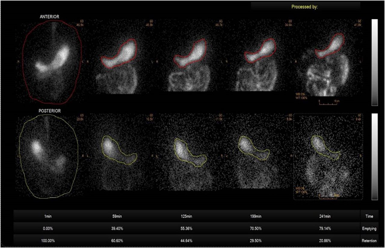
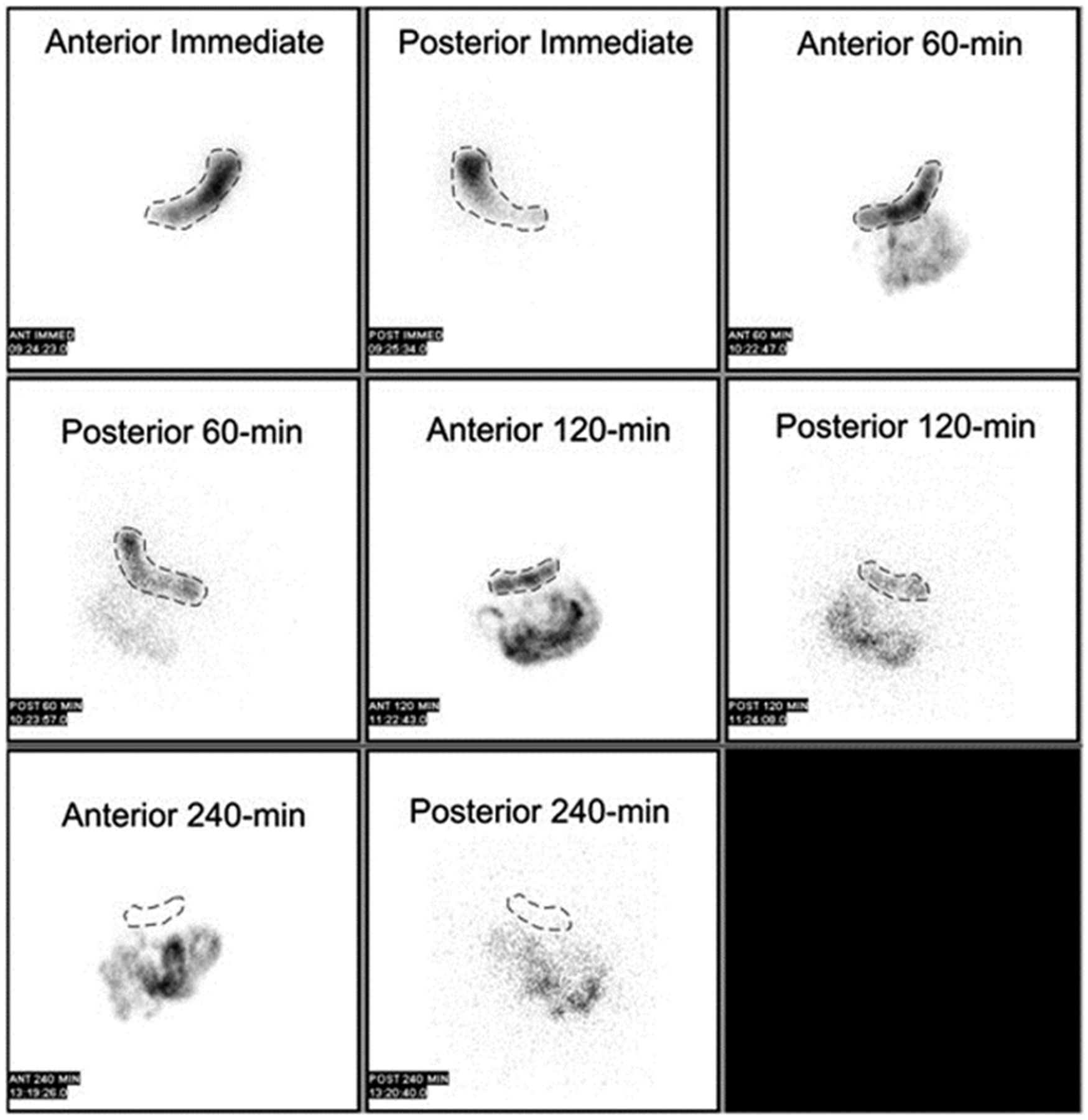
- FIGURE 6.
Abnormal solid gastric emptying study delayed emptying with 20.9% retention at 4 hours. At top, anterior images; at bottom, posterior images. Images courtesy of Jon A. Baldwin, MD, University of Alabama at Birmingham, Birmingham, AL.
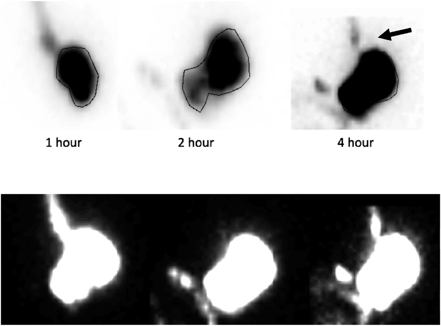
- FIGURE 7.
Abnormal gastric emptying study demonstrating delayed emptying and esophageal reflux (arrow). At top: normal intensity display. At bottom: inverted images with increased intensity. Images courtesy of Lorraine M. Fig, MD, FACNM VA Ann Arbor Healthcare System, Ann Arbor, MI.
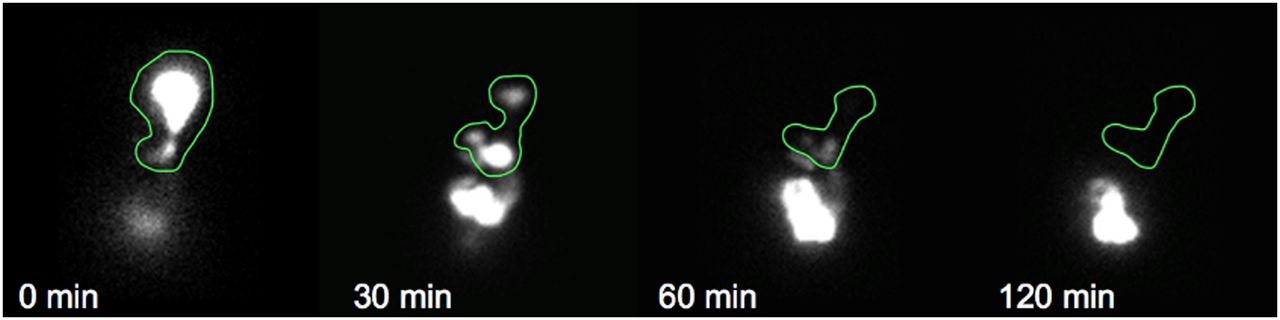
- FIGURE 8.
Abnormal gastric emptying study demonstrating rapid gastric emptying or dumping syndrome. The percentage retention at 30 minutes, 60 minutes, and 120 minutes was 40%, 5%, and 1%, respectively. Images courtesy of Lorraine M. Fig, MD, FACNM VA Ann Arbor Healthcare System, Ann Arbor, MI.
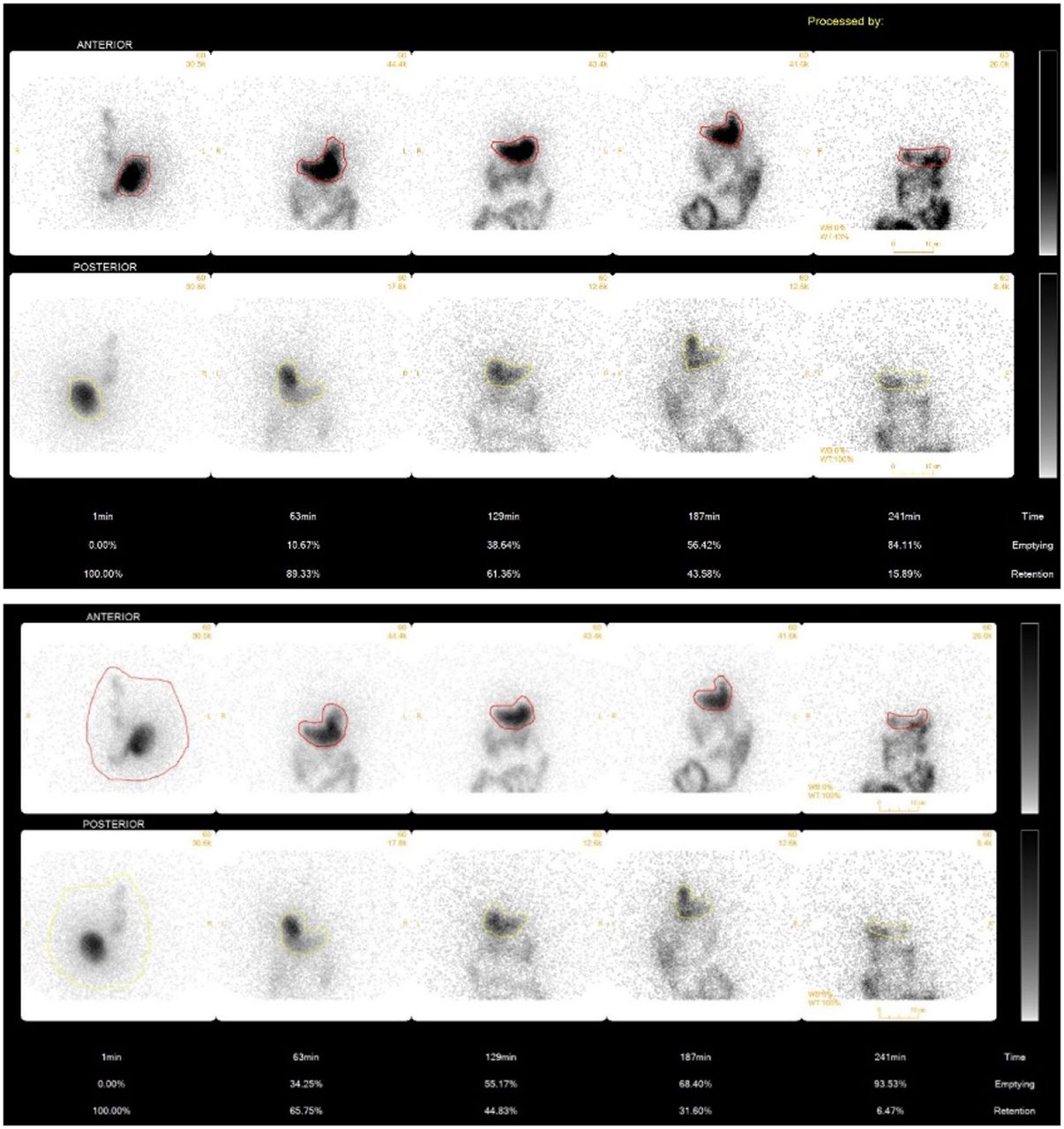
- FIGURE 9.
Solid gastric emptying study demonstrating the effects of region of interest placement (top). On the immediate image, the region of interest is incorrectly drawn around only the fundus. Residual activity in the esophagus and activity in the antrum is not included. The percent remaining in the stomach at later times is falsely elevated because fewer initial counts (denominator) were used in the calculation. At 4 hours, the percent remaining in the stomach is abnormal at 15.9% (bottom). The region of interest is correctly drawn including the esophagus and antrum. When the counts at 4 hours are divided by this higher total activity, the percentage of meal remaining in the stomach is now normal at 6.4%. Images courtesy of Jon A. Baldwin, MD, University of Alabama at Birmingham, Birmingham, AL.


Tables
Factors that Increase the Rate of Emptying Factors that Decrease the Rate of Emptying Liquids Solids Small particle size Large particle size Low fiber or low residue High fiber Proteins and carbohydrates Fats Low calorie High calorie Large volume Small volume Alkaline Acidic Hot food Cool food Early in the day Late in the day Activity Sedentary Upright Lying down Absence of pain Pain Lying on right side Lying on left side Male Female Prokinetic, erythromycin Narcotics, anticholinergic Reserpine, anticholinesterases, guanethidine, cholinergic agents (Atropine), tricyclic antidepressants, phenothiazines 120 g (4 oz.) of liquid egg whites (99% real eggs, cholesterol free, fat-free and low calorie) 2 slices of white bread 30 g strawberry jam 120 ml (4 oz.) water 18.5–37 MBq (0.5–1.0 mCi) 99mTc-SC Imaging Time Lower Normal Limita Upper Normal Limitb 0 minutes 0.5 hours 70% 1 hour 30% 90% 2 hours 60% 3 hours 30% 4 hours 10% ↵a For the lower normal limit, lower values suggest rapid gastric emptying.
↵b For upper normal limit values, a greater value suggests delayed gastric emptying.
Reprinted with permission from Abell TL, Camilleri M, Donohoe K, et al. Consensus recommendations for gastric emptying scintigraphy: a joint report of the American Neurogastroenterology and Motility Society and the Society of Nuclear Medicine. J Nucl Med Technol. 2008;36:44–54.


