
Main menu

  • Home
  • Content
    • Current
      • JNMT Supplement
    • Ahead of print
    • Past Issues
    • Continuing Education
    • JNMT Podcast
    • SNMMI Annual Meeting Abstracts
  • Subscriptions
    • Subscribers
    • Rates
    • Journal Claims
    • Institutional and Non-member
  • Authors
    • Submit to JNMT
    • Information for Authors
    • Assignment of Copyright
    • AQARA Requirements
  • Info
    • Reviewers
    • Permissions
    • Advertisers
    • Corporate & Special Sales
  • About
    • About Us
    • Editorial Board
    • Contact Information
  • More
    • Alerts
    • Feedback
    • Help
    • SNMMI Journals
  • SNMMI
    • JNMT
    • JNM
    • SNMMI Journals
    • SNMMI

User menu

  • Subscribe
  • My alerts
  • Log in
  • Log out
  • My Cart

Search

  • Advanced search
Journal of Nuclear Medicine Technology
  • SNMMI
    • JNMT
    • JNM
    • SNMMI Journals
    • SNMMI
  • Subscribe
  • My alerts
  • Log in
  • Log out
  • My Cart
Journal of Nuclear Medicine Technology

Advanced Search

  • Home
  • Content
    • Current
    • Ahead of print
    • Past Issues
    • Continuing Education
    • JNMT Podcast
    • SNMMI Annual Meeting Abstracts
  • Subscriptions
    • Subscribers
    • Rates
    • Journal Claims
    • Institutional and Non-member
  • Authors
    • Submit to JNMT
    • Information for Authors
    • Assignment of Copyright
    • AQARA Requirements
  • Info
    • Reviewers
    • Permissions
    • Advertisers
    • Corporate & Special Sales
  • About
    • About Us
    • Editorial Board
    • Contact Information
  • More
    • Alerts
    • Feedback
    • Help
    • SNMMI Journals
  • Watch or Listen to JNMT Podcast
  • Visit SNMMI on Facebook
  • Join SNMMI on LinkedIn
  • Follow SNMMI on Twitter
  • Subscribe to JNMT RSS feeds
Review ArticleTeaching Case Studies

Hepatobiliary Iminodiacetic Acid Scanning Detects Previously Unknown Hepatic Metastases from Colorectal Adenocarcinoma

Jonathan Chapman, Edward Sutherland and Jane Palka
Journal of Nuclear Medicine Technology March 2019, 47 (1) 83-84; DOI: https://doi.org/10.2967/jnmt.117.206748
Jonathan Chapman
William Beaumont School of Medicine, Beaumont Health System, Oakland University, Royal Oak, Michigan
  • Find this author on Google Scholar
  • Find this author on PubMed
  • Search for this author on this site
Edward Sutherland
William Beaumont School of Medicine, Beaumont Health System, Oakland University, Royal Oak, Michigan
  • Find this author on Google Scholar
  • Find this author on PubMed
  • Search for this author on this site
Jane Palka
William Beaumont School of Medicine, Beaumont Health System, Oakland University, Royal Oak, Michigan
  • Find this author on Google Scholar
  • Find this author on PubMed
  • Search for this author on this site
  • Article
  • Figures & Data
  • Info & Metrics
  • PDF
Loading

Abstract

A case of previously unknown metastatic colorectal adenocarcinoma presented as focal photopenia on hepatobiliary iminodiacetic acid scintigraphy. Differential diagnosis of focal photopenia and heterogenous hepatic radiotracer uptake on hepatobiliary scintigraphy is reviewed.

  • colorectal adenocarcinoma
  • HIDA scan
  • gastrointestinal

Colorectal adenocarcinoma is the third most common type of cancer worldwide, with approximately 1.36 million new cases diagnosed each year (1). Approximately 30%–50% of patients with colorectal cancer develop liver metastases (2). This report presents a case of metastatic colorectal adenocarcinoma first discovered on a hepatobiliary iminodiacetic acid scan, which to our knowledge has not previously been described in the literature, and reviews the differential diagnosis of focal photopenia and heterogeneous hepatic radiotracer uptake on hepatobiliary scintigraphy.

CASE REPORT

A 50-y-old man with no significant past medical history presented to his primary care physician for 1 mo of worsening aching and burning in the right upper quadrant and suprapubic pain with associated dysuria, nausea, and weight loss. Urinalysis performed in the office revealed trace hematuria. The clinician hypothesized the pain to be related to separate etiologies: GERD versus cholelithiasis to explain the right upper quadrant pain and nephrolithiasis causing the suprapubic pain. Abdominal ultrasound and a hepatobiliary scan were ordered. In addition, the patient was prescribed famotidine and advised to drink more water.

Hepatobiliary scanning was performed, before ultrasound, with 198 MBq (5.35 mCi) of 99mTc-disofenin. Anterior and left-anterior-oblique images of the abdomen were obtained in the supine position at 45 and 50 min after radiotracer administration, respectively. At 60 min after radiotracer administration, the patient was injected with 2 μg of cholecystokinin infused over 60 min, during which a dynamic acquisition was performed to calculate gallbladder ejection fraction, which was normal.

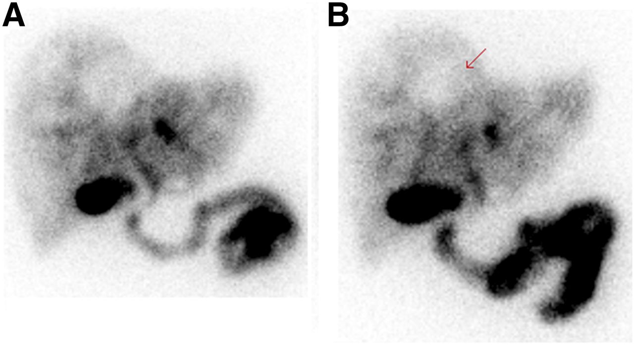
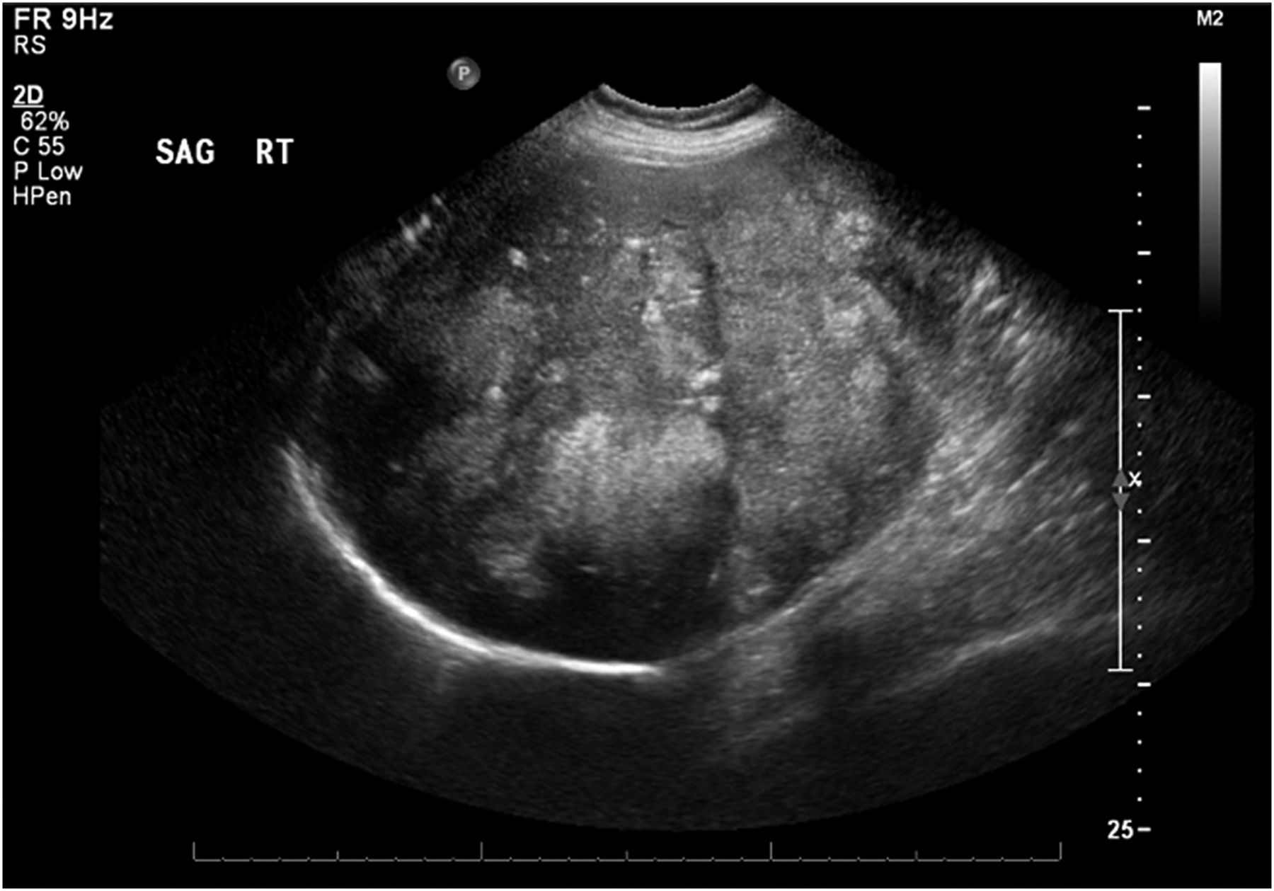
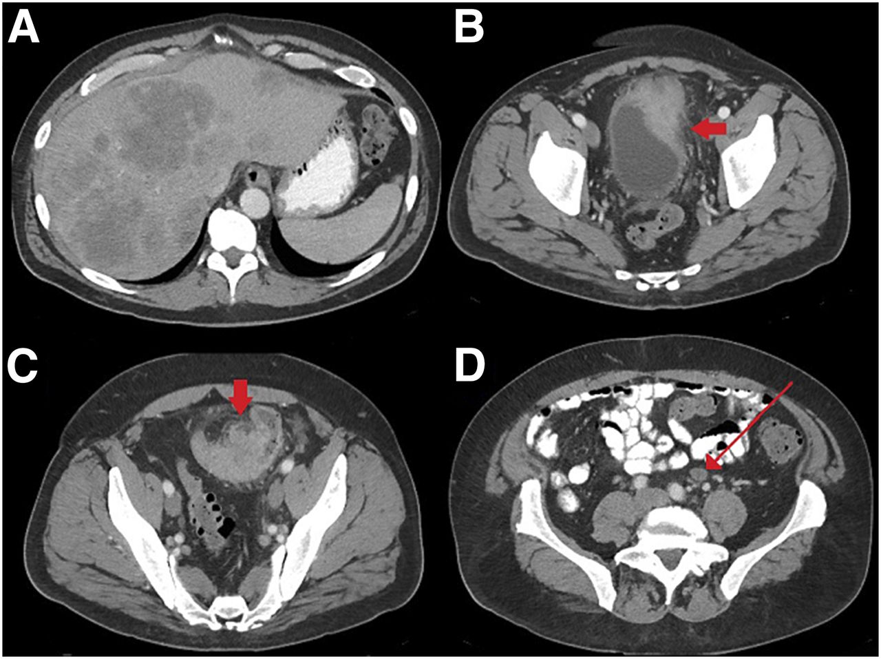
Images obtained at 45 and 50 min after 99mTc-disofenin administration demonstrated heterogeneous liver uptake with multiple scattered photopenic areas (Fig. 1). Given that these findings were suspected of being liver masses, abdominal ultrasound was recommended. Ultrasound showed multiple echogenic masses throughout the liver (Fig. 2). Subsequent laboratory tests yielded an elevated carcinoembryonic antigen level of 1,738 and a lactate dehydrogenase level of 804. Contrast-enhanced CT of the abdomen and pelvis demonstrated hypodense masses throughout the liver, lymphadenopathy, and a primary sigmoid colon mass invading the bladder (Fig. 3). Core liver biopsy revealed metastatic colorectal adenocarcinoma.

FIGURE 1.
  • Download figure
  • Open in new tab
  • Download powerpoint
FIGURE 1.

Anterior (A) and left anterior oblique (B) spot images obtained 45 and 50 min after 99mTc-disofenin administration show heterogeneous distribution of radiotracer activity throughout liver as well as large photopenic defect (arrow).

FIGURE 2.
  • Download figure
  • Open in new tab
  • Download powerpoint
FIGURE 2.

Sagittal ultrasound of right hepatic lobe shows numerous echogenic masses.

FIGURE 3.
  • Download figure
  • Open in new tab
  • Download powerpoint
FIGURE 3.

(A) Axial portal-vein–phase contrast-enhanced CT images of abdomen show hypodense masses scattered throughout liver. (B–D) Also seen is primary sigmoid colon mass (arrow, C) invading bladder (arrow, B) and lymphadenopathy (arrow, D).

DISCUSSION

In the normal liver, hepatocytes demonstrate uptake of iminodiacetic acid, leading to homogeneous radiotracer distribution. Theoretically, any process that replaces or displaces normal hepatocytes would be expected to cause focal photopenic areas or heterogenous hepatic radiotracer uptake. The potential causes are many but include benign hepatic cysts, metastasis, primary liver masses, and less common processes such as focal confluent hepatic fibrosis and Caroli disease (3). Focal hepatic fibrosis would demonstrate wedge-shaped areas of decreased radiotracer activity, whereas hepatic cysts and metastases are more likely to demonstrate randomly distributed photopenic defects. Caroli disease is known to initially show multiple focal photopenic defects that later fill with radiotracer on subsequent delayed imaging (3). In the setting of cirrhosis, a heterogeneous scintigraphic appearance of the liver can be seen because of the replacement of normally functioning hepatocytes with hepatocytes that exhibit either increased or decreased function, such as is seen with regenerative nodules or with the development of poorly differentiated hepatocellular carcinoma (4).

CONCLUSION

Focal photopenia on hepatobiliary iminodiacetic acid scanning is suggestive of metastatic or primary neoplasia. In addition to correlating the findings with patient history and prior imaging, one should consider further evaluation with other imaging modalities.

DISCLOSURE

No potential conflict of interest relevant to this article was reported.

Footnotes

  • Published online Aug. 3, 2018.

REFERENCES

  1. 1.↵
    Cancer fact sheets: colorectal cancer. Global Cancer Observatory website. http://gco.iarc.fr/today/fact-sheets-cancers?cancer=6&type=0&sex=0. Accessed September 6, 2018.
  2. 2.↵
    1. Leufkens AM,
    2. van den Bosch M,
    3. van Leeuwen M,
    4. Siersema P
    . Diagnostic accuracy of computed tomography for colon cancer staging: a systematic review. Scand J Gastroenterol. 2011;46:887–894.
    OpenUrlCrossRefPubMed
  3. 3.↵
    1. Shinto AS,
    2. Selvakumar J
    . Unexpectedly diagnosed Caroli’s disease on HIDA scintigraphy in a patient with calculous cholecystitis. Nucl Med Mol Imaging. 2010;44:304–306.
    OpenUrl
  4. 4.↵
    1. Kotzerke J,
    2. Schwarzrock R,
    3. Krischek O,
    4. Wiese H,
    5. Hundeshagen H
    . Technetium-99m DISIDA hepatobiliary agent in diagnosis of hepatocellular carcinoma, adenoma, and focal nodular hyperplasia. J Nucl Med. 1989;30:1278–1280.
    OpenUrlFREE Full Text
  • Received for publication April 30, 2018.
  • Accepted for publication June 29, 2018.
PreviousNext
Back to top

In this issue

Journal of Nuclear Medicine Technology: 47 (1)
Journal of Nuclear Medicine Technology
Vol. 47, Issue 1
March 1, 2019
  • Table of Contents
  • About the Cover
  • Index by author
Print
Download PDF
Article Alerts
Sign In to Email Alerts with your Email Address
Email Article

Thank you for your interest in spreading the word on Journal of Nuclear Medicine Technology.

NOTE: We only request your email address so that the person you are recommending the page to knows that you wanted them to see it, and that it is not junk mail. We do not capture any email address.

Enter multiple addresses on separate lines or separate them with commas.
Hepatobiliary Iminodiacetic Acid Scanning Detects Previously Unknown Hepatic Metastases from Colorectal Adenocarcinoma
(Your Name) has sent you a message from Journal of Nuclear Medicine Technology
(Your Name) thought you would like to see the Journal of Nuclear Medicine Technology web site.
Citation Tools
Hepatobiliary Iminodiacetic Acid Scanning Detects Previously Unknown Hepatic Metastases from Colorectal Adenocarcinoma
Jonathan Chapman, Edward Sutherland, Jane Palka
Journal of Nuclear Medicine Technology Mar 2019, 47 (1) 83-84; DOI: 10.2967/jnmt.117.206748

Citation Manager Formats

  • BibTeX
  • Bookends
  • EasyBib
  • EndNote (tagged)
  • EndNote 8 (xml)
  • Medlars
  • Mendeley
  • Papers
  • RefWorks Tagged
  • Ref Manager
  • RIS
  • Zotero
Share
Hepatobiliary Iminodiacetic Acid Scanning Detects Previously Unknown Hepatic Metastases from Colorectal Adenocarcinoma
Jonathan Chapman, Edward Sutherland, Jane Palka
Journal of Nuclear Medicine Technology Mar 2019, 47 (1) 83-84; DOI: 10.2967/jnmt.117.206748
Twitter logo Facebook logo LinkedIn logo Mendeley logo
  • Tweet Widget
  • Facebook Like
  • Google Plus One
Bookmark this article

Jump to section

  • Article
    • Abstract
    • CASE REPORT
    • DISCUSSION
    • CONCLUSION
    • DISCLOSURE
    • Footnotes
    • REFERENCES
  • Figures & Data
  • Info & Metrics
  • PDF

Related Articles

  • No related articles found.
  • PubMed
  • Google Scholar

Cited By...

  • No citing articles found.
  • Google Scholar

More in this TOC Section

  • High-Sensitivity Troponin Elevation in a Young Woman with Typical Chest Pain: The Heart of the Matter
  • Pulmonary Adenocarcinoma Revealed by Parathyroid Scintigraphy: An Incidental Case to Remember
  • Prominent Right Ventricular Tracer Uptake: A Harbinger of Multivessel Coronary Artery Disease
Show more Teaching Case Studies

Similar Articles

Keywords

  • colorectal adenocarcinoma
  • HIDA scan
  • gastrointestinal
SNMMI

© 2025 SNMMI

Powered by HighWire