


Article Figures & Data
Figures
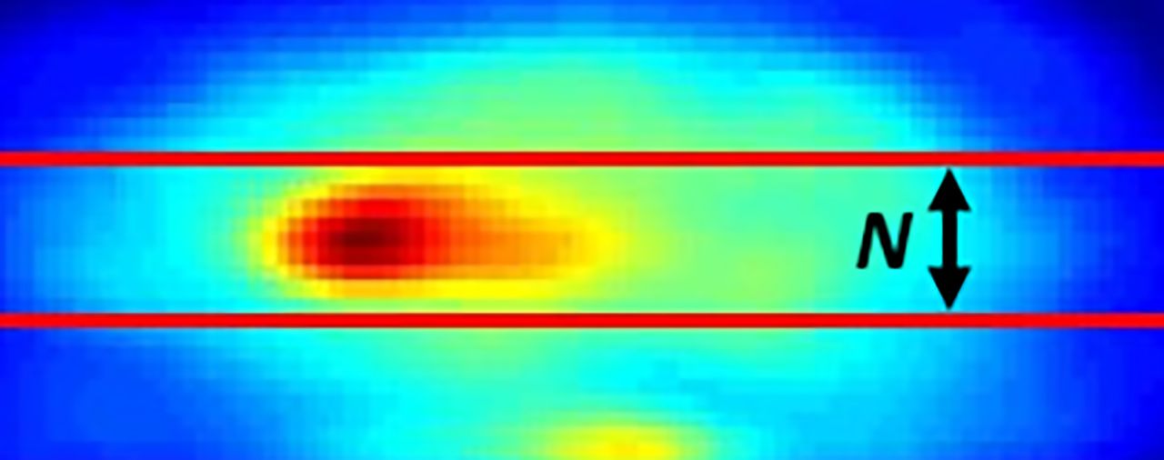
- FIGURE 1.
Summed sagittal slices of brain volume showing selection of transverse slices (between red lines); total slices added for analysis is N.
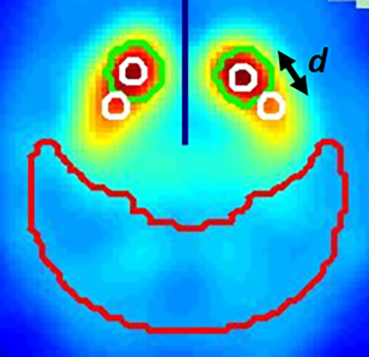
- FIGURE 2.
Summed transverse slices of brain volume showing ROI placements (white circles) for calculating SBRs. Distance between caudate and putamen ROIs is d, measured in pixels. Black line is automated location of center line between left and right striata. Green circle is SBRquant initial location of left and right caudates and is used to place ROIs onto center of caudate; putamen ROI is placed relative to caudate ROI. Reference ROI is red crescent in occipital region.
- FIGURE 3.
Average annual change between repeat scans for 51 subjects is plotted on y-axis as function of 2 parameters: distance in pixels between caudate and putamen ROIs, d, on upper x-axis and number of central striatal summed transverse slices, N, on lower x-axis. Average annual changes are graphed for 4 parameters: lowest putamen (Lowest P), average of both caudates and putamina (Avg C & P), average of caudates (Avg C), and average of putamina (Avg P). Largest average annual change (red circle) is obtained with lowest putamen parameter when summing 11 transverse slices (N = 11) and placing putamen ROI 3 pixels from caudate ROI (d = 3). Largest average annual change for group was fractional change of 0.112 (11.2%).


Jump to section
Related Articles
Cited By...
- No citing articles found.