


Article Figures & Data
Figures
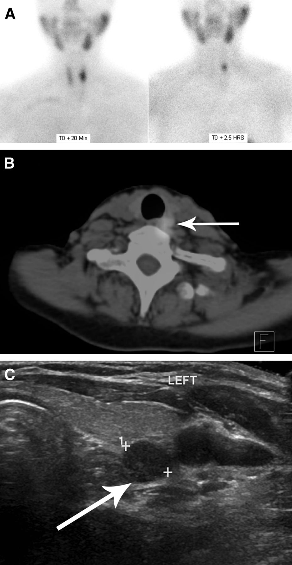
- FIGURE 1.
Multimodality approach to parathyroid localization. Left superior parathyroid adenoma is diagnosed using both 99mTc-sestamibi SPECT/CT and ultrasound. (A) Planar radionuclide imaging performed after 99mTc-sestamibi injection at 20 min and 2.5 h after injection. Planar image demonstrates tracer retention after 2.5 h. (B) SPECT/CT confirming localization of adenoma (arrow) posterior to left lobe of thyroid. (C) Ultrasound showing tracer uptake corresponding to single hypoechoic nodule (arrow) posterior to left thyroid lobe in midgland region. Parathyroid adenomas have characteristic feeding artery that can often be seen on Doppler examination (not shown on this image).
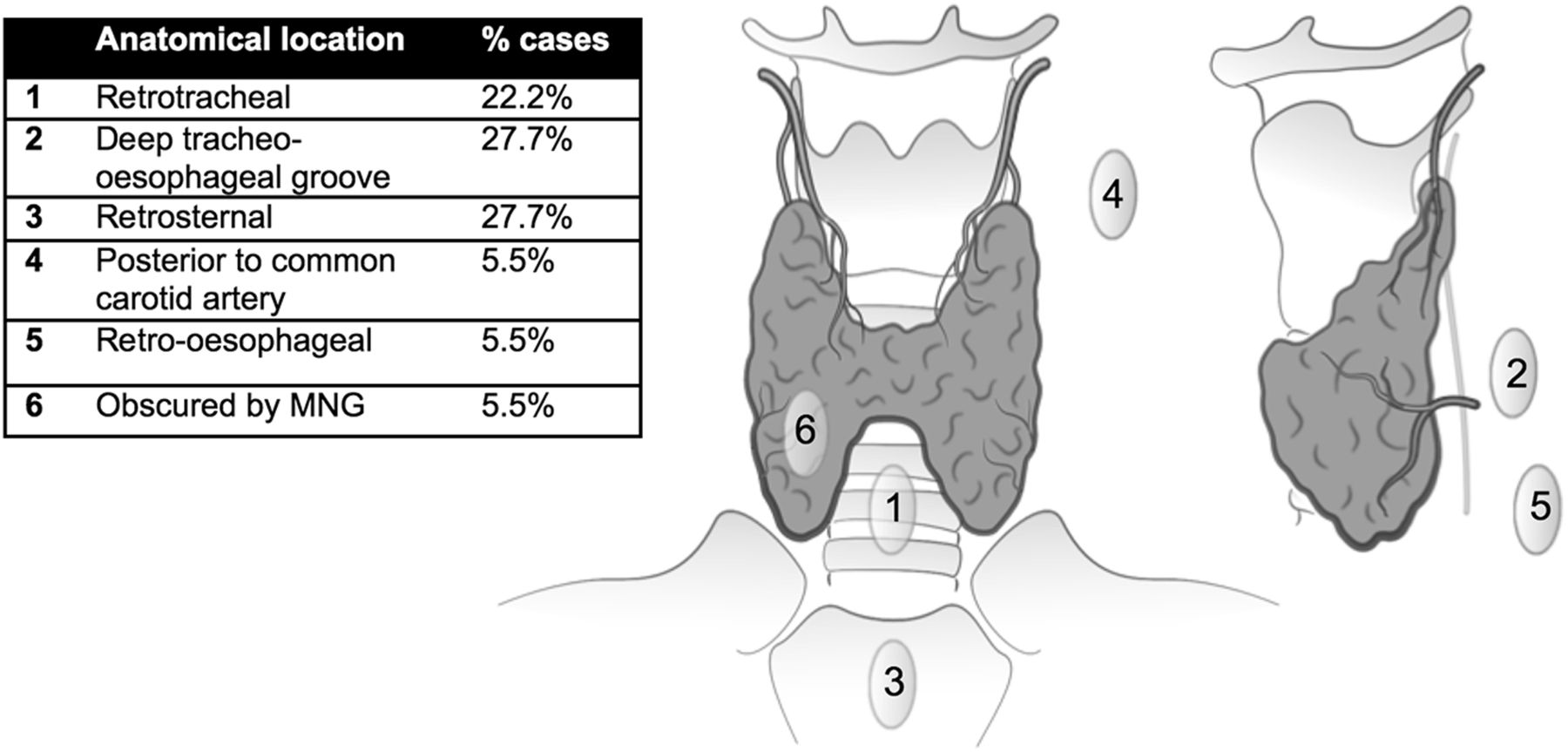
- FIGURE 2.
Adenomas not identified on ultrasound categorized by anatomic location. MNG = multinodular goiter.
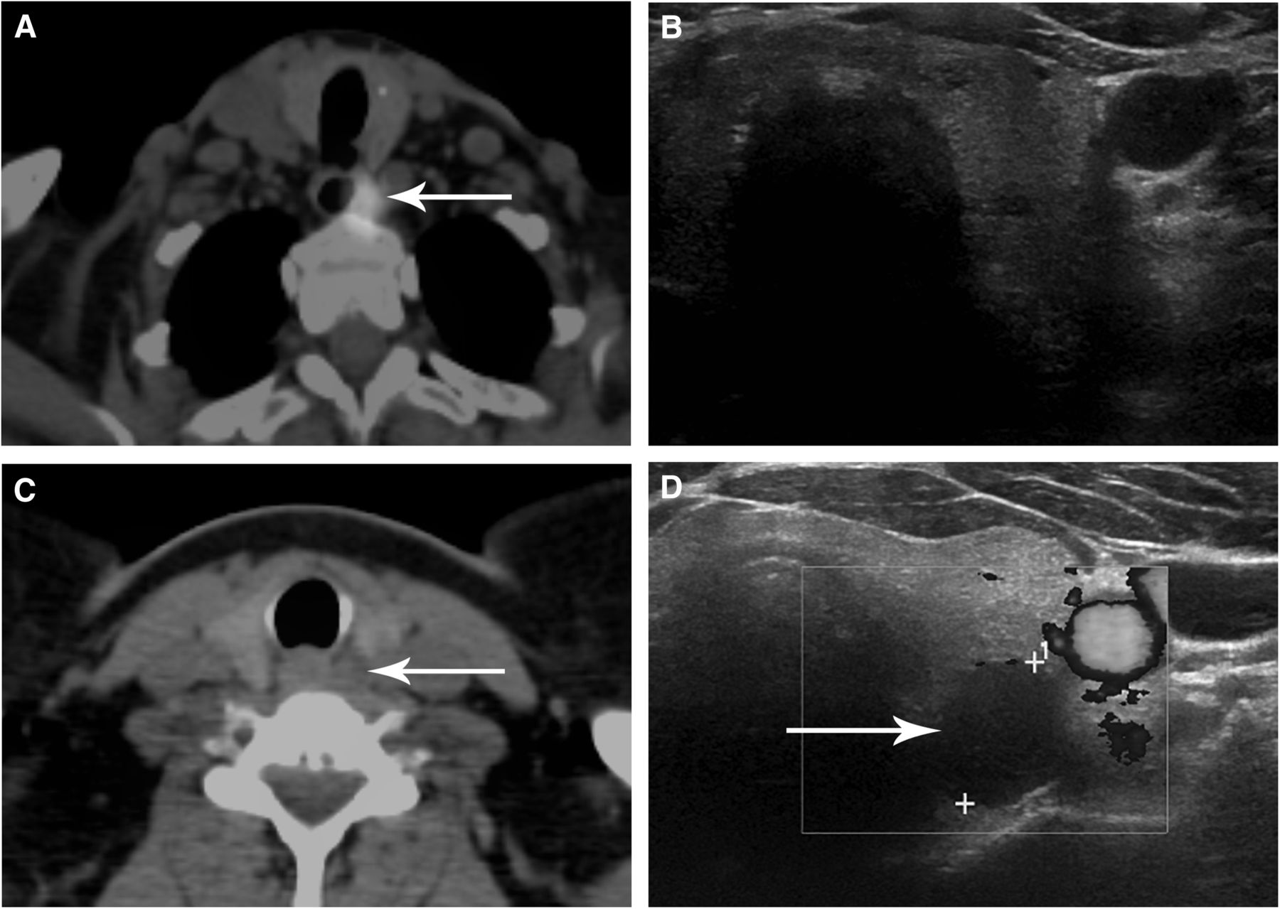
- FIGURE 3.
Series of images demonstrating discrepancies between modalities. (A) Avid tracer uptake is seen on SPECT/CT within left superior parathyroid adenoma (arrow), which is located deep in tracheoesophageal groove. (B) This finding is not visible on corresponding ultrasound because of acoustic shadowing from trachea but was subsequently located at surgery. (C) SPECT/CT reveals no tracer retention in nodule posterior to left lobe of thyroid (arrow). (D) On ultrasound, this nodule (arrow) appears to be characteristic of parathyroid adenoma, which has exhibited early washout with 99mTc-sestamibi. Diagnosis was confirmed at surgery.


Tables
Number of diseased glands Frequency Single 128 (88%) Multigland (≥2) 10 (7%) Negative exploration 8 (5%) Total 146 - TABLE 2
Sensitivity, Specificity, Negative Predictive Value, and Positive Predictive Value According to Analysis and Modality
Analysis Sensitivity Specificity Negative predictive value Positive predictive value Laterality and gland number (n = 146) SPECT/CT plus ultrasound 83 (75.5–88.3) 96 (91.1–98.4) 84 (77.5–89.3) 95 (90.2–98.3) SPECT/CT* 75 (66.8–81.5) 96 (91.3–98.5) 79 (72.4–84.8) 95 (89.0–98.1) Quadrant and gland number (n = 135) SPECT/CT plus ultrasound 77 (68.9–83.4) 97 (94.5–98.3) 92 (89.1–94.5) 89 (82.5–94.2) SPECT/CT* 70 (61.5–77.1) 97 (94.8–98.4) 90 (86.7–92.7) 89 (81.9–94.3) ↵* Analyzed by disregarding ultrasound component of examination.
Data are percentage followed by 95% confidence interval in parentheses.
- TABLE 3
Effect of Thyroid Disease on Localization of Parathyroid Adenomas with Combined SPECT/CT and Ultrasound
Finding Sensitivity Specificity Negative predictive value Positive predictive value Normal (n = 86) 82% 97% 85% 96% Atrophic (n = 5) 100% (0.383) 100% (0.847) 100% 100% Benign nodules (no glandular enlargement) (n = 20) 76% (0.194) 100% (0.556) 79% 100% Hashimoto thyroiditis (n = 4) 100% (0.462) 100% (0.875) 100% 100% Multinodular goiter (n = 60) 94% (0.069) 96% (0.429) 93% 97% Data in parentheses are probability values.


Jump to section
Related Articles
Cited By...
- No citing articles found.