


Article Figures & Data
Figures
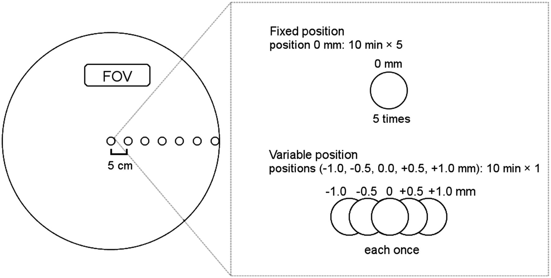
- FIGURE 1.
Setting of point sources. Seven point sources were placed on x-axis (interval, 5 cm). For fixed-position images, data were acquired for 10 min, 5 times serially. For variable-position images, data were acquired for 10 min each with point sources at 0, ±0.5, and ±1.0 mm in x-axis direction to simulate minimum of repositioning.
- FIGURE 2.
Images of point sources at fixed position reconstructed by OSEM and OSEM+PSF with 5 different matrix sizes. Images of point sources far from center became faint and broad. Hot spots were smaller and denser on OSEM+PSF images than on OSEM images.
- FIGURE 3.
Images of point sources at variable positions reconstructed by OSEM and OSEM+PSF with 400 × 400 matrix. Density and shape of point sources differed among positions.
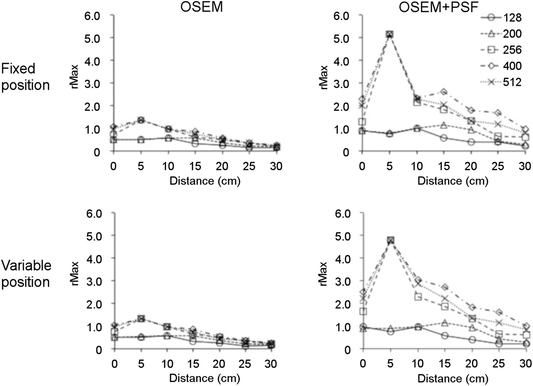
- FIGURE 4.
rMax,i at both fixed and variable positions on OSEM and OSEM+PSF images. At both positions, rMax,i decreased at positions far from center. rMax,i was higher on OSEM+PSF images than on OSEM images.
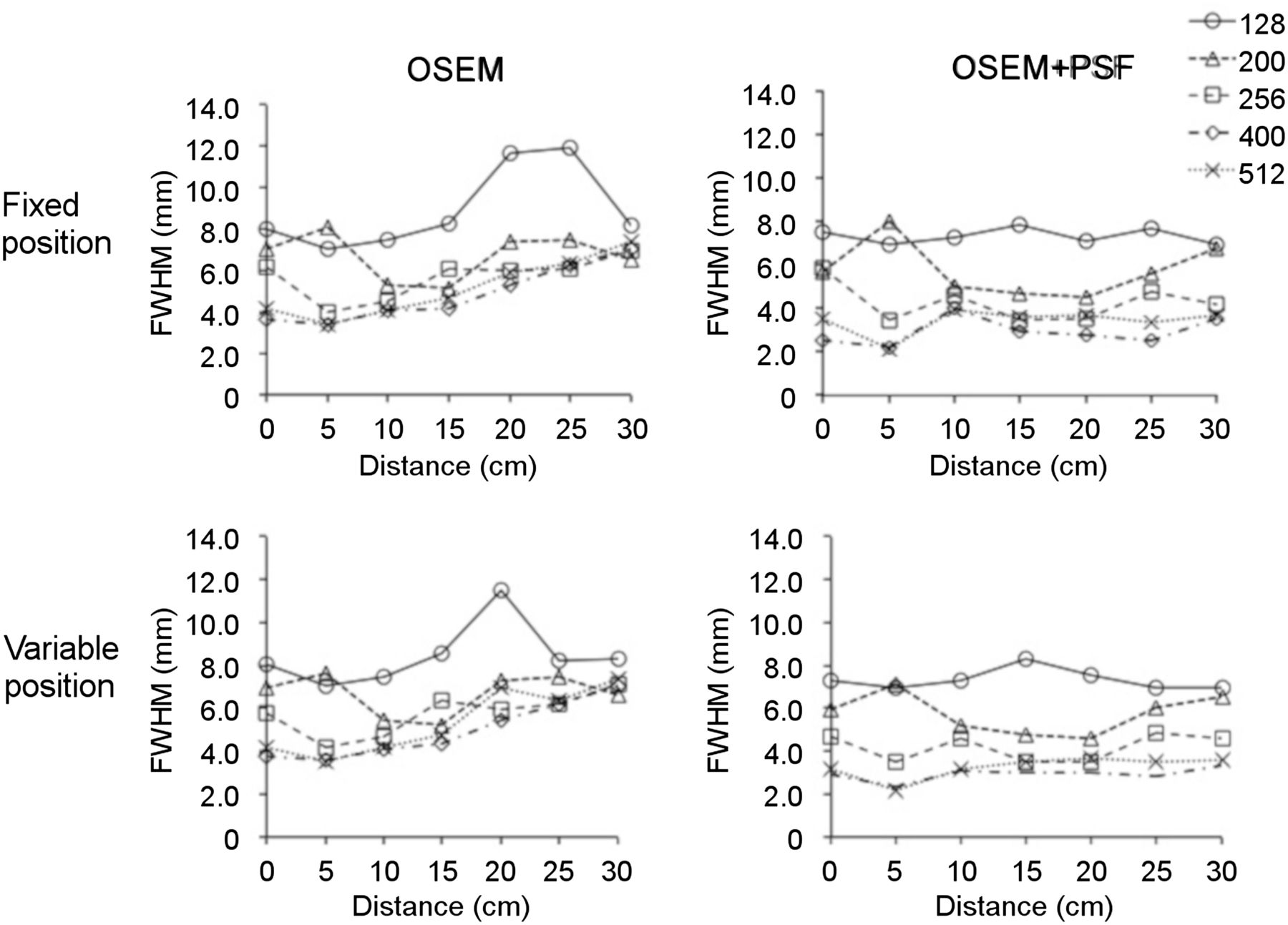
- FIGURE 5.
FWHM,i at both fixed and variable positions on OSEM and OSEM+PSF images. FWHM,i increased at positions far from center on OSEM images. FWHM,i of OSEM+PSF images was stable at all positions.


Tables
Position (cm) Matrix 0 5 10 15 20 25 30 128 × 128 Fixed 0.39% 0.31% 0.31% 0.56% 1.30% 1.03% 0.52% Variable 7.06% 0.70% 3.68% 4.90% 12.5% 11.1% 5.40% 200 × 200 Fixed 0.39% 0.49% 0.31% 0.49% 0.32% 1.04% 1.02% Variable 7.06% 5.84% 3.68% 0.64% 7.80% 5.61% 1.86% 256 × 256 Fixed 0.69% 0.67% 0.46% 0.75% 0.41% 0.88% 1.73% Variable 10.9% 3.91% 7.68% 6.35% 5.48% 1.14% 3.17% 400 × 400 Fixed 0.49% 0.67% 0.46% 0.98% 0.87% 0.84% 1.18% Variable 9.13% 3.91% 5.96% 2.58% 2.38% 1.60% 3.29% 512 × 512 Fixed 0.28% 0.67% 0.46% 0.85% 0.80% 0.31% 0.68% Variable 4.81% 3.91% 6.31% 1.71% 2.07% 0.75% 2.46% Position (cm) Matrix 0 5 10 15 20 25 30 128 × 128 Fixed 0.44% 0.84% 0.56% 0.47% 0.33% 1.22% 1.39% Variable 9.89% 0.92% 10.4% 7.09% 6.83% 18.7% 5.76% 200 × 200 Fixed 0.46% 0.85% 0.50% 0.57% 0.30% 1.09% 1.32% Variable 9.89% 10.21% 10.4% 7.09% 6.64% 18.7% 10.89% 256 × 256 Fixed 1.55% 0.73% 0.84% 1.00% 1.04% 1.99% 0.92% Variable 25.1% 5.47% 31.7% 6.97% 5.66% 8.85% 22.1% 400 × 400 Fixed 1.27% 0.73% 0.49% 1.48% 0.69% 2.76% 1.39% Variable 18.8% 5.47% 16.3% 16.6% 21.0% 14.8% 13.9% 512 × 512 Fixed 0.39% 0.73% 0.41% 1.48% 1.04% 0.88% 1.03% Variable 12.4% 5.47% 13.7% 12.3% 10.3% 5.33% 12.6%


Jump to section
Related Articles
Cited By...
- No citing articles found.