


Article Figures & Data
Figures
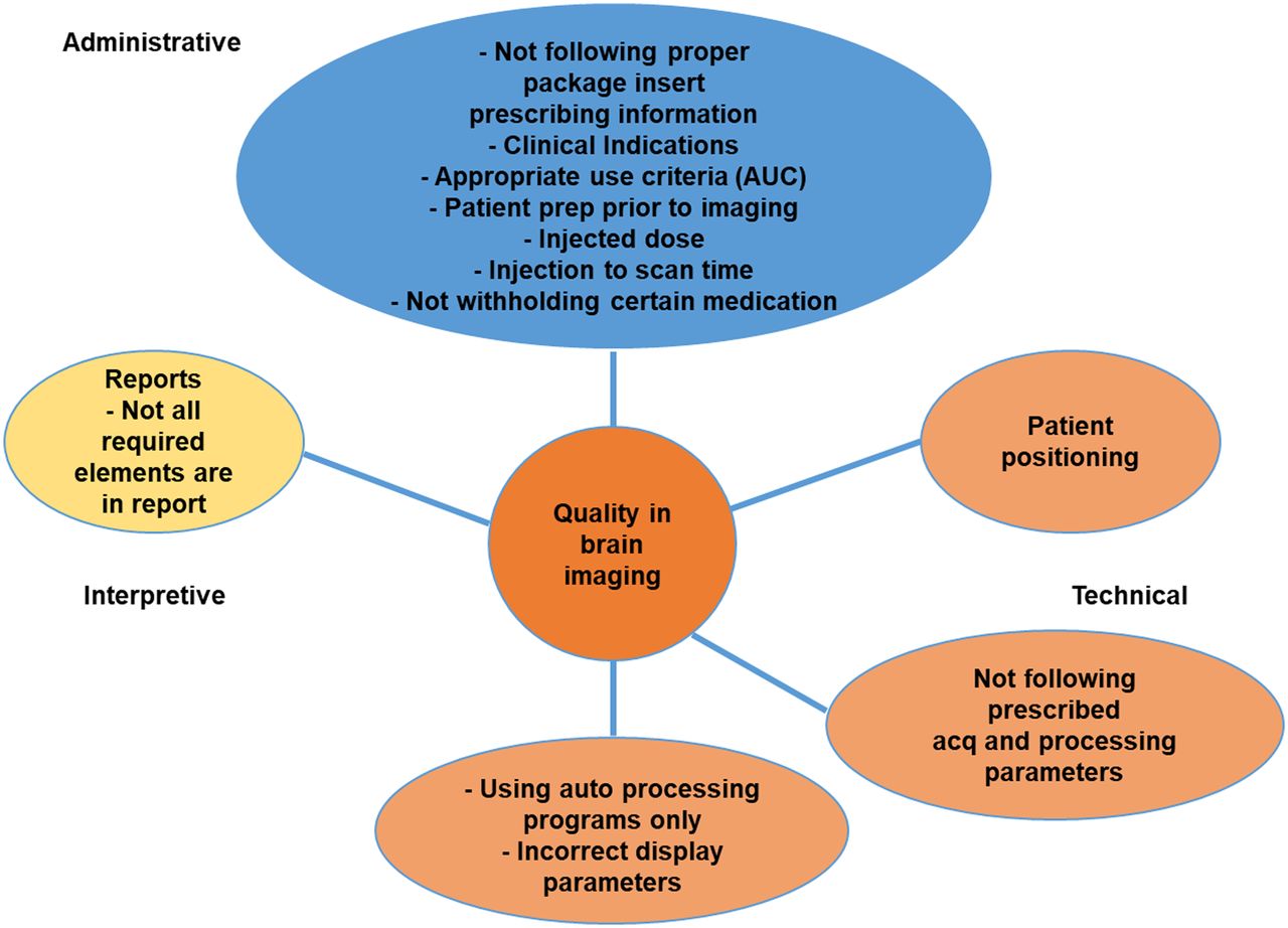
- FIGURE 1.
Three areas of quality in brain imaging: administrative, technical, and interpretative.
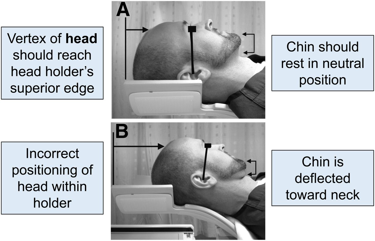
- FIGURE 2.
(A) Proper positioning in head holder. Canthomeatal line is imaginary line that runs from lateral corner of eye to ear canal. (B) Improper positioning. Canthomeatal line is not vertical and perpendicular to imaging table. (Reprinted from (8).)
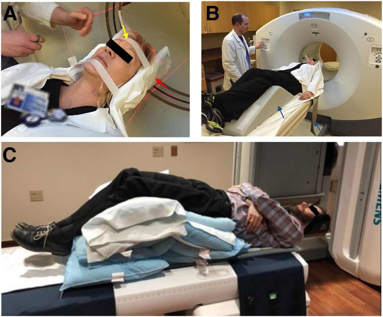
- FIGURE 3.
(A) Proper positioning on imaging table using head strap, chin strap, and laser lights. Laser light denoted by yellow arrow is positioned at center of nose and forehead to prevent head rotation. Laser light denoted by red arrow, at level of ear, positions table height so that head is in middle of field of view. (Courtesy of Virtua Health, Voorhees, NJ.) (B) Knee cushion and body strap denoted by arrows. (Courtesy of Virtua Health, Voorhees, NJ.) (C) Challenge of positioning patients with kyphosis, severe neck pain, or severe back pain. Multiple pillows under legs, buttocks, and lower back help patient lie flat. Use of body straps to secure patient to table is important.
- FIGURE 4.
Key DaTscan positioning considerations. (A) Location of caudate nucleus and putamen within brain on MRI. Blue denotes right caudate, red denotes right putamen, and arrows denote left caudate and left putamen. (B) Transverse DaTscan image of caudate and putamen. (Courtesy of Holy Name Hospital, Teaneck, NJ.) (C) Effect of increasing camera radius on DaTscan image quality. As radius increases, quality decreases. Putamen looks shorter and less defined with 20-cm radius than with 12.8-cm radius. (Courtesy of Liz Clarke.) (D) Camera head must be as close as possible to nose for best image quality and must clear shoulders. (Courtesy of Holy Name Hospital, Teaneck, NJ.)
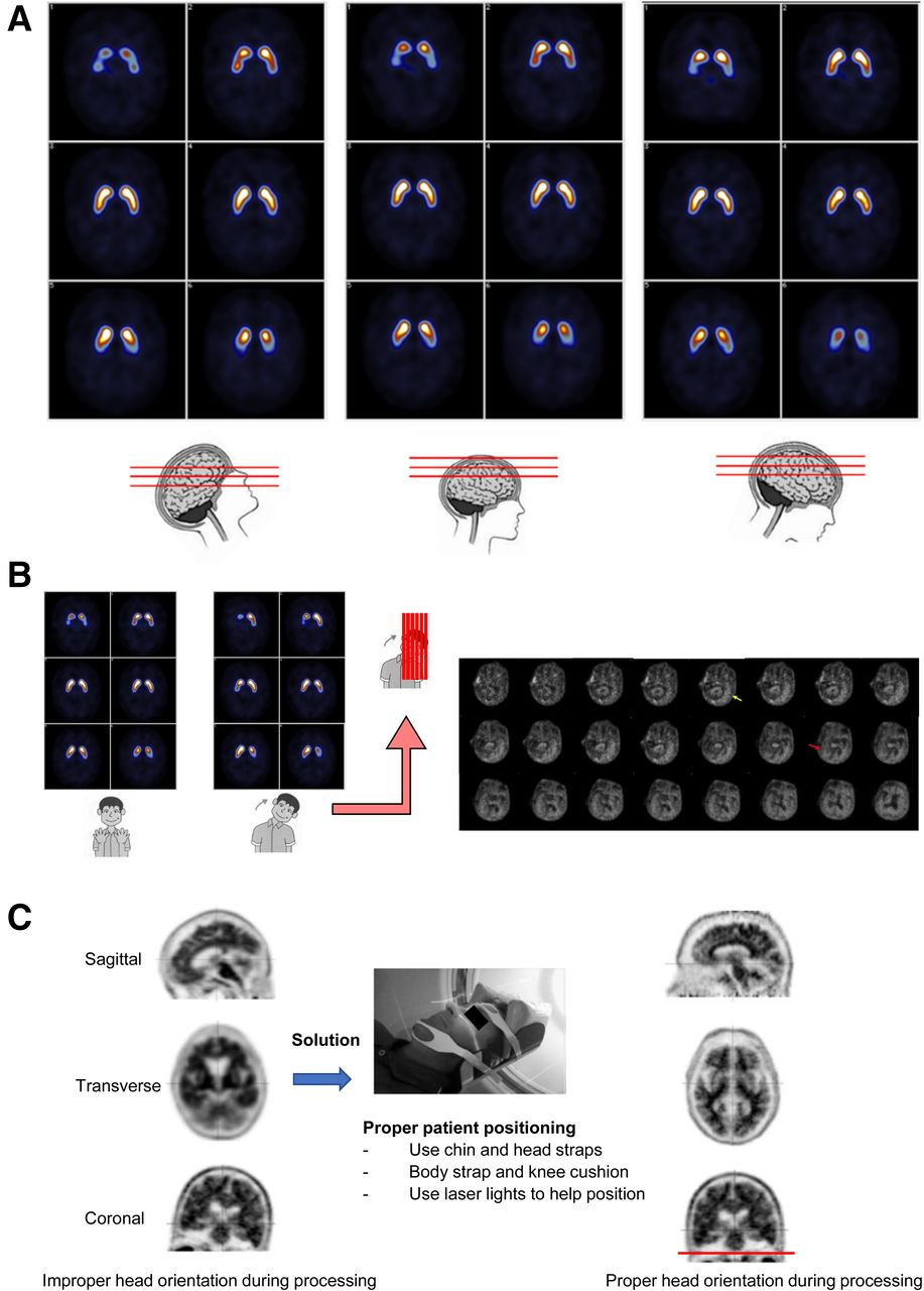
- FIGURE 5.
Effect of head orientation on DaTscan imaging. (A) Forward, backward, or sagittal head tilt has no effect on image quality. (Courtesy of Glasgow Southern Hospital.) (B) Lateral head tilt shows one putamen before the other, complicating interpretation. On amyloid imaging (right), yellow arrow denotes image on which left lateral lobe is first seen, and red arrow denotes image on which right lateral temporal lobe is first seen, almost 10 frames later. (Courtesy of Piramal Imaging.) (C) Head is tilted too far forward in sagittal slice at left and too far right in coronal slice. Although head position can be corrected during processing, best solution is to correctly position patient prior to image acquisition. Red line shows positioning that will result in properly oriented images after processing.
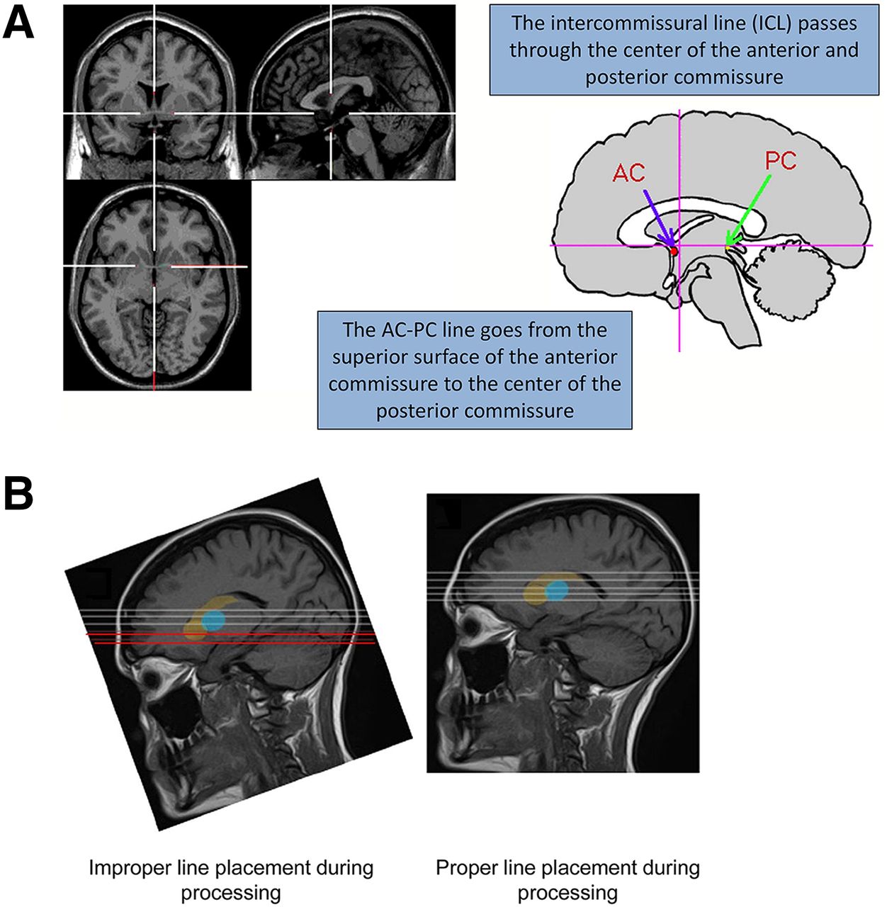
- FIGURE 6.
(A) Location of intercommissural line and AC–PC line. (B) MRI study showing improper and proper placement of processing lines. Forward head tilt results in slices (red lines) through caudate head that do not include putamen. On DaTscan studies, false-positive findings can result from incorrect positioning of processing lines. (Reprinted from (8).)
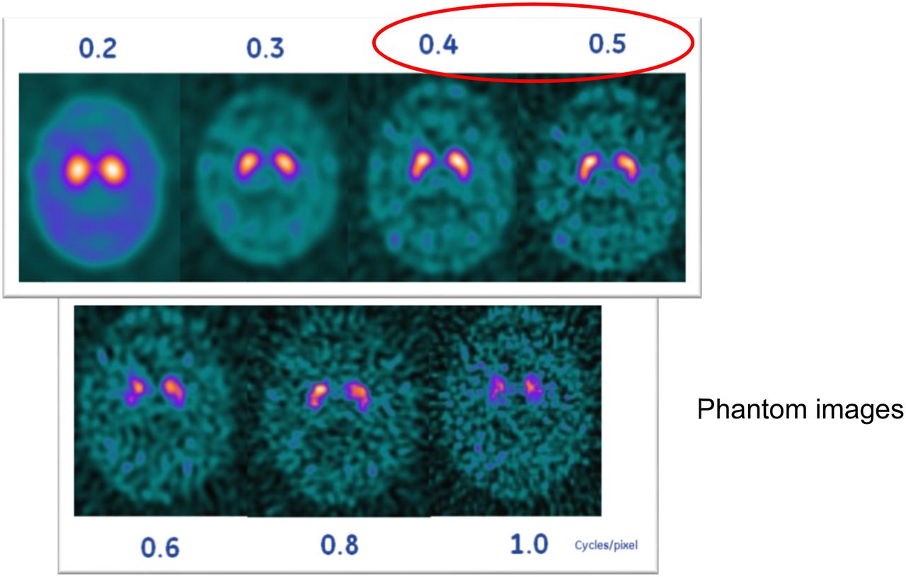
- FIGURE 7.
DaTscan study with different filter settings. Cutoff of 0.4–0.5 is usually good, depending of type of SPECT camera. Once interpreting physician chooses filter setting, it should be applied consistently for each study. (Courtesy of Liz Clarke.)


Tables
- TABLE 1
Examples of Health-Care Organizations That Implemented Lean Six Sigma Projects (17)
Hospital Project Effect Boston Medical Center Project to emphasize diagnostic imaging Cost savings and revenue increases of more than $2.2 million Rapides Regional Medical Center Project to increase efficiency of emergency department Drop in wait times; increase in patients seen; annual savings of more than $950,000 Valley Baptist Health System Project to reduce surgery cycle time Increase in annual capacity by 1,100 more cases, for potential income of $1.3 million Yale–New Haven Medical Center Multiple projects in surgical intensive care unit 75% decrease in bloodstream infections, for annual savings of ∼$1.2 million Project Purpose Analysis of report generation times (time physician ordered study to time physician received final report) Decrease waiting time to receive reports Analysis of patient escort times to and from nuclear medicine department Ensure proper staffing of escort department; increase on-time patient arrivals; increase number of patients scanned 5S Kaizen events Decrease waste and improve efficiency in imaging department


Jump to section
- Article
- Abstract
- DEFINITION OF QUALITY
- THREE AREAS OF QUALITY IN IMAGING
- ACQUISITION OF GOOD-QUALITY BRAIN IMAGES
- PATIENT POSITIONING
- HEAD ORIENTATION
- PROCESSING AND DISPLAY PARAMETERS
- CONSIDERATIONS AND ADJUSTMENTS
- IMAGE INTERPRETATION AND REPORTING
- CHECKLIST TO ACHIEVE OPTIMAL BRAIN IMAGES
- CONCLUSION
- DISCLOSURE
- Acknowledgments
- Footnotes
- REFERENCES
- Figures & Data
- Info & Metrics