


Article Figures & Data
Figures
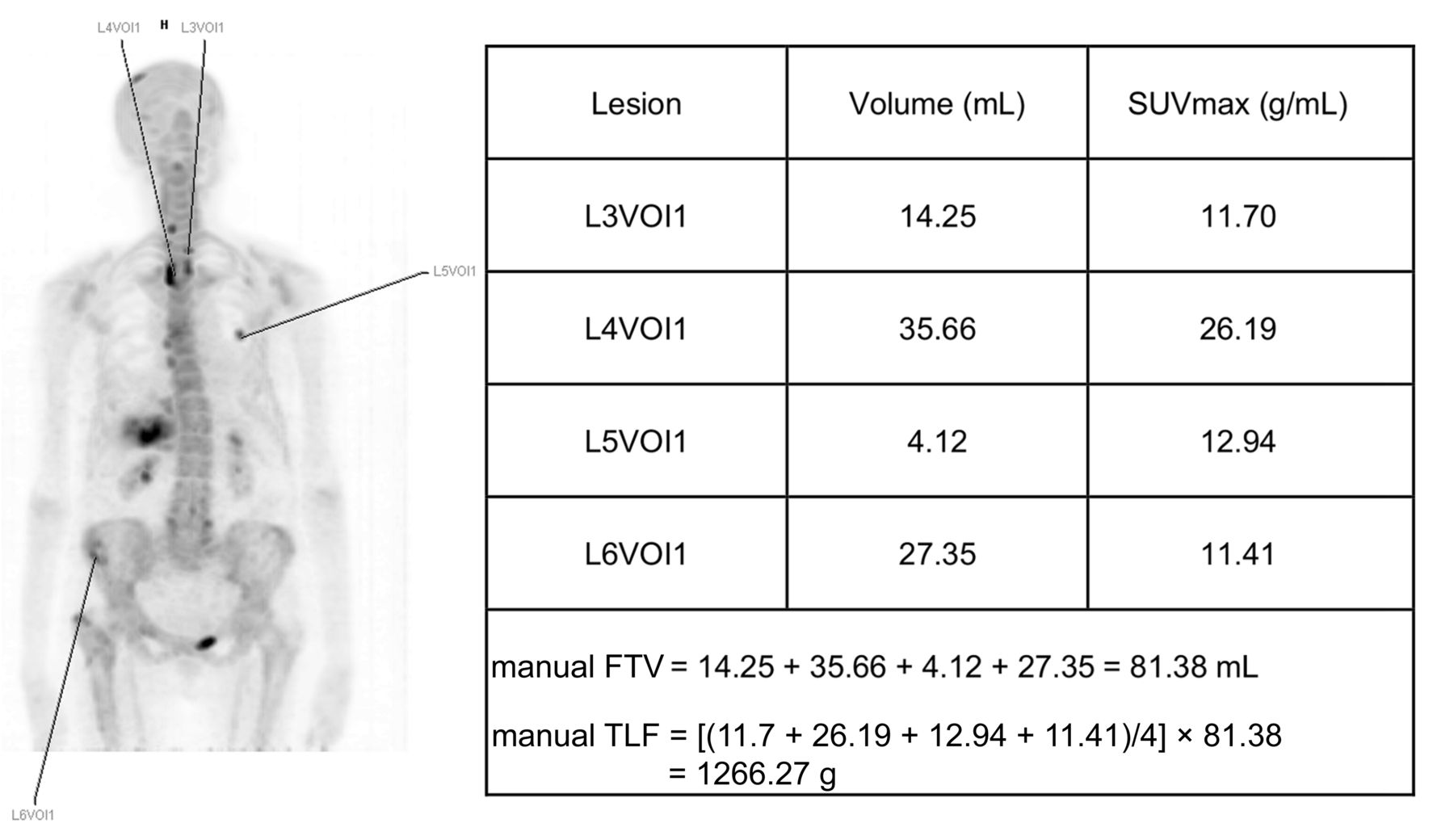
- FIGURE 1.
Example of manual quantification for one patient, with mFTV10 corresponding to sum of all VOIs (81.38 mL) and mTLF10 corresponding to mFTV10 multiplied by average of all SUVmax (1,266.27 g).
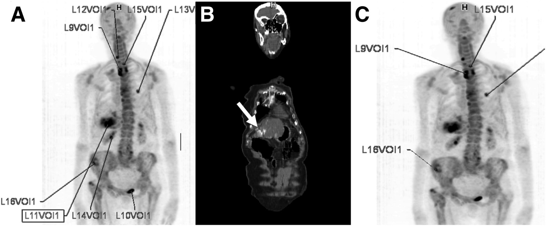
- FIGURE 2.
(A) After threshold selection (SUVmax of 10), several VOIs are automatically created. (B) On visual inspection, VOIs not related to metastases are deleted. In this patient, as indicated by white arrow in coronal section on CT scan, uptake was noted in calcified portion of liver, which was a sequela of radioablation (L11VOI1); this lesion was manually excluded. (C) Patient’s final saTLF10 was 3,969 g.
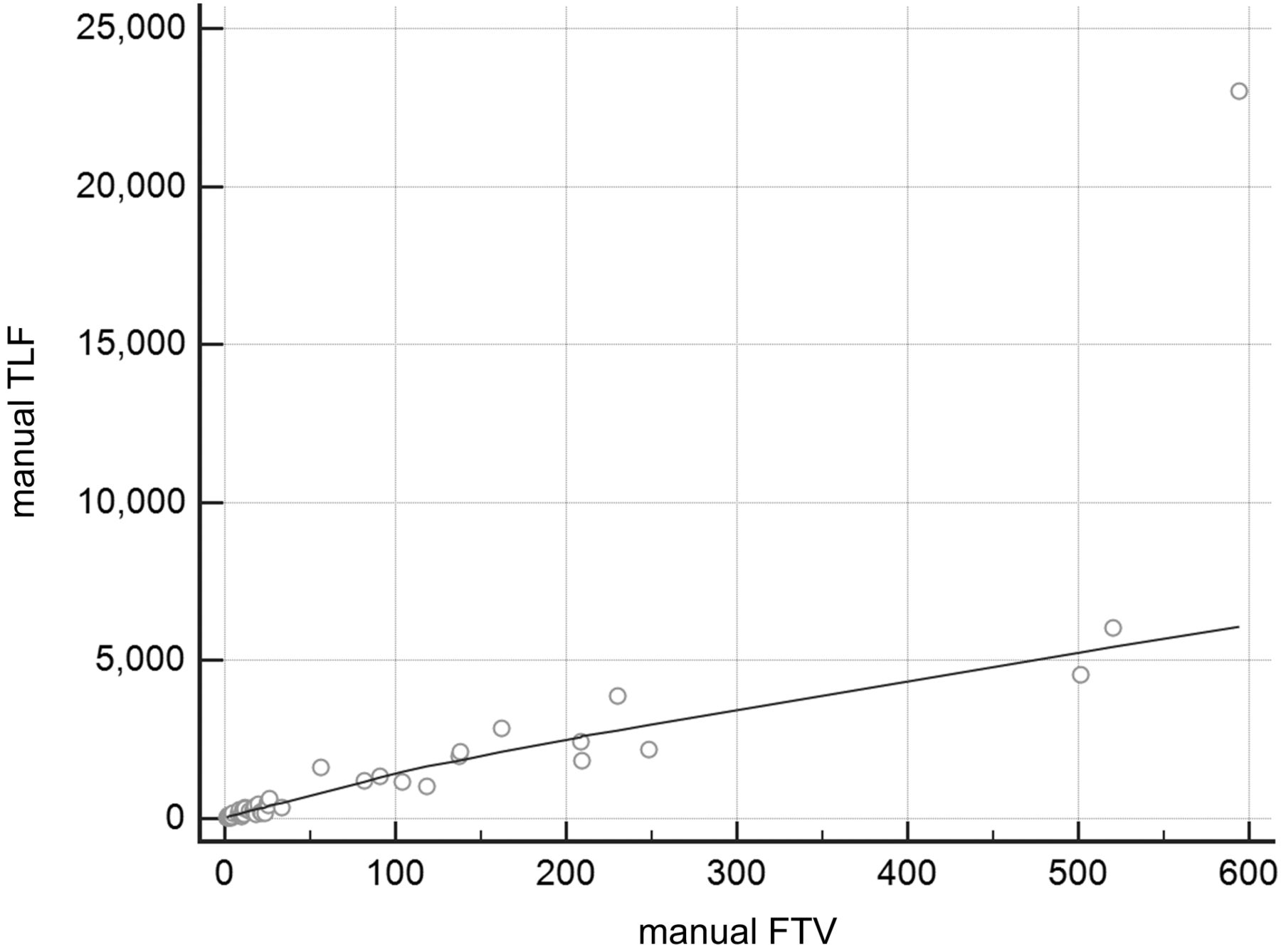
- FIGURE 3.
Graph showing strong correlation between values for mTLF10 and mFTV10 (ρ = 0.8117; P < 0.0001; 95% confidence interval, 0.6905–0.8885).
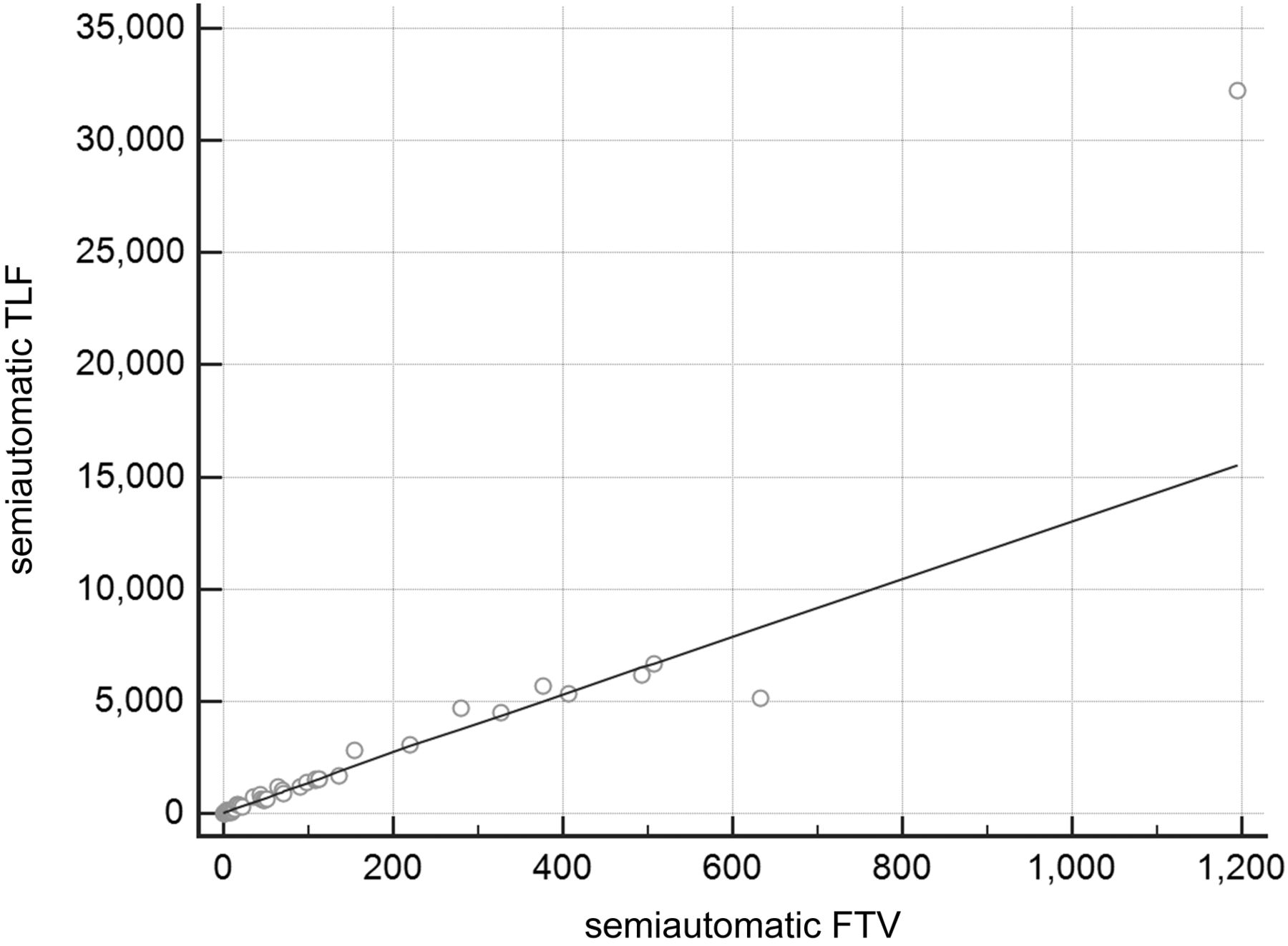
- FIGURE 4.
Graph showing strong correlation between values for saTLF10 and saFTV10 (ρ = 0.9234; P < 0.0001; 95% confidence interval, 0.8690–0.9558).
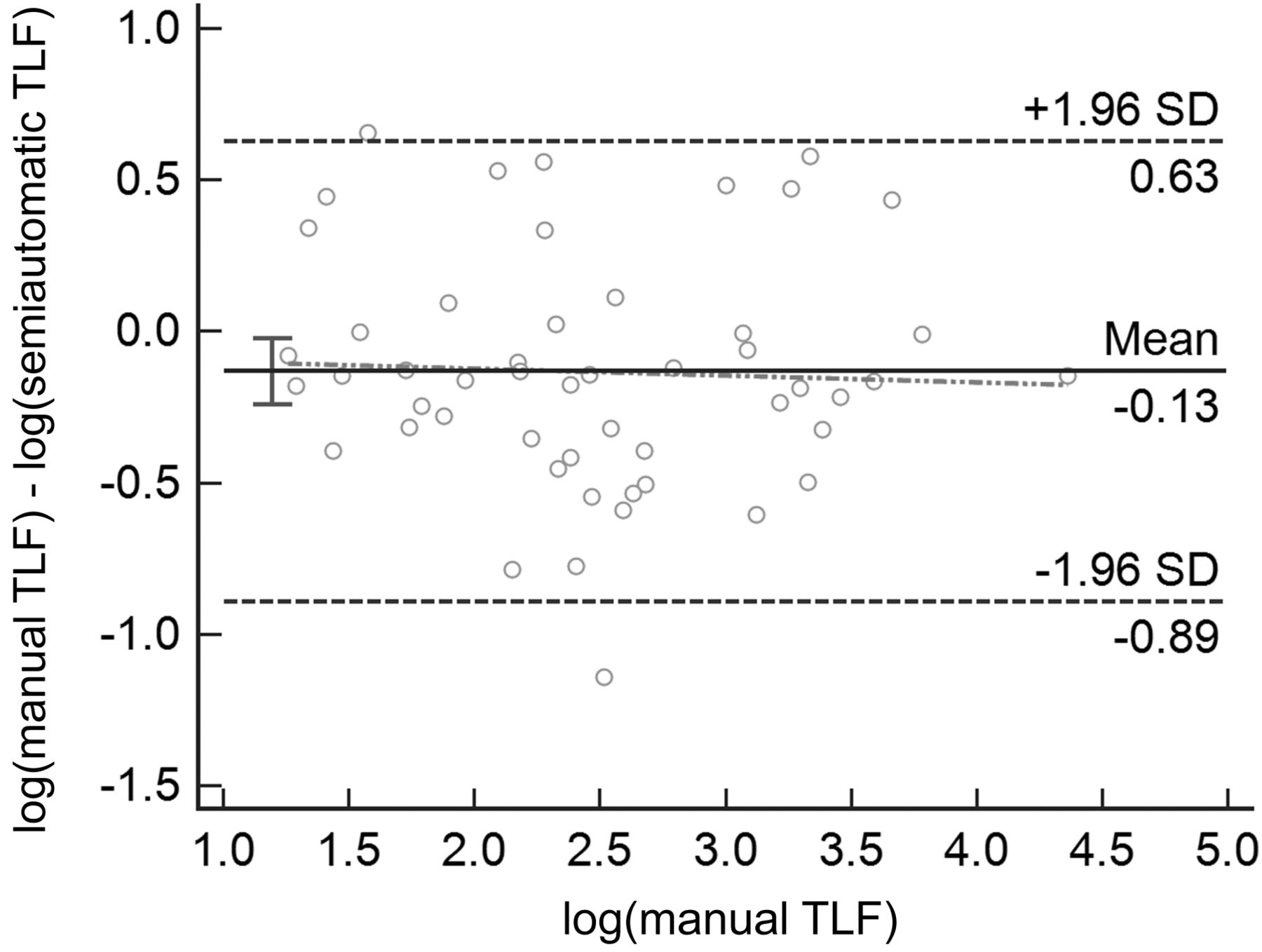
- FIGURE 5.
Bland–Altman test showing similarity between manual and semiautomatic quantifications.
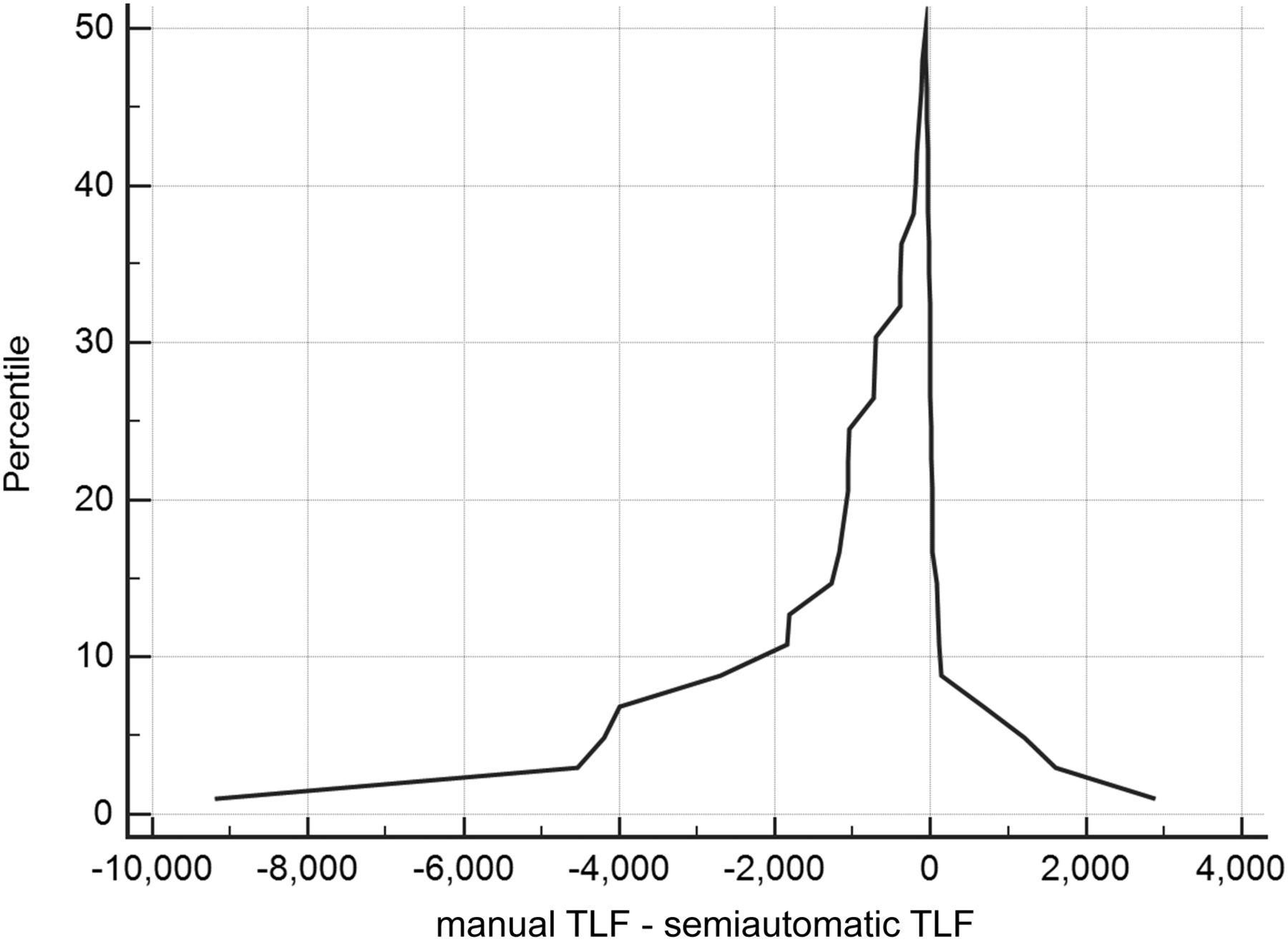
- FIGURE 6.
Mountain plot showing similarity between manual and semiautomatic quantifications, with peak near zero and small left tail.


Tables
- TABLE 1
Correlation of Clinical and Imaging Variables to Overall Survival and Time to Progression on Multivariable Analysis
Variable 95%CI P Overall survival saTLF10 1.0003–1.0009 0.0001 Visceral metastases 1.8334–24.6637 0.0041 Age 1.0042–1.1148 0.0342 Time to progression saTLF10 1.0001–1.0002 0.0006 Age 0.9359–0.9949 0.0342 CI = confidence interval.


Jump to section
Related Articles
Cited By...
- Validation of Convolutional Neural Networks for Fast Determination of Whole-Body Metabolic Tumor Burden in Pediatric Lymphoma
- Whole-Skeleton SUVmean Measured on 18F-NaF PET/CT Studies as a Prognostic Indicator in Patients with Breast Cancer Metastatic to Bone
- Validation of a Multifocal Segmentation Method for Measuring Metabolic Tumor Volume in Hodgkin Lymphoma