


Article Figures & Data
Figures
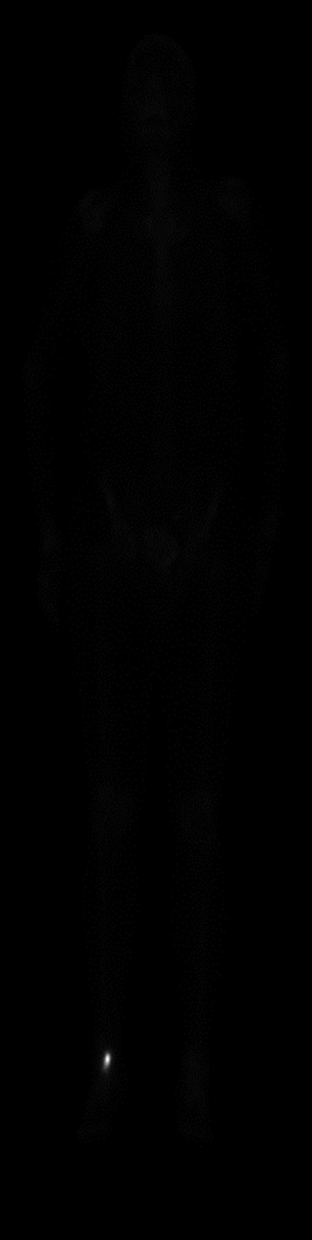
- FIGURE 1.
Appearance of whole-body 99mTc-MDP bone scintigraphy image when dynamic range of input image exceeds dynamic range of monitor.
- FIGURE 2.
Characteristic curves for linear and IT function: linear transformation curve and IT curves with slopes (e) equaling 1, 4, and 10. In each case, threshold was 127.
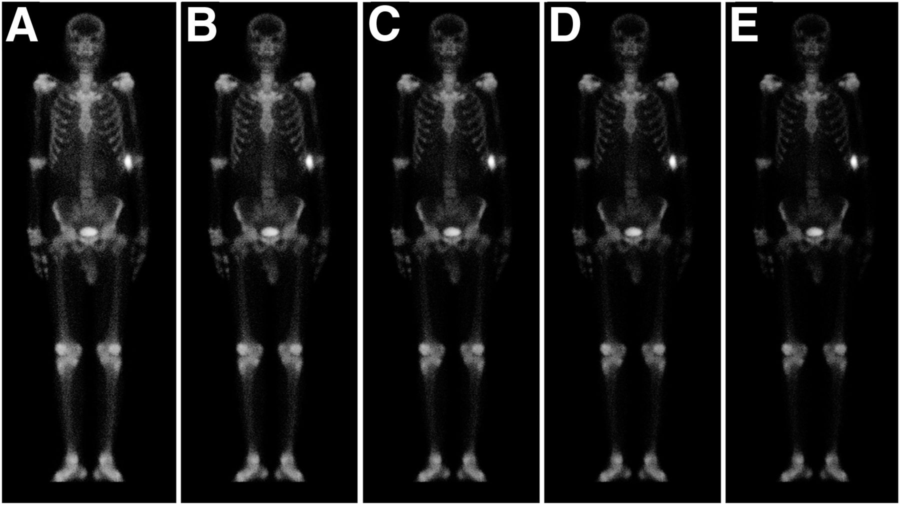
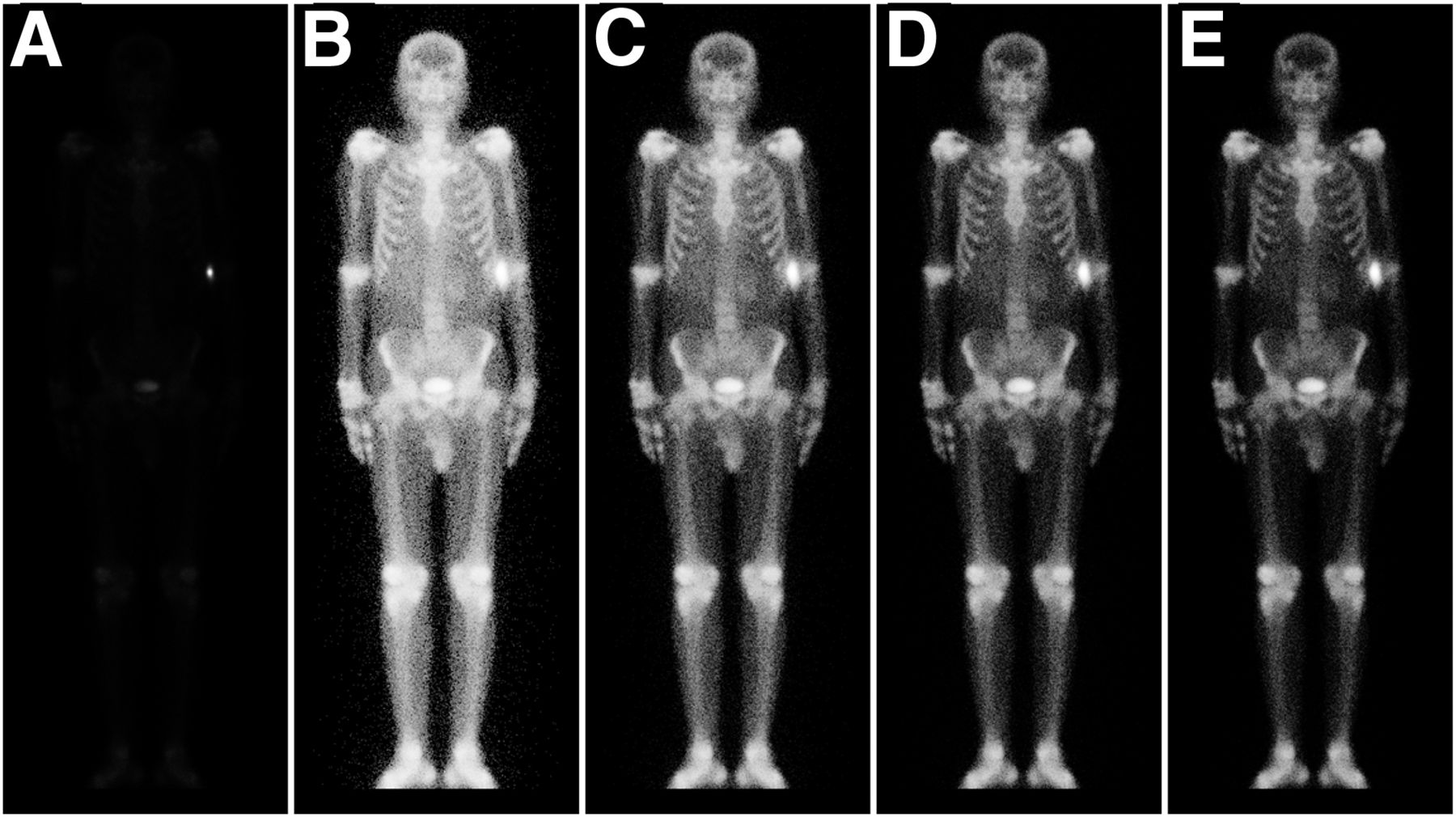
- FIGURE 3.
Whole-body 99mTc-MDP bone scintigraphy images: input image (A) and output images with slopes of 1–4 (B–E).
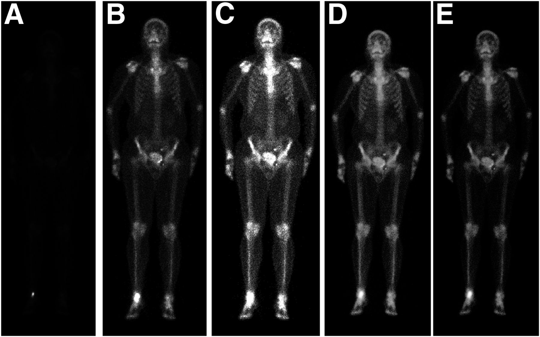
- FIGURE 4.
Processed whole-body 99mTc-MDP bone scintigraphy images with slopes of 5–9 (A–E) and threshold equal to mean count in respective input image.
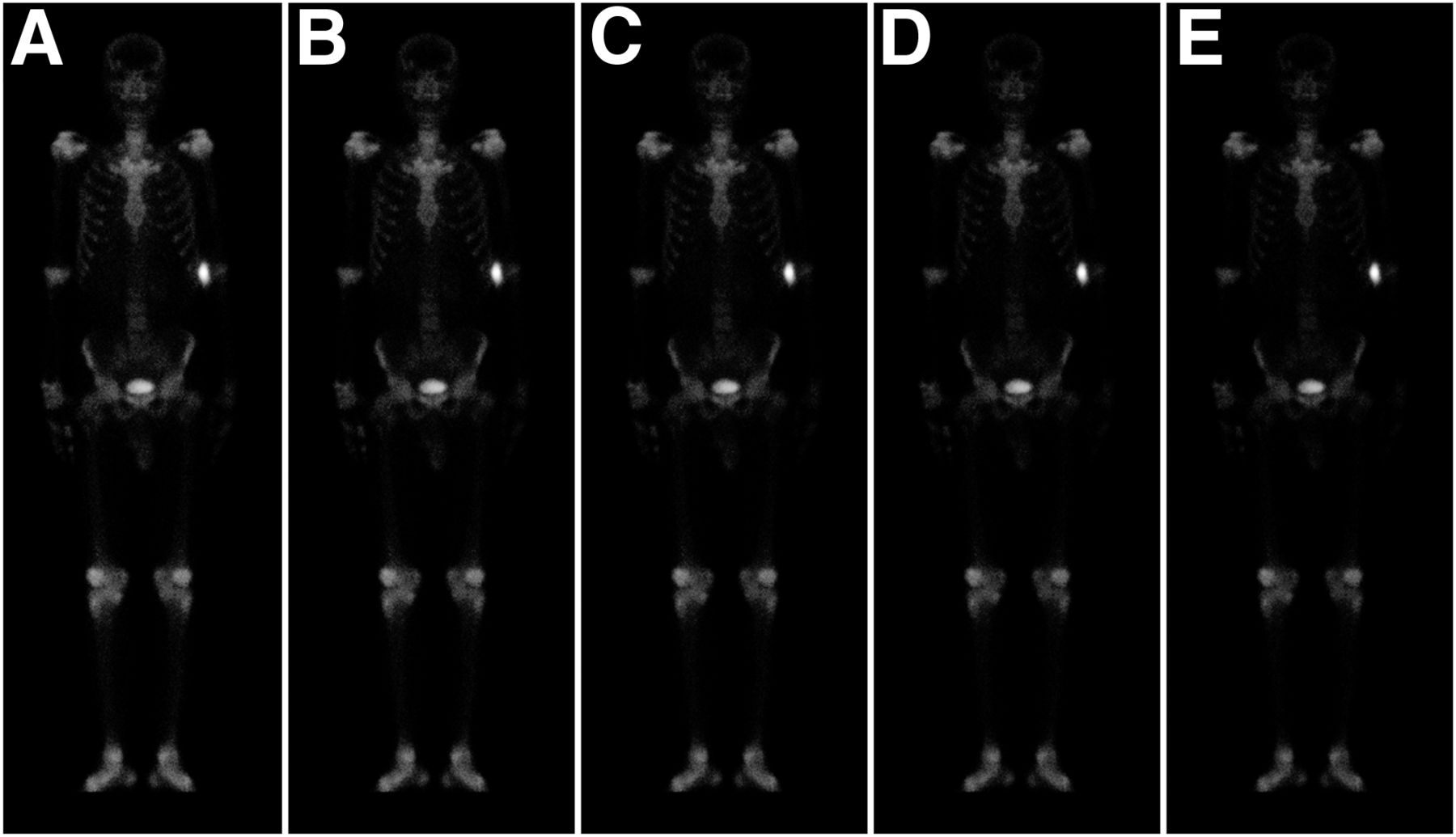
- FIGURE 5.
Processed whole-body 99mTc-MDP bone scintigraphy images with slopes of 10–14 (A–E) and threshold equal to mean count in respective input image.
- FIGURE 6.
(A) Input image. (B and C) Two images processed using window-level contrast-adjustment tool. (D and E) Two images processed using IT function.
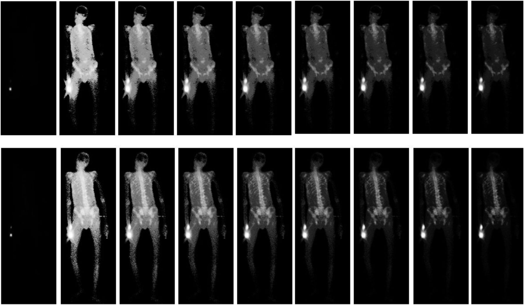
- FIGURE 7.
Two sets of whole-body 99mTc-MDP bone scintigraphy images that did not score on the higher end of the scale.


Tables
Slope n 2 7 3 20 4 25 5 20 6 19 7 28 8 29 9 18 10 9 11 4 12 3 13 1 14 2 ↵* Of 40 total.
Observer 1 Observer 2 Image no. Input score Output score Difference Input score Output score Difference 1 4 4 0 3 4 1 2 3 5 2 2 5 3 3 4 4 0 4 5 1 4 3 5 2 4 5 1 5 4 5 1 3 5 2 6 1 5 4 1 4 3 7 3 5 2 2 5 3 8 1 4 3 1 3 2 9 1 4 3 1 4 3 10 3 4 1 2 4 2 11 1 5 4 1 5 4 12 3 4 1 2 4 2 13 2 5 3 2 5 3 14 2 4 2 1 4 3 15 1 4 3 1 4 3 16 2 5 3 2 5 3 17 3 5 2 2 5 3 18 3 5 2 3 5 2 19 1 4 3 1 4 3 20 1 5 4 1 5 4 21 4 5 1 3 5 2 22 2 3 1 2 4 2 23 1 4 3 1 3 2 24 3 5 2 2 5 3 25 3 5 2 3 5 2 26 2 5 3 2 5 3 27 2 5 3 2 5 3 28 2 5 3 2 5 3 29 3 5 2 2 5 3 30 3 5 2 2 5 3 31 1 3 2 1 3 2 32 1 3 2 1 3 2 33 1 2 1 1 2 1 34 1 2 1 1 2 1 35 1 4 3 1 4 3 36 2 4 2 2 5 3 37 1 4 3 2 5 3 38 1 3 2 1 3 2 39 1 3 2 1 3 2 40 1 4 3 2 5 3 Difference ≥ 0 indicates output image same as or better than input image.
Input image Output image Score Observer 1 Observer 2 Average Observer 1 Observer 2 Average 1 17 16 16.5 0 0 0 2 8 17 12.5 2 2 2 3 11 5 8 5 6 5.5 4 4 2 3 14 10 12 5 0 0 0 19 22 20.5


Jump to section
Related Articles
Cited By...
- No citing articles found.