


Article Figures & Data
Figures
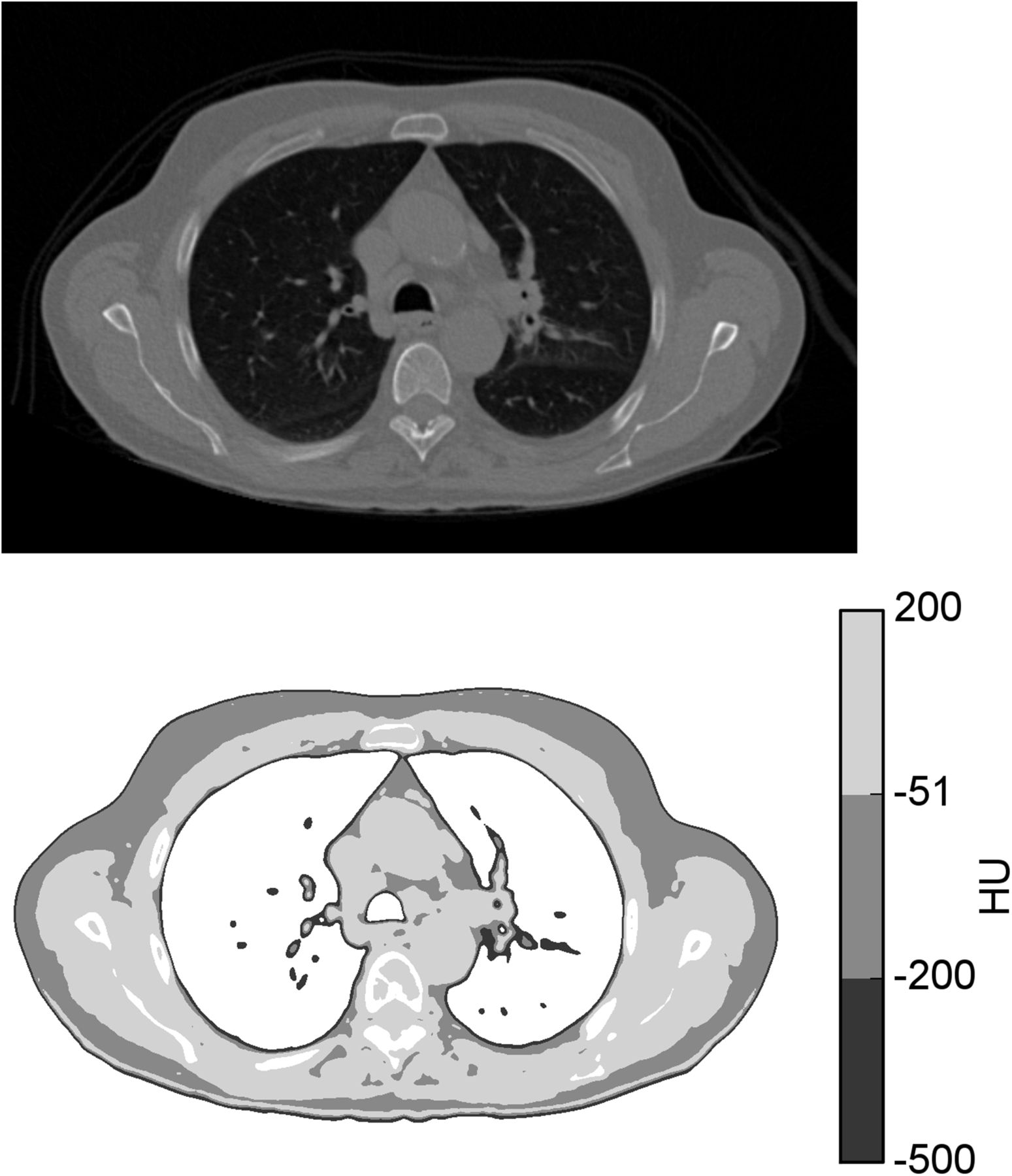
- FIGURE 1.
Tissue segmentation in accordance with CT-based method described by Hamil et al. (18). (Top) CT thorax image in axial projection. (Bottom) Regions segmented as skin, fat, and LBM in Hounsfield units (HU) of −500 to −201, −200 to −51, and −50 to 200, respectively.
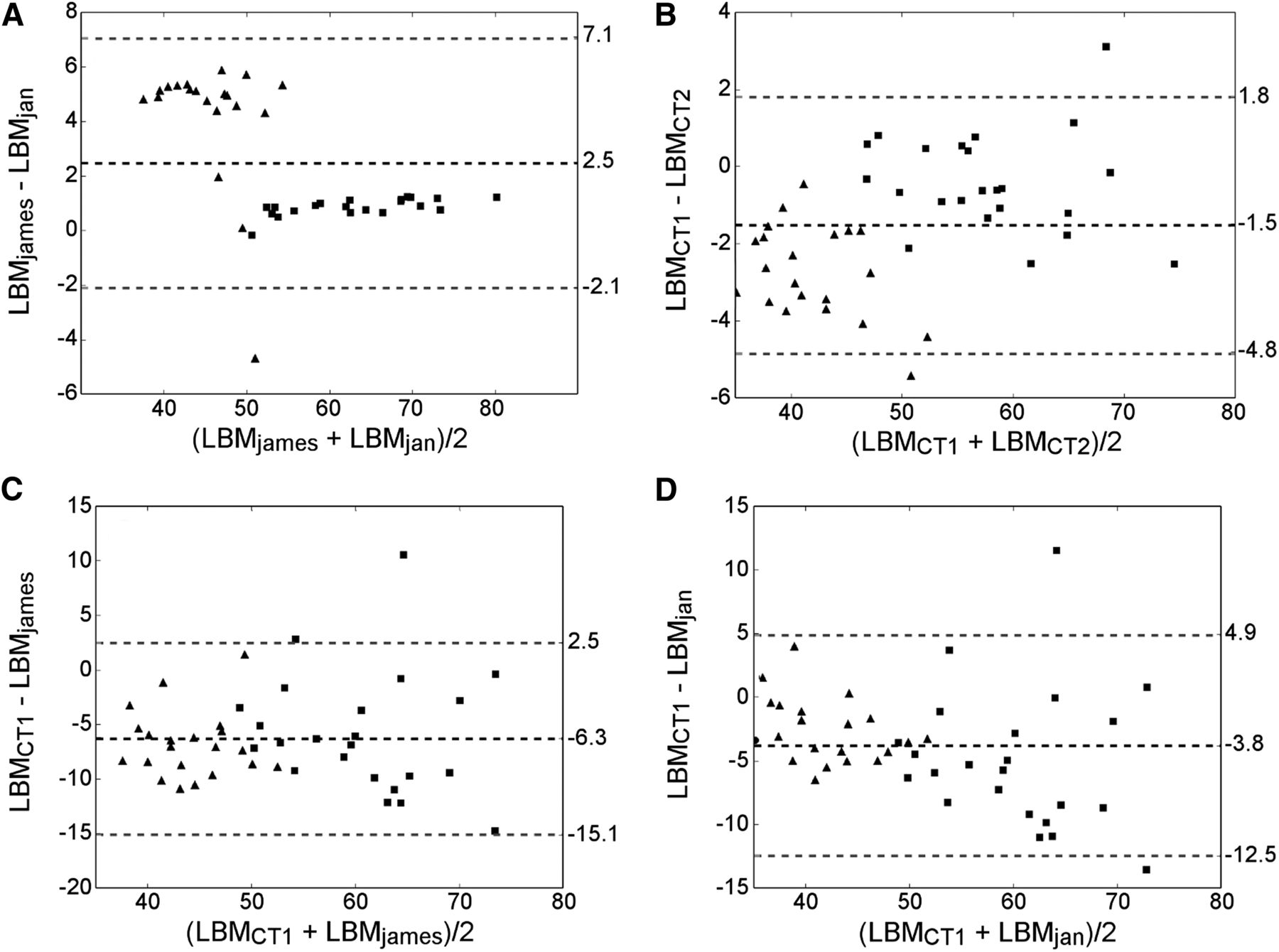
- FIGURE 2.
Bland–Altman plots illustrating differences between
 and (A), and (B), and (C), and and (D). Dashed black lines denote mean difference, whereas gray dashed lines denote limits of agreement. Squares and triangles denote male and female patients, respectively.
 - FIGURE 3.
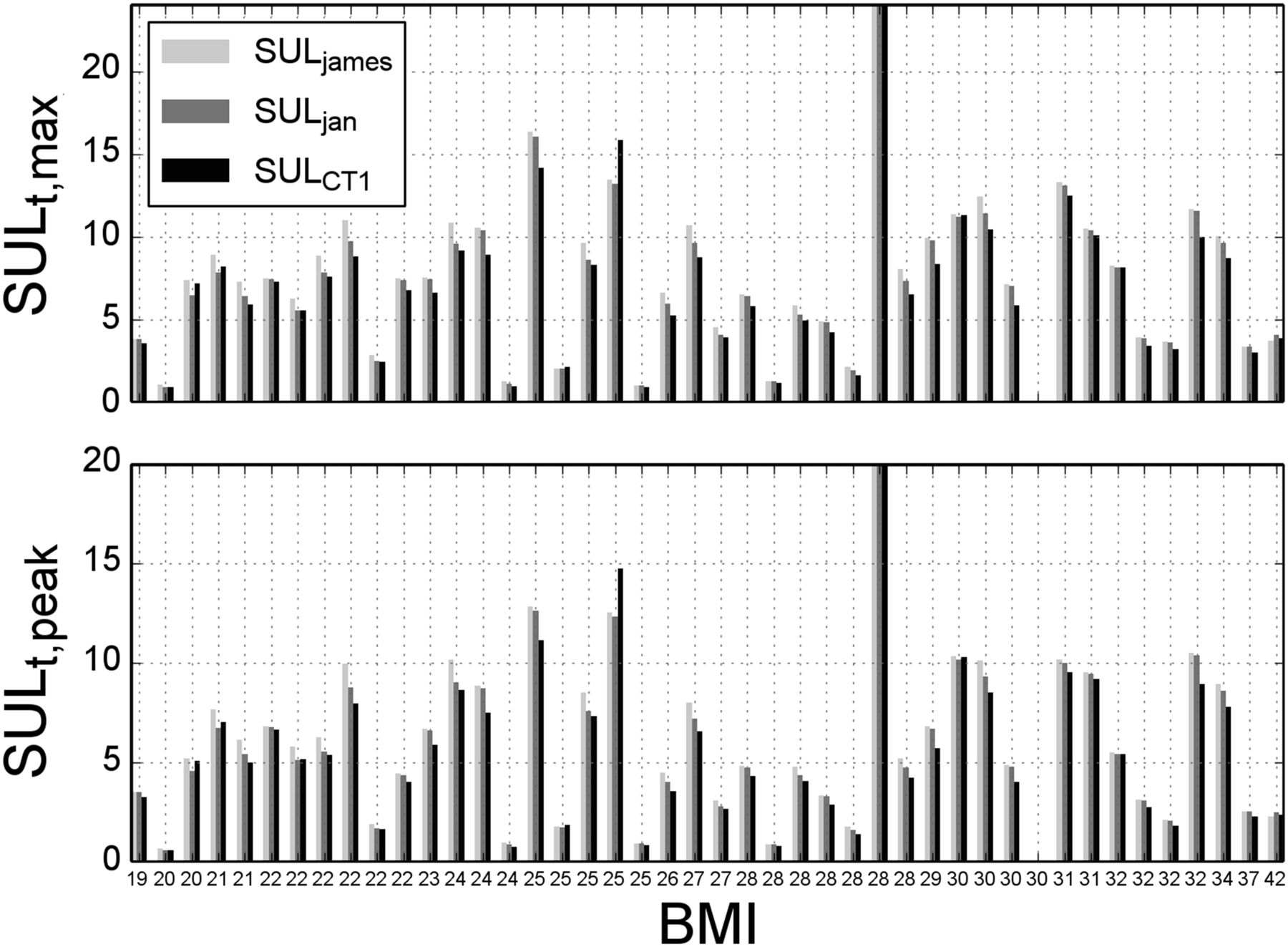
Bar plots showing how variations in LBM calculation can affect maximum and peak SUL for individual tumors. SUL of a single tumor is given for each patient, with patients being sorted by increasing BMI.
- FIGURE 4.
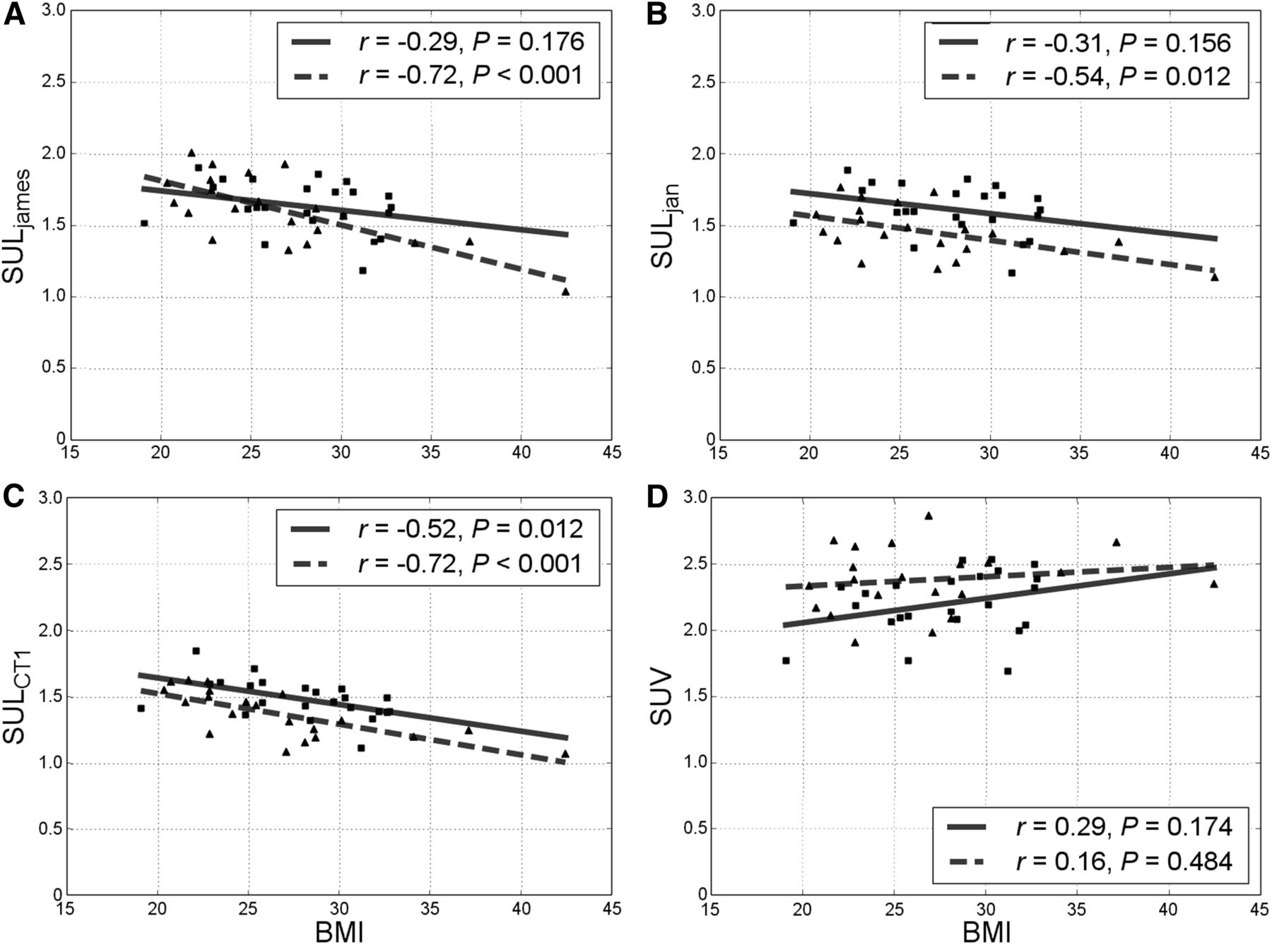
Liver SUL vs. BMI for
(A), , (B) (C), and BMI (D). Squares and triangles denote male and female patients, respectively, with associated solid regression lines.


Tables
BMI range Characteristic 18.5–24.9 25.0–29.9 30.0–∞ Subjects (n) 15 16 13 Measured BMI 22.4 ±± 1.6 27.3 ±± 1.4 32.7 ±± 4.1 Height (m) 1.66 ±± 0.06 1.71 ±± 0.07 1.73 ±± 0.10 Weight (kg) 62.2 ±± 6.1 80.1 ±± 9.0 98.0 ±± 8.8 Sex (n) Male 5 9 9 Female 10 7 4 Method LBM ICC 95% confidence interval P 55.8 ±± 10.2 0.77 0.11–0.91 53.3 ±± 11.7 0.87 0.60–0.94 51.0 ±± 9.6 0.98 0.89–0.99 49.5 ±± 10.4 1 —


