


Article Figures & Data
Figures
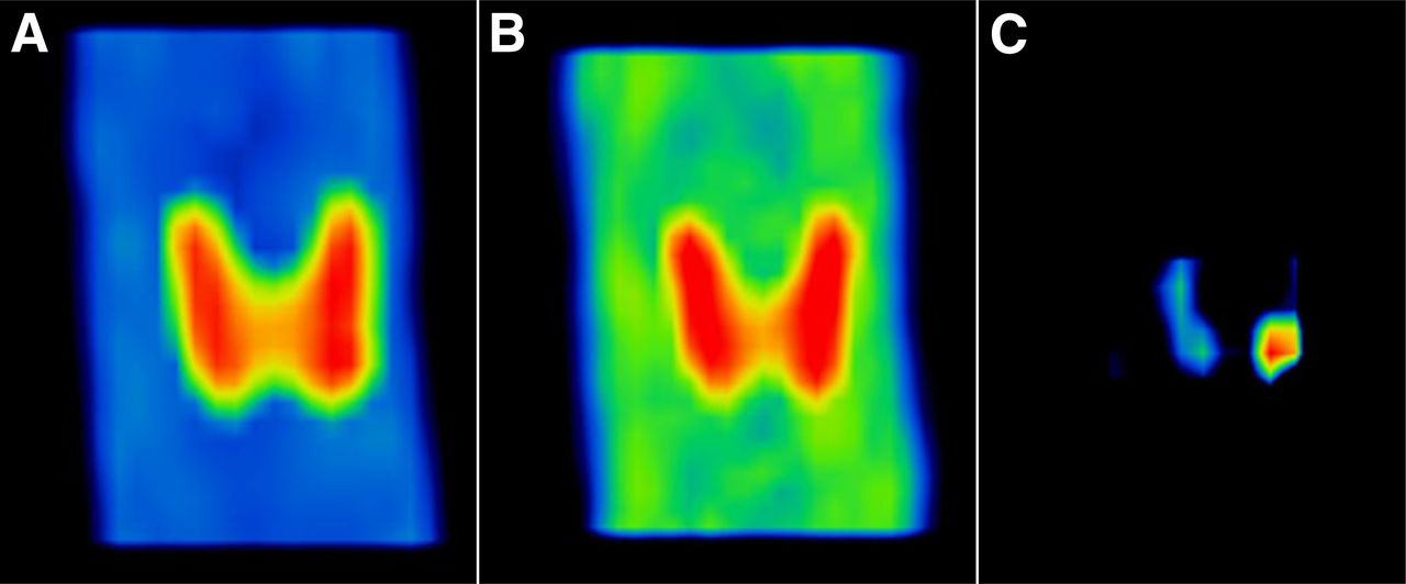
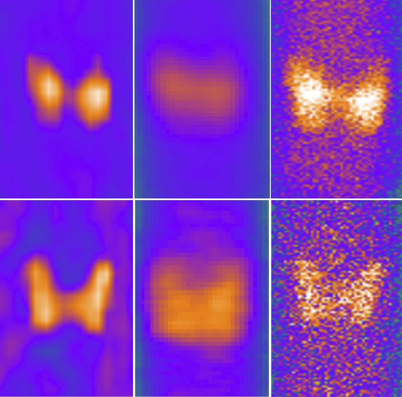
- FIGURE 1.
Typical thyroid images using 99mTc (top) and 123I (bottom) tracers. Shown are MIP images (left), PE images (middle), and NaI planar images (right).
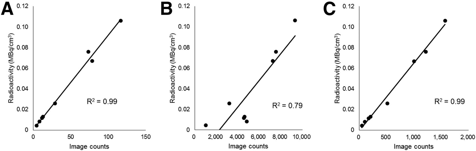
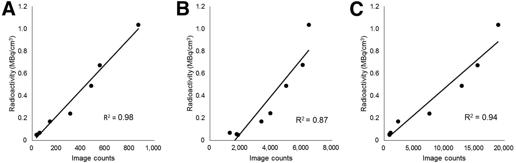
- FIGURE 2.
Relationship between image counts and radioactivity of 99mTc-containing thyroid phantoms: NaI planar image (A), MIP image (B), and PE image (C).
- FIGURE 3.
Relationship between image counts and radioactivity of 123I thyroid phantoms: NaI planar image (A), MIP image (B), and PE image (C).
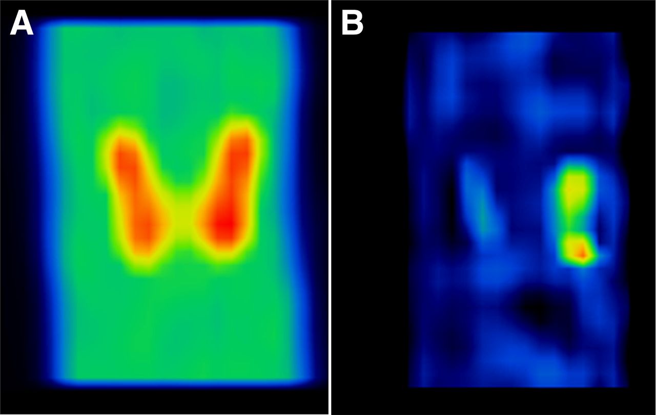
- FIGURE 4.
Typical early (A) and delayed (B) images from dual-phase 99mTc-sestamibi protocol using MIP. Clear accumulation of tracer is visible in lower quadrant of left thyroid lobe in delayed image.
- FIGURE 5.
Typical single-tracer (A), dual-tracer (B), and subtracted (C) images from 99mTc-sestamibi/99mTc-pertechnetate protocol using MIP. After single-tracer image was subtracted from dual-tracer image, solitary parathyroid adenoma was clearly visible.
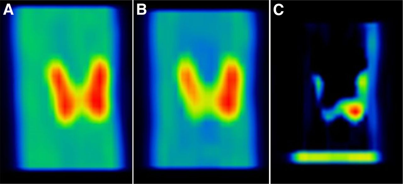
- FIGURE 6.
Representative 99mTc photopeak (A), 123I photopeak (B), and subtracted (C) parathyroid images from 99mTc-sestamibi/123I-iodine protocol using MIP. 99mTc tracer is seen in thyroid and parathyroid glands, and 123I is distributed in thyroid tissue. Parathyroid adenoma was then clearly visualized by subtracting 123I image from 99mTc image.


Tables
Protocol Thyroid (MBq) Parathyroid (MBq) Background (MBq/mL) Dual-phase 99mTc-sestamibi Early phase (99mTc) 4.0 0.1 0.02 Delayed phase (99mTc) 0.5 0.07 0.014 Dual-tracer 99mTc-sestamibi/99mTc-pertechnetate Single tracer (99mTc) 4.0 — 0.02 Dual tracer (99mTc) 8.0 0.1 0.04 Dual-tracer 99mTc-sestamibi/123I-iodine 99mTc 4.0 0.1 0.02 123I 0.74 — 0.006 Single-tracer acquisition Window CZT (keV) NaI (keV) Dual-tracer acquisition (CZT [keV]) 99mTc 140.5% ± 10% 140.5% ± 15% 140.5% ± 5% 123I 159% ± 10% 159% ± 15% 159% ± 5%


Jump to section
Related Articles
Cited By...
- No citing articles found.