


Article Figures & Data
Figures
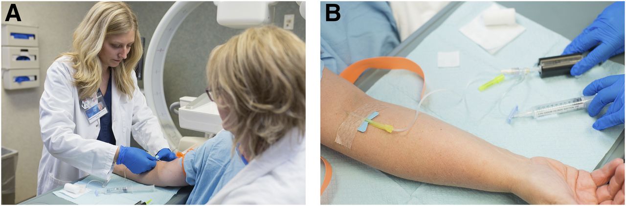
- FIGURE 1.
(A) Intravenous injection of 99mTc-sestamibi via butterfly needle. (B) Radiotracer in low-adhesion syringe (encased in syringe shield) connected to 10-mL saline flush for injection.
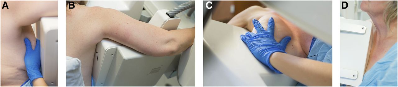
- FIGURE 2.
Proper breast positioning for craniocaudal view. (A) Patient is seated in chair with shoulder relaxed and arms resting in lap. While breast is guided to camera, IMF is lifted until nipple extends perpendicular to chest wall. (B) With nipple extending perpendicular to chest wall, 2 hands are used to lift breast onto detector. (C) Lateral aspect of breast is pulled and anchored into FOV while compression is slowly applied. (D) Compression is applied in this craniocaudal view. (E) Patient’s back is supported with pillows to increase comfort and prevent motion.
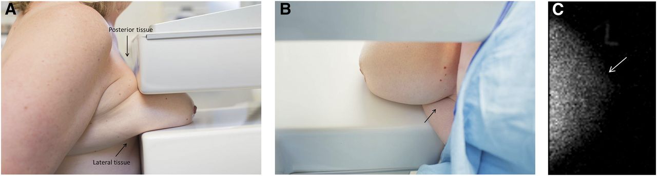
- FIGURE 3.
Improper breast positioning for craniocaudal view. (A) Lateral and posterior breast tissue is not fully included in FOV. (B) Camera is lower than IMF, creating fold underneath breast and causing nipple to point inferiorly. (C) Motion during craniocaudal (CC) acquisition creates appearance of blurred edge to breast tissue on resultant image.
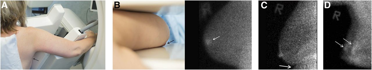
- FIGURE 4.
Proper breast positioning for MLO view. (A) Detector angle matches angle of pectoral muscle. (B) Patient’s arm is on top of detector, with no gap (arrow) between detector and chest. (C) IMF (arrow) is opened by lifting breast up and away from chest wall. (D) In a medial view of the MLO with compression applied, there is no gap between upper detector and chest wall, and corner of detector rests below clavicle.
- FIGURE 5.
Improper breast positioning for MLO view. (A) Gap (arrow) between detector and chest indicates improper positioning. (B) MLO view of breast with nipple not positioned in profile (black arrow) creates appearance of pseudo lesion (white arrow) on resultant image. Radioactive “R” marker indicates laterality. (C) In patient with larger body habitus in whom IMF is not open, abdominal tissue is included in FOV (arrow). (D) Motion on MLO view creates appearance of 2 outlines of breast.
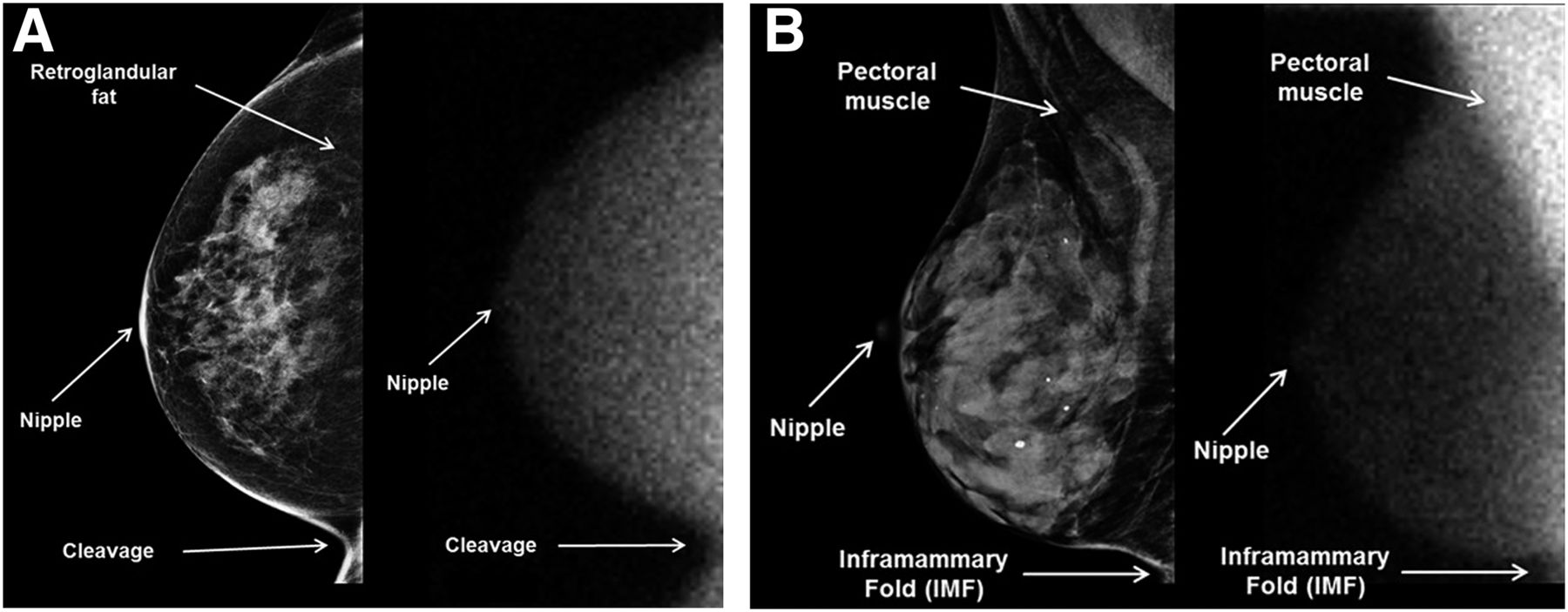
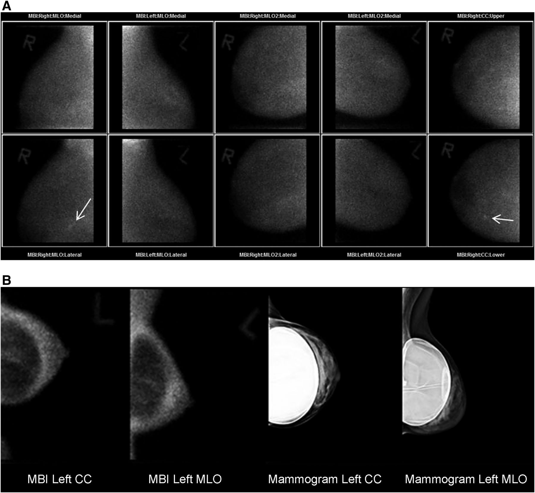
- FIGURE 6.
Proper breast positioning on mammography (left) and MBI (right). (A) Presence of cleavage and all lateral breast tissue while nipple is kept in profile indicates properly positioned breast for craniocaudal view. (B) Presence of pectoral muscle, nipple in profile, and open IMF indicate properly positioned breast for MLO view.
- FIGURE 7.
Common imaging variants. (A) Tiled MLO views were performed on patient whose breast size exceeded MBI FOV, with additional craniocaudal (CC) view being acquired to assess area of uptake (arrow). (B) Breast implants appear photopenic on MBI views.


Tables
- TABLE 1
Guidelines for Waiting Periods Between Imaging Procedures Performed Before MBI Examination
Modality Procedure Administered dose Recommended waiting period CT With or without contrast None MRI With or without contrast None PET 18F-FDG 555 MBq (15 mCi) >12 h 11C-choline 370–740 MBq (10–20 mCi) >2 h 13N-ammonia 370–740 MBq (10–20 mCi) >1 h Nuclear medicine 99mTc-sestamibi for parathyroid or cardiac studies 370–1,110 MBq (10–30 mCi) None* 99mTc-radiopharmaceuticals, intravenous >740 MBq (20 mCi) >24 h >370 MBq (10 mCi) >18 h >185 MBq (5 mCi) >12 h <185 MBq (5 mCi) >6 h 99mTc-radiopharmaceuticals, oral <185 MBq (5 mCi) None 123I-based radiopharmaceuticals >370 MBq (10 mCi) >2 d 111In- and 67Ga-based radiopharmaceuticals 185–370 MBq (5–10 mCi) >3 d <37 MBq (1 mCi) >24 h These are general guidelines and in some cases may overestimate waiting period needed, depending on biologic distribution of radiopharmaceutical.
↵* In cases of prior administration of 99mTc-sestamibi for cardiac or parathyroid studies, reduced (or no dose) of 99mTc-sestamibi may be used, depending on residual activity from prior study.
- TABLE 2
Guidelines for Waiting Periods Between MBI Examination and Other Imaging Procedures
Modality Procedure Administered dose Recommended waiting period CT With or without contrast None MRI With or without contrast None PET All radiopharmaceuticals None Nuclear medicine 99mTc-sestamibi for parathyroid (or cardiac rest only) studies 370–1,110 MBq (10–30 mCi) None* 99mTc-radiopharmaceuticals, intravenous or oral >740 MBq (20 mCi) >12 h >370 MBq (10 mCi) >24 h >185 MBq (5 mCi) >24 h <185 MBq (5 mCi) >36 h 99mTc-sulfur colloid (sentinel node) <19 MBq (500 μCi) >24 h 123I-based radiopharmaceuticals >370 MBq (10 mCi) None <37 MBq (1 mCi) >24 h 111In- and 67Ga-based radiopharmaceuticals None These are general guidelines and in some cases may overestimate waiting period needed, depending on biologic distribution of radiopharmaceutical.
↵* May consider combining dose administration. In cases of prior administration of 99mTc-sestamibi for cardiac or parathyroid studies, reduced (or no dose) of 99mTc-sestamibi may be used, depending on residual activity from prior study.


