


Article Figures & Data
Figures
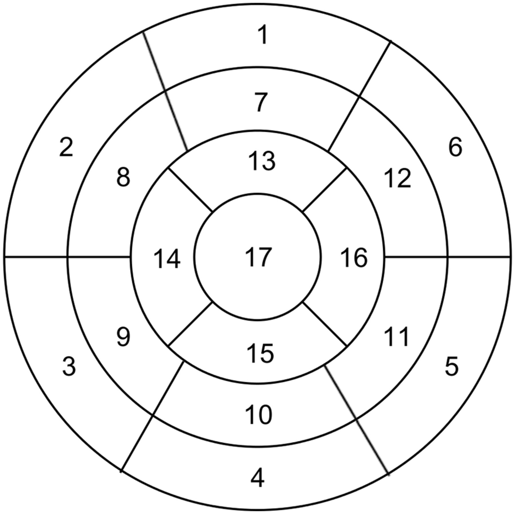
- FIGURE 1.
17-segment classification of polar map.
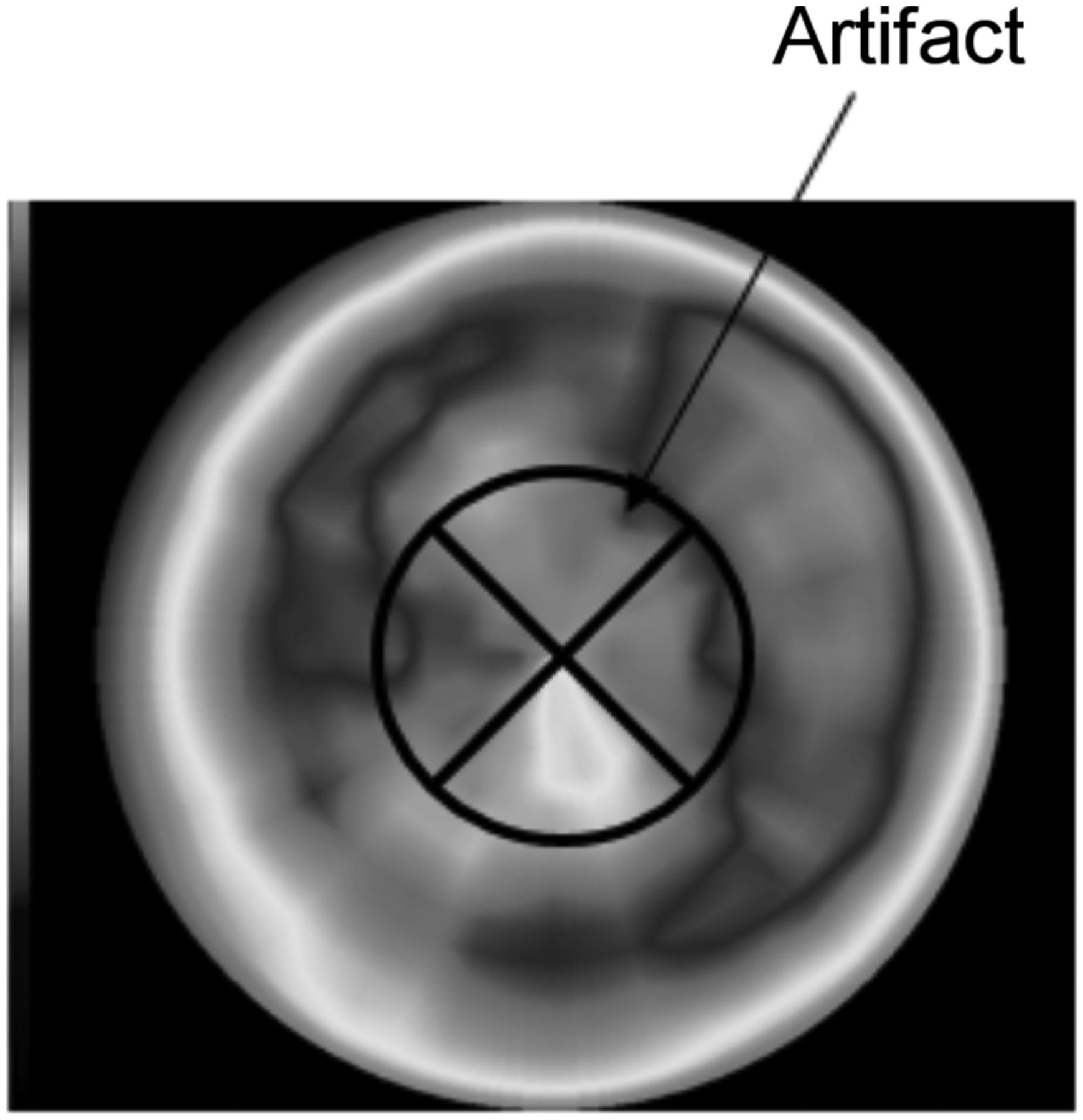
- FIGURE 2.
Apical artifact appearing with upright D-SPECT imaging.
- FIGURE 3.
Percentage uptake ratios for prone and upright images. Mann–Whitney U test was used to show significant differences between imaging methods. (A) Segment 3/2. (B) Segment 4/1. (C) Segment 5/6. (D) Segment 9/8. (E) Segment 10/7. (F) Segment 11/12. (G) Segment 15/13.
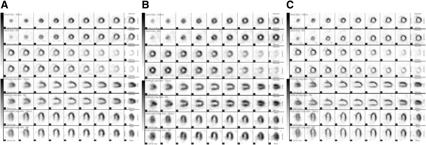
- FIGURE 4.
Supine (A), prone (B), and upright (C) SPECT images in same patient. Top row of each set is stress image, and bottom row is resting image.


Tables
Characteristic Prone group (n = 23) Upright group (n = 22) Age (y) 69 ± 10 74 ± 4 Male (n) 23 (100%) 22 (100%) Height (m) 1.67 ± 0.05 1.66 ± 0.06 Body weight (kg) 64.5 ± 6.02 64.5 ± 9.19 Body mass index 23.2 ± 1.79 23.3 ± 2.37 Coronary risk factors Smoking 14 (60.8%) 18 (78.2%) Hypertension 18 (78.3%) 18 (78.2%) Hyperlipidemia 16 (69.6%) 18 (78.2%) Diabetes mellitus 10 (43.5%) 7 (30.4%) Familial hypercholesterolemia 4 (17.4%) 8 (34.7%) Impaired glucose tolerance 0 (0%) 1 (4.3%) Other factors Blood glucose 128.7 ± 63.2 120.3 ± 36.2 Hemoglobin A1c 6.30 ± 0.87 6.57 ± 0.72 High-density-lipoprotein cholesterol 54.6 ± 11.6 49.9 ± 14.4 Low-density-lipoprotein cholesterol 90.9 ± 24.4 87.9 ± 19.4 Study protocol Exercise time 8 min 9 s 7 min 26 s Maximum stress (watt [%]) 121.7 ± 24.8 110.2 ± 24.6 (Target heart rate/maximum heart rate) × 100 (%) 91.7 ± 11.4 87.9 ± 19.4 Patients reaching target heart rate (n) 8 (34.7%) 7 (30.4%) Maximum blood pressure (mm Hg) 205.8 ± 25.8 196.7 ± 28.5 Patients in whom ST changed on electrocardiography (n) 3 (13.0%) 2 (8.70%) There were no significant differences between the prone and upright groups.


