


Article Figures & Data
Figures
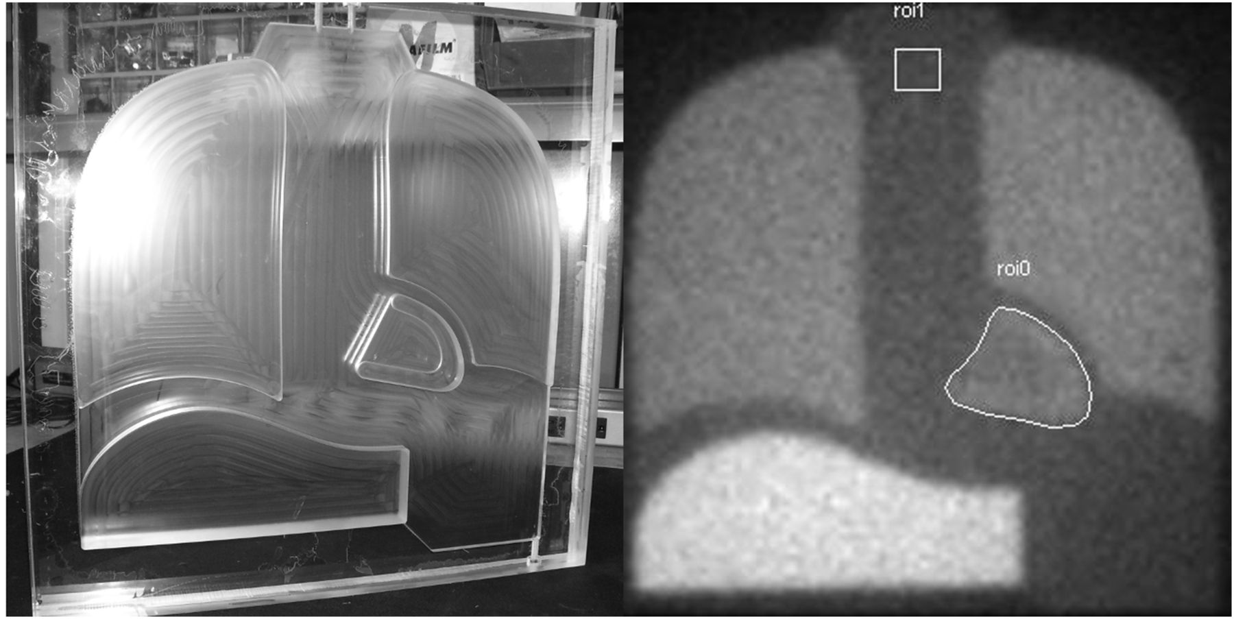
- FIGURE 1.
Single-chamber 123I-MIBG thorax phantom. Photograph of unfilled phantom (A) and example planar acquisition after filling with 123I (B). Polygonal heart and square mediastinum ROIs of standardized image analysis are shown.
- FIGURE 2.
Box-and-whisker plot of head 1 H/M ratio. Bottom and top of box are first and third quartiles, respectively. Horizontal line in box is median. + in box represents mean. Ends of the whiskers are minimum and maximum. Cam1 = GE Healthcare Millennium MG/LEHR (n = 10); Cam2 = GE Healthcare Discovery/LEHR (n = 8); Cam3 = GE Healthcare Infinia/LEHR (n = 10); Cam4 = Philips Brightview/LEHR (n = 2); Cam5 = Philips Cardio 60/LEHR (n = 2); Cam6 = Philips Cardio MD/LEHR (n = 4); Cam7 = Philips Vertex/VXGP (vertex general purpose) or VXHR (vertex high resolution) (n = 8); Cam8 = Siemens e.cam/LEHR (n = 8); Cam9 = Siemens Symbia/LEHR (n = 8). VXGP = vertex general purpose; VXHR = vertex high resolution.
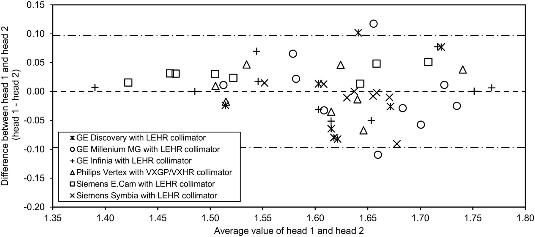
- FIGURE 3.
Bland–Altman plot for reader 1. Mean H/M (i.e., values for both heads) for each camera is plotted against difference between head 1 H/M and head 2 H/M. Results from reader 2 were similar to those shown here. VXGP = vertex general purpose; VXHR = vertex high resolution.


Tables
Manufacturer Model LEHR collimator type Mean septal thickness (mm) GE Healthcare Millennium MG Cast 0.20 GE Healthcare Infinia and Discovery NM series Foil 0.20 Siemens e.cam and Symbia Foil 0.16 ADAC/Philips Vertex Foil 0.152 (26) Philips Brightview Foil 0.156 (27) - TABLE 2
Summary of Camera Models, Collimator Types, and Sample Size Per Model Included in Study
Camera model Collimator type Sample size GE Healthcare, Millennium MG LEHR 10 GE Healthcare, Discovery LEHR 8 GE Healthcare, Infinia LEHR 10 Philips, Vertex VXGP/VXHR 8 Siemens, e.cam LEHR 8 Siemens, Symbia LEHR 8 Philips, Brightview LEHR 2 Philips, Cardio 60 LEHR 2 Philips, Cardio MD LEHR 4 VXGP = vertex general purpose; VXHR = vertex high resolution.
Measurement GE Healthcare, Millennium MG/LEHR (n = 10) GE Healthcare, Discovery/LEHR (n = 8) GE Healthcare, Infinia/LEHR (n = 10) Philips, Brightview/LEHR (n = 2) Philips, Cardio 60/LEHR (n = 2) Philips, Cardio MD/LEHR (n = 4) Philips, Vertex/VXGP or VXHR (n = 8) Siemens, e.cam/LEHR (n = 8) Siemens, Symbia/LEHR (n = 8) Mean ± SD 1.648 ± 0.0776 1.624 ± 0.0849 1.607 ± 0.1193 1.665 (0.0257) 1.677 ± 0.0302 1.342 ± 0.0262 1.611 ± 0.0866 1.566 ± 0.1118 1.635 ± 0.0455 Median 1.65 (range, 1.52–1.76) 1.62 (range, 1.51–1.75) 1.59 (range, 1.39–1.78) 1.66 (range, 1.65–1.68) 1.68 (range, 1.66–1.70) 1.34 (range, 1.31–1.37) 1.63 (range, 1.49–1.77) 1.53 (range, 1.41–1.73) 1.63 (range, 1.55–1.69) 95% CI 1.592–1.703 1.553–1.695 1.521–1.692 1.434–1.895 1.406–1.949 1.300–1.384 1.539–1.683 1.472–1.659 1.597–1.673 Analysis is based on average of H/M ratios from 2 independent readers on camera head 1 for each camera model.
n = sample size for each camera/collimator combination; VXGP = vertex general purpose; VXHR = vertex high resolution.
Camera/collimator Sample size H/M LSM ± SE* LSM difference ± SE* P* GE Healthcare, Millennium MG/LEHR 10 1.645 ± 0.0286 GE Healthcare, Discovery/LEHR 8 1.621 ± 0.0319 0.024 ± 0.0429 0.5804 GE Healthcare, Infinia/LEHR 10 1.609 ± 0.0286 0.036 ± 0.0404 0.3824 Philips, Vertex/VXGP or VXHR 8 1.604 ± 0.0319 0.040 ± 0.0429 0.3496 Siemens, e.cam/LEHR 8 1.564 ± 0.0319 0.081 ± 0.0429 0.0655 Siemens, Symbia/LEHR 8 1.631 ± 0.0319 0.014 ± 0.0429 0.7496 ↵* Obtained from mixed model with repeated measures on H/M ratios for first head tested as dependent variable and camera/collimator combination as independent variable. Each camera model is compared with reference camera Millennium MG (GE Healthcare).
LSM = least-squares mean; VXGP = vertex general purpose; VXHR = vertex high resolution.
Only camera/collimator categories with a sample size of at least 8 are included in model, as specified in statistical analysis plan.


Jump to section
Related Articles
Cited By...
- No citing articles found.