


Article Figures & Data
Figures
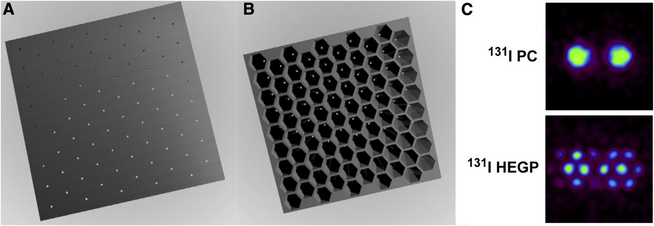
- FIGURE 1.
Face (A) and back (B) of parallel-cone (PC) collimator. (C) Double-point-source Monte Carlo simulations obtained for 131I on PC collimator and high-energy general-purpose (HEGP) collimator. PC collimator can detect the 2 sources separately, whereas HEGP collimator cannot. (Reprinted from (8).)
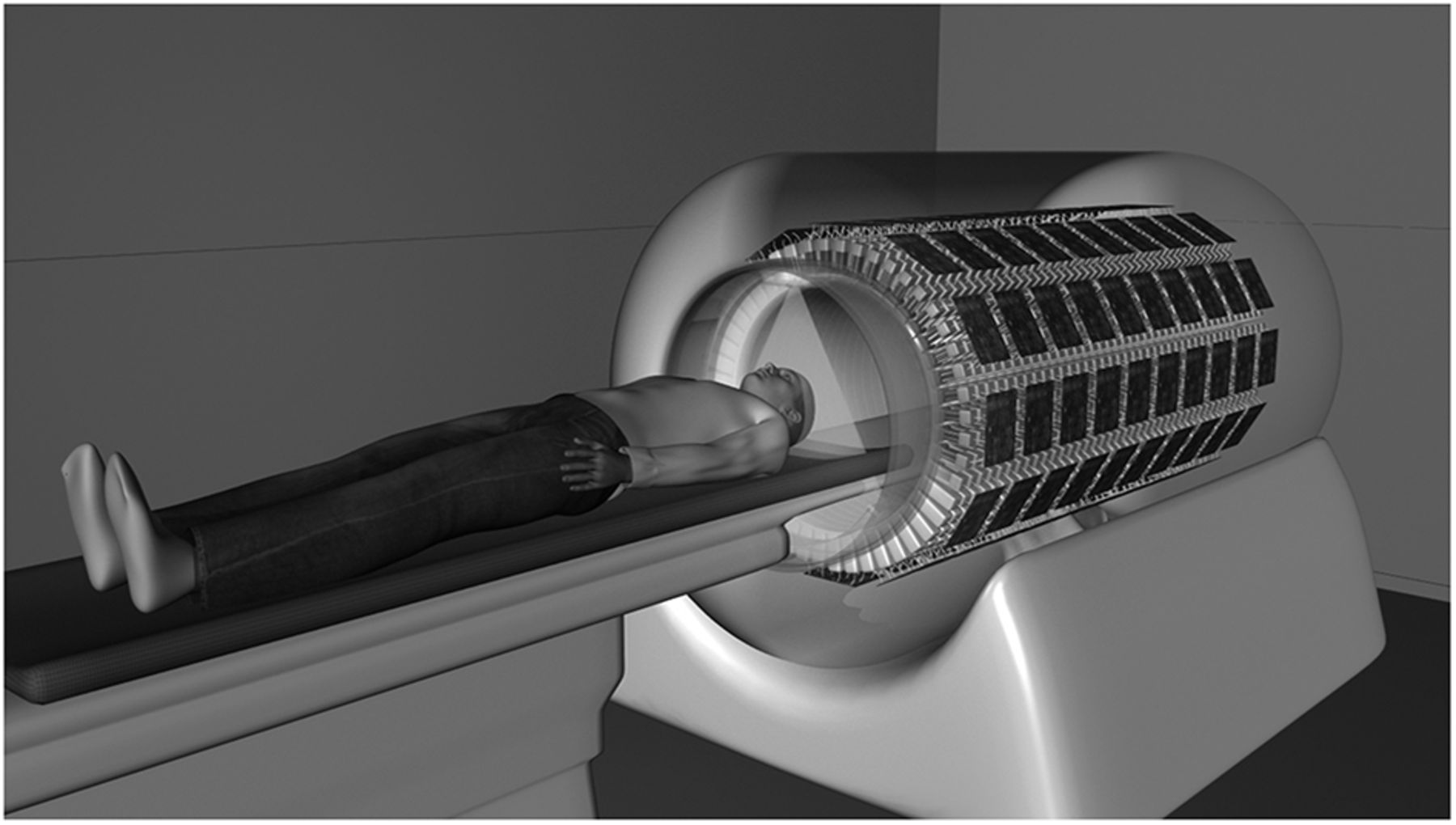
- FIGURE 2.
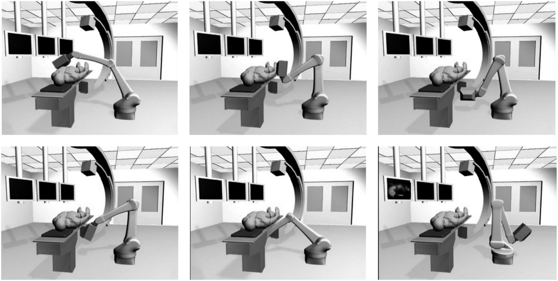
Interventional pinhole SPECT camera mounted on 6-axis arm robot (58). The images show how the robotic gantry is able to acquire tomographic images from multiple angles.
- FIGURE 3.
Explorer total-body PET scanner. (Courtesy of Drs. Simon R. Cherry and Ramsey D. Badawi, University of California, Davis.)
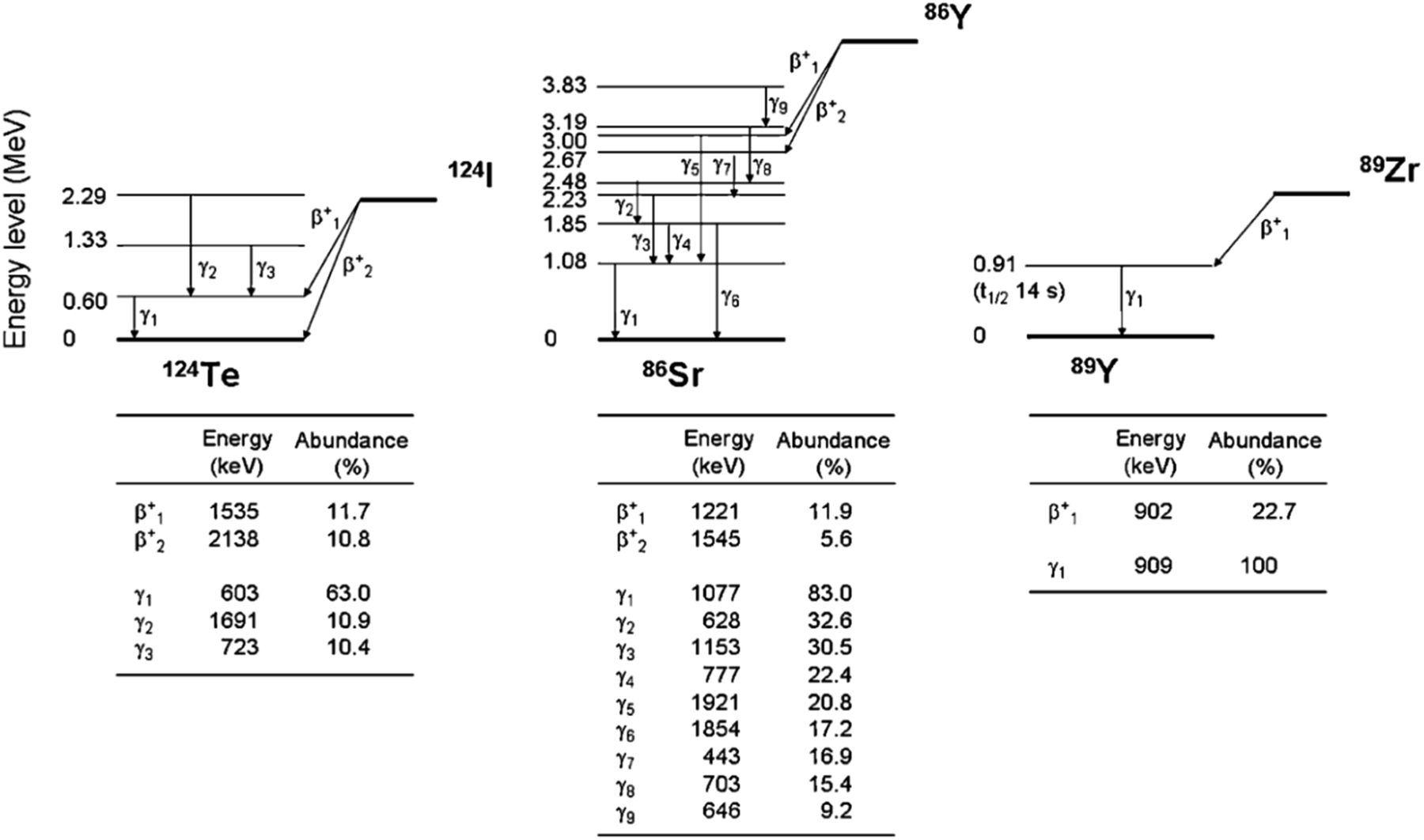
- FIGURE 4.
Decay scheme of several isotopes that emit prompt γ-rays in cascade with positron emission (59).
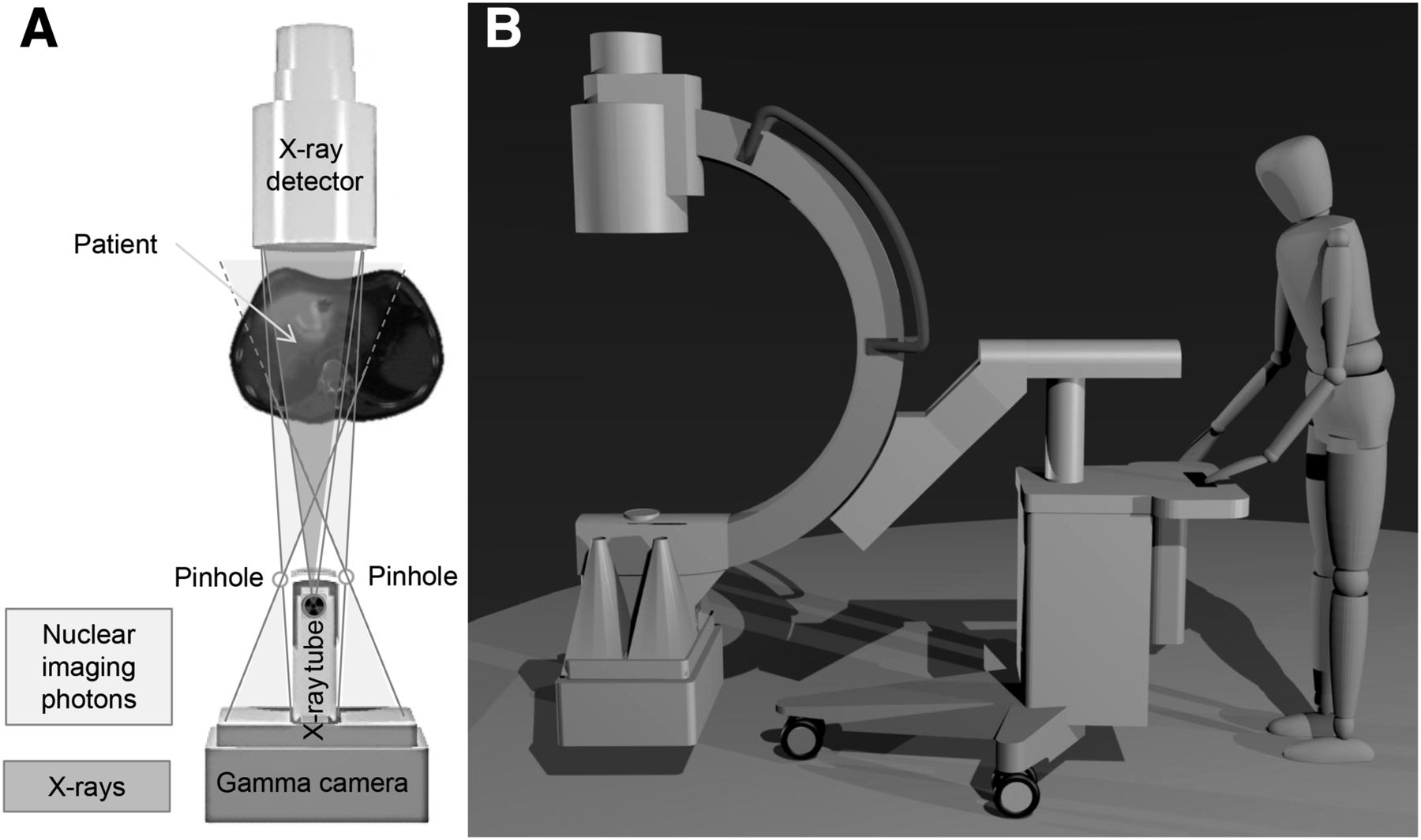
- FIGURE 5.
(A) Hybrid C-arm showing field of view of pinhole collimator and x-ray photons. (B) Entire system (50).
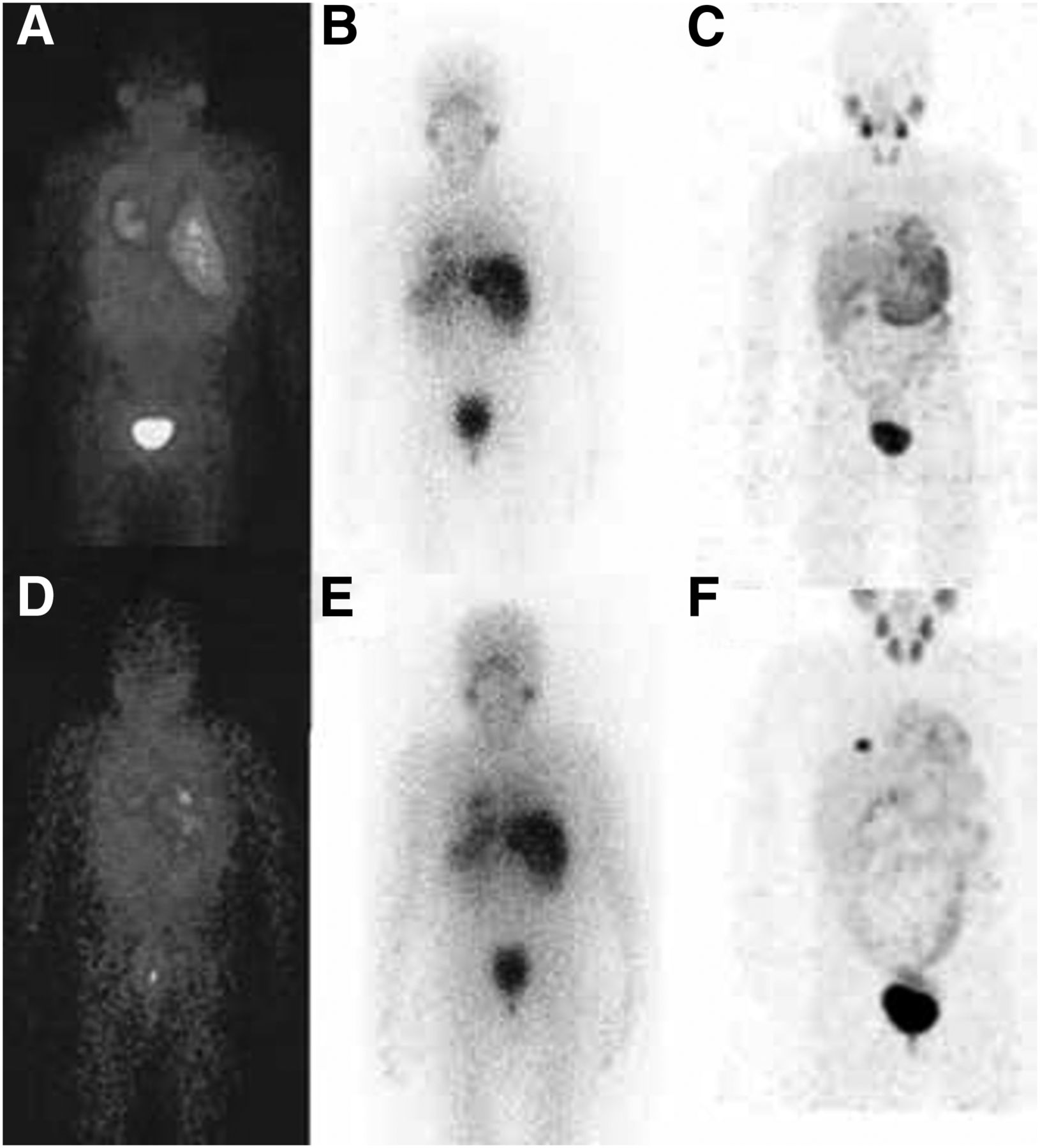
- FIGURE 6.
Diagnostic 123I-MIBG planar acquisitions at 4 h (A) and 24 h (D) after injection, posttherapy 131I-MIBG planar acquisitions at 24 h (B) and 48 h (E) after injection, and 124I-MIBG maximum-intensity-projection acquisitions at 24 h (C) and 48 h (F) after injection (51). Resolution advantage of 124I PET over 123I and 131I SPECT is clear.
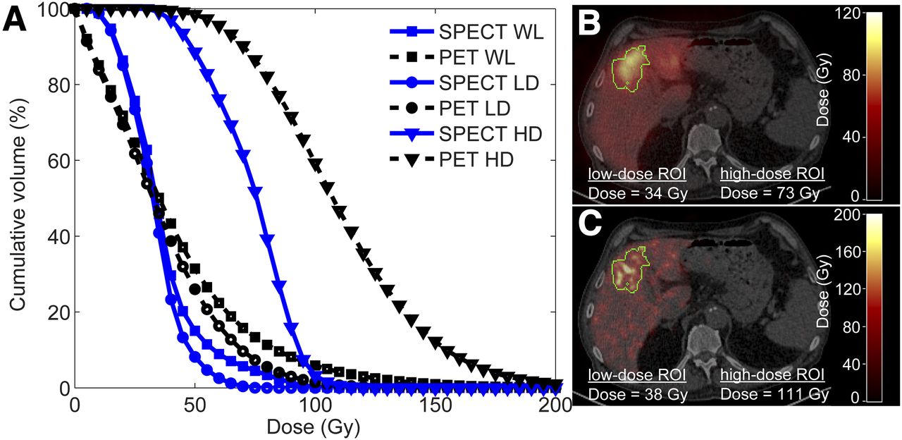
- FIGURE 7.
Comparison of dose estimates based on 90Y PET and 90Y SPECT images after radioembolization procedure. (A) Graph shows cumulative dose–volume histogram of whole liver (WL), low-dose region of interest (LD), and high-dose region of interest (HD). (B and C) Same transversal slice through SPECT-based dose map fused with CT (B) and through PET-based dose map (C) (60) shows advantage of PET over SPECT in terms of resolution and effect on calculated dose distribution.


Jump to section
Related Articles
Cited By...
- No citing articles found.