
Main menu

  • Home
  • Content
    • Current
      • JNMT Supplement
    • Ahead of print
    • Past Issues
    • SNMMI Annual Meeting Abstracts
  • Subscriptions
    • Subscribers
    • Rates
    • Journal Claims
    • Institutional and Non-member
  • Authors
    • Submit to JNMT
    • Information for Authors
    • Assignment of Copyright
    • AQARA Requirements
  • Info
    • Permissions
    • Continuing Education
    • Advertisers
    • Corporate & Special Sales
  • About
    • About Us
    • Editorial Board
    • Editorial Contact
  • More
    • Alerts
    • Feedback
    • Help
    • SNMMI Journals
  • SNMMI
    • JNMT
    • JNM
    • SNMMI Journals
    • SNMMI

User menu

  • Subscribe
  • My alerts
  • Log in
  • My Cart

Search

  • Advanced search
Journal of Nuclear Medicine Technology
  • SNMMI
    • JNMT
    • JNM
    • SNMMI Journals
    • SNMMI
  • Subscribe
  • My alerts
  • Log in
  • My Cart
Journal of Nuclear Medicine Technology

Advanced Search

  • Home
  • Content
    • Current
    • Ahead of print
    • Past Issues
    • SNMMI Annual Meeting Abstracts
  • Subscriptions
    • Subscribers
    • Rates
    • Journal Claims
    • Institutional and Non-member
  • Authors
    • Submit to JNMT
    • Information for Authors
    • Assignment of Copyright
    • AQARA Requirements
  • Info
    • Permissions
    • Continuing Education
    • Advertisers
    • Corporate & Special Sales
  • About
    • About Us
    • Editorial Board
    • Editorial Contact
  • More
    • Alerts
    • Feedback
    • Help
    • SNMMI Journals
  • Follow SNMMI on Twitter
  • Visit SNMMI on Facebook
  • Join SNMMI on LinkedIn
  • Subscribe to JNMT RSS feeds
Review ArticleTeaching Case Studies

Nuclear Imaging of a Cardiac Paraganglioma

Nada Almenieir, Shawn Karls, Vilma Derbekyan and Robert Lisbona
Journal of Nuclear Medicine Technology September 2017, 45 (3) 247-248; DOI: https://doi.org/10.2967/jnmt.116.182212
Nada Almenieir
Division of Nuclear Medicine, Royal Victoria Hospital, Montreal, Quebec, Canada
  • Find this author on Google Scholar
  • Find this author on PubMed
  • Search for this author on this site
Shawn Karls
Division of Nuclear Medicine, Royal Victoria Hospital, Montreal, Quebec, Canada
  • Find this author on Google Scholar
  • Find this author on PubMed
  • Search for this author on this site
Vilma Derbekyan
Division of Nuclear Medicine, Royal Victoria Hospital, Montreal, Quebec, Canada
  • Find this author on Google Scholar
  • Find this author on PubMed
  • Search for this author on this site
Robert Lisbona
Division of Nuclear Medicine, Royal Victoria Hospital, Montreal, Quebec, Canada
  • Find this author on Google Scholar
  • Find this author on PubMed
  • Search for this author on this site
  • Article
  • Figures & Data
  • Info & Metrics
  • PDF
Loading

Abstract

We report a case of a cardiac paraganglioma in the right atrioventricular groove in which the use of different nuclear medicine studies aided in the diagnosis.

  • paraganglioma
  • cardiac
  • succinate dehydrogenase/deficiency

Nuclear imaging is useful for the evaluation of suspected paragangliomas. 123I/131I-metaiodobenzylguanidine (MIBG) scintigraphy is the initial recommended study but is associated with a high false-negative rate, as the sensitivity is 18%–50% (1). Additional nuclear imaging with 111In-pentetreotide can be used, with a sensitivity and specificity of 94% and 75%, respectively (2). Also, studies show that 18F-FDG PET is as specific as 123/131I-MIBG for detection of the primary tumor and metastases and even more sensitive than 123I-MIBG and CT/MRI for detection of metastatic disease (3). Mutations in the SDHx genes are associated with paragangliomas. All hereditary SDHx paraganglioma syndromes have an autosomal dominant inheritance pattern with varying penetrance (4).

CASE REPORT

A 25-y-old woman was referred to the cardiology department because she was experiencing palpitations. An echocardiogram showed a large mass anterior to the right ventricle, measuring 6 × 5 cm. The mass appeared to be compressing the right ventricle in the right atrioventricular groove. Chest CT (Fig. 1A) and cardiac MRI were performed. CT showed the mass to be heterogeneously hyperenhancing, with a central stellate “scar” centered on the right atrium; the imaging characteristics were suggestive of a cardiac hemangioma. Cardiac paraganglioma was included in the differential diagnosis. Cardiac MRI showed the lesion to be predominantly solid, with several small cystic components, but the scan did not otherwise contribute to the characterization of the lesion.

FIGURE 1.
  • Download figure
  • Open in new tab
  • Download powerpoint
FIGURE 1.

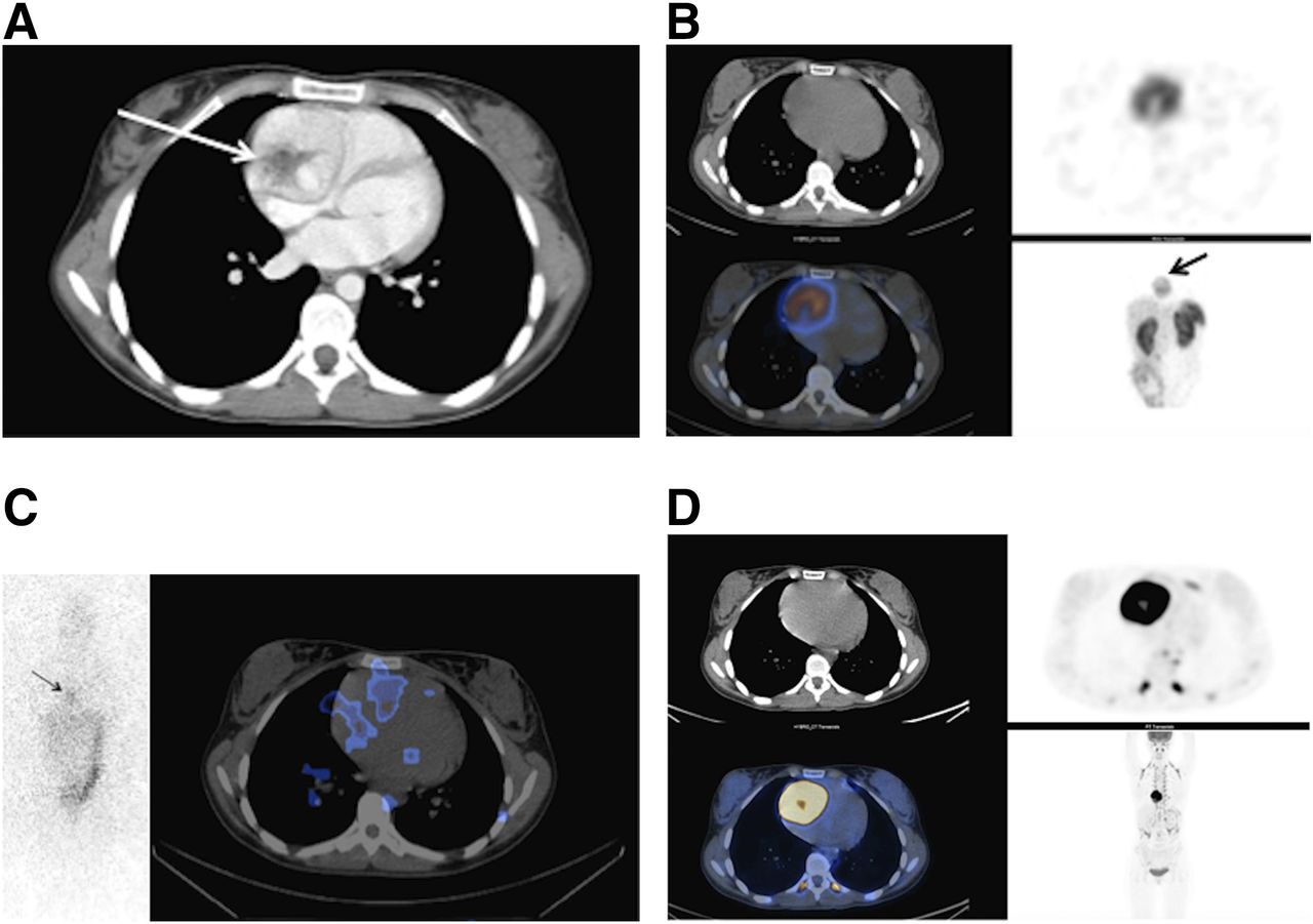
(A) Chest CT image showing hyperenhancing mass with hypoenhancing thin rim and central stellate “scar.” (B) 111In-pentetreotide SPECT/CT images showing focal, intense uptake by mass. (C) 131I-MIBG anterior planar and SPECT/CT images showing only mild heterogeneous radiotracer accumulation in mass. (D) 18F-FDG PET/CT images showing mass to be intensely hypermetabolic with central necrosis.

The symptomology was suggestive of a neuroendocrine tumor, with elevated levels of serum chromogranin A, 24-h urinary norepinephrine, dopamine, and normetanephrine. 111In-pentetreotide scanning was performed and showed intense increased uptake by the mass (Fig. 1B). To evaluate treatment options, 131I-MIBG scanning was performed and showed only a mild heterogeneous accumulation of radiotracer in the mass (Fig. 1C). 18F-FDG PET/CT was performed to rule out metastasis (Fig. 1D). It showed the mass to be intensely hypermetabolic with central necrosis, an SUVmax of 31, and no metastasis. A multigated acquisition was obtained to evaluate the cardiac dynamics and showed no impairment (left and right ventricular ejection fractions, 64% and 52%, respectively).

The mass was surgically resected and the right ventricle repaired with a bovine pericardial patch. Because the presurgical coronary angiogram showed the proximal right coronary artery to be dilated, with a large branch feeding the mass, bypass grafting of the right coronary artery was performed. Pathologic examination confirmed the mass to be a paraganglioma, with a Ki-67 labeling index of 16%. Chromogranin A decreased from 380.7 to 26 ng/mL; 24-h urinary norepinephrine, from 2,249 to 101 nmol/d; dopamine, from 26,096 to 535 nmol/d; and normetanephrine, from 1,266 to 66 nmol/d. Immunohistochemistry showed cytoplasmic loss of expression of succinate dehydrogenase complex, iron–sulfur subunit B. The patient is currently undergoing genetic testing.

DISCUSSION

In this case, an intracardiac tumor produced a diagnostic dilemma. Although radiographic studies provided structural information, 111In-pentetreotide scanning confirmed the clinical suspicion of a neuroendocrine tumor. 18F-FDG PET can also be useful and brings the additional value of quantitative SUV. Although the link between the malignant potential of paragangliomas and Ki-67 has not been fully established, in our case the high Ki-67 expression was associated with a high SUVmax.

CONCLUSION

123I-MIBG scans are associated with a high rate of false-negative findings in paragangliomas. 111In-octreotide scintigraphy and 18F-FDG PET are useful secondary scans.

DISCLOSURE

No potential conflict of interest relevant to this article was reported.

Footnotes

  • Published online Jun. 13, 2017.

REFERENCES

  1. 1.↵
    1. Taïeb D,
    2. Neumann H,
    3. Rubello D,
    4. et al
    . Modern nuclear imaging for paragangliomas: beyond SPECT. J Nucl Med. 2012;53:264–274.
    OpenUrlAbstract/FREE Full Text
  2. 2.↵
    1. Telischi FF,
    2. Bustillo A,
    3. Whiteman ML,
    4. et al
    . Octreotide scintigraphy for the detection of paragangliomas. Otolaryngol Head Neck Surg. 2000;122:358–362.
    OpenUrlCrossRefPubMed
  3. 3.↵
    1. Timmers HJ,
    2. Chen CC,
    3. Carrasquillo JA,
    4. et al
    . Staging and functional characterization of pheochromocytoma and paraganglioma by 18F-fluorodeoxyglucose (18F-FDG) positron emission tomography. J Natl Cancer Inst. 2012;104:700–708.
    OpenUrlCrossRefPubMed
  4. 4.↵
    1. Welander J,
    2. Söderkvist P,
    3. Gimm O
    . Genetics and clinical characteristics of hereditary pheochromocytomas and paragangliomas. Endocr Relat Cancer. 2011;18:R253–R276.
    OpenUrlAbstract/FREE Full Text
  • Received for publication March 13, 2017.
  • Accepted for publication May 10, 2017.
PreviousNext
Back to top

In this issue

Journal of Nuclear Medicine Technology: 45 (3)
Journal of Nuclear Medicine Technology
Vol. 45, Issue 3
September 1, 2017
  • Table of Contents
  • About the Cover
  • Index by author
Print
Download PDF
Article Alerts
Sign In to Email Alerts with your Email Address
Email Article

Thank you for your interest in spreading the word on Journal of Nuclear Medicine Technology.

NOTE: We only request your email address so that the person you are recommending the page to knows that you wanted them to see it, and that it is not junk mail. We do not capture any email address.

Enter multiple addresses on separate lines or separate them with commas.
Nuclear Imaging of a Cardiac Paraganglioma
(Your Name) has sent you a message from Journal of Nuclear Medicine Technology
(Your Name) thought you would like to see the Journal of Nuclear Medicine Technology web site.
Citation Tools
Nuclear Imaging of a Cardiac Paraganglioma
Nada Almenieir, Shawn Karls, Vilma Derbekyan, Robert Lisbona
Journal of Nuclear Medicine Technology Sep 2017, 45 (3) 247-248; DOI: 10.2967/jnmt.116.182212

Citation Manager Formats

  • BibTeX
  • Bookends
  • EasyBib
  • EndNote (tagged)
  • EndNote 8 (xml)
  • Medlars
  • Mendeley
  • Papers
  • RefWorks Tagged
  • Ref Manager
  • RIS
  • Zotero
Share
Nuclear Imaging of a Cardiac Paraganglioma
Nada Almenieir, Shawn Karls, Vilma Derbekyan, Robert Lisbona
Journal of Nuclear Medicine Technology Sep 2017, 45 (3) 247-248; DOI: 10.2967/jnmt.116.182212
Twitter logo Facebook logo LinkedIn logo Mendeley logo
  • Tweet Widget
  • Facebook Like
  • Google Plus One
Bookmark this article

Jump to section

  • Article
    • Abstract
    • CASE REPORT
    • DISCUSSION
    • CONCLUSION
    • DISCLOSURE
    • Footnotes
    • REFERENCES
  • Figures & Data
  • Info & Metrics
  • PDF

Related Articles

  • No related articles found.
  • PubMed
  • Google Scholar

Cited By...

  • No citing articles found.
  • Google Scholar

More in this TOC Section

  • Confirmation of Intracranial Neuroendocrine Metastasis Using 68Ga-DOTATATE PET/CT
  • 99mTc Pertechnetate in Diagnosis of Meckel’s Diverticulum in an Adult Patient: Case report
  • RARE CASE OF PHYLLODES TUMOUR OF BREAST WITH CARDIAC AND PANCREATIC METASTASES. FINDINGS ON FDG PET-CT.
Show more Teaching Case Studies

Similar Articles

Keywords

  • Paraganglioma
  • Cardiac
  • Succinate dehydrogenase/deficiency
SNMMI

© 2023 Journal of Nuclear Medicine Technology

Powered by HighWire