
Main menu

  • Home
  • Content
    • Current
      • JNMT Supplement
    • Ahead of print
    • Past Issues
    • Continuing Education
    • JNMT Podcast
    • SNMMI Annual Meeting Abstracts
  • Subscriptions
    • Subscribers
    • Rates
    • Journal Claims
    • Institutional and Non-member
  • Authors
    • Submit to JNMT
    • Information for Authors
    • Assignment of Copyright
    • AQARA Requirements
  • Info
    • Reviewers
    • Permissions
    • Advertisers
    • Corporate & Special Sales
  • About
    • About Us
    • Editorial Board
    • Contact Information
  • More
    • Alerts
    • Feedback
    • Help
    • SNMMI Journals
  • SNMMI
    • JNMT
    • JNM
    • SNMMI Journals
    • SNMMI

User menu

  • Subscribe
  • My alerts
  • Log in
  • My Cart

Search

  • Advanced search
Journal of Nuclear Medicine Technology
  • SNMMI
    • JNMT
    • JNM
    • SNMMI Journals
    • SNMMI
  • Subscribe
  • My alerts
  • Log in
  • My Cart
Journal of Nuclear Medicine Technology

Advanced Search

  • Home
  • Content
    • Current
    • Ahead of print
    • Past Issues
    • Continuing Education
    • JNMT Podcast
    • SNMMI Annual Meeting Abstracts
  • Subscriptions
    • Subscribers
    • Rates
    • Journal Claims
    • Institutional and Non-member
  • Authors
    • Submit to JNMT
    • Information for Authors
    • Assignment of Copyright
    • AQARA Requirements
  • Info
    • Reviewers
    • Permissions
    • Advertisers
    • Corporate & Special Sales
  • About
    • About Us
    • Editorial Board
    • Contact Information
  • More
    • Alerts
    • Feedback
    • Help
    • SNMMI Journals
  • Watch or Listen to JNMT Podcast
  • Visit SNMMI on Facebook
  • Join SNMMI on LinkedIn
  • Follow SNMMI on Twitter
  • Subscribe to JNMT RSS feeds
Review ArticleTeaching Case Studies

18F-FDG PET/CT Finding of Drop Metastases from Germ Cell Tumor of Pineal Gland

Tarun K. Jain, Rajender K. Basher, Ashwani Sood, Bhagwant R. Mittal, Gaurav Prakash and Anmol Bhatia
Journal of Nuclear Medicine Technology June 2017, 45 (2) 114-115; DOI: https://doi.org/10.2967/jnmt.116.185850
Tarun K. Jain
1Department of Nuclear Medicine, Postgraduate Institute of Medical Education and Research, Chandigarh, India
  • Find this author on Google Scholar
  • Find this author on PubMed
  • Search for this author on this site
Rajender K. Basher
1Department of Nuclear Medicine, Postgraduate Institute of Medical Education and Research, Chandigarh, India
  • Find this author on Google Scholar
  • Find this author on PubMed
  • Search for this author on this site
Ashwani Sood
1Department of Nuclear Medicine, Postgraduate Institute of Medical Education and Research, Chandigarh, India
  • Find this author on Google Scholar
  • Find this author on PubMed
  • Search for this author on this site
Bhagwant R. Mittal
1Department of Nuclear Medicine, Postgraduate Institute of Medical Education and Research, Chandigarh, India
  • Find this author on Google Scholar
  • Find this author on PubMed
  • Search for this author on this site
Gaurav Prakash
2Department of Internal Medicine, Postgraduate Institute of Medical Education and Research, Chandigarh, India; and
  • Find this author on Google Scholar
  • Find this author on PubMed
  • Search for this author on this site
Anmol Bhatia
3Department of Radiodiagnosis, Postgraduate Institute of Medical Education and Research, Chandigarh, India
  • Find this author on Google Scholar
  • Find this author on PubMed
  • Search for this author on this site
  • Article
  • Figures & Data
  • Info & Metrics
  • PDF
Loading

Article Figures & Data

Figures

  • FIGURE 1.
    • Download figure
    • Open in new tab
    • Download powerpoint
    FIGURE 1.

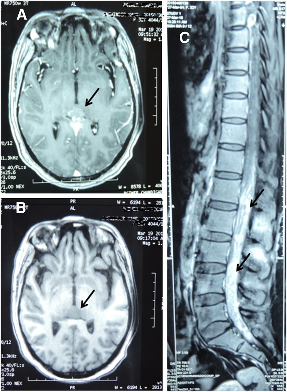
    (A and B) On T1-weighted axial CT image, lesion in pineal gland region was isointense before contrast administration (A) and enhanced afterward (B). (C) Sagittal images of dorsolumbar spine after contrast administration showed intensely enhancing sheetlike soft tissue in epidural space.
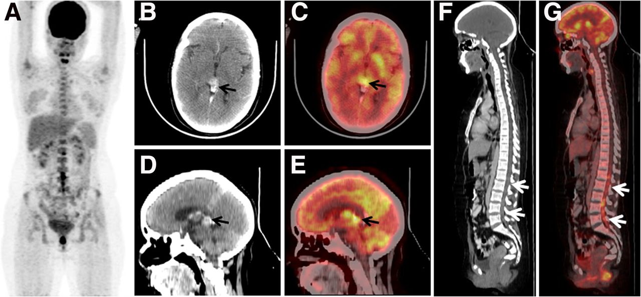
  • FIGURE 2.
    • Download figure
    • Open in new tab
    • Download powerpoint
    FIGURE 2.

    (A) 18F-FDG PET/CT maximum-intensity-projection image revealed abnormal uptake in lumbar spine region. (B–E) Transaxial (top) and sagittal (bottom) CT (left) and PET/CT (right) images of head revealed avidly enhancing nodular lesion (∼1.4 × 1.3 cm) in pineal gland (arrows). (F and G) Sagittal CT (left) and PET/CT (right) images revealed increased uptake in ill-defined densities in spinal canal extending from L1 to L5 (arrows).

PreviousNext
Back to top

In this issue

Journal of Nuclear Medicine Technology: 45 (2)
Journal of Nuclear Medicine Technology
Vol. 45, Issue 2
June 1, 2017
  • Table of Contents
  • About the Cover
  • Index by author
Print
Download PDF
Article Alerts
Sign In to Email Alerts with your Email Address
Email Article

Thank you for your interest in spreading the word on Journal of Nuclear Medicine Technology.

NOTE: We only request your email address so that the person you are recommending the page to knows that you wanted them to see it, and that it is not junk mail. We do not capture any email address.

Enter multiple addresses on separate lines or separate them with commas.
18F-FDG PET/CT Finding of Drop Metastases from Germ Cell Tumor of Pineal Gland
(Your Name) has sent you a message from Journal of Nuclear Medicine Technology
(Your Name) thought you would like to see the Journal of Nuclear Medicine Technology web site.
Citation Tools
18F-FDG PET/CT Finding of Drop Metastases from Germ Cell Tumor of Pineal Gland
Tarun K. Jain, Rajender K. Basher, Ashwani Sood, Bhagwant R. Mittal, Gaurav Prakash, Anmol Bhatia
Journal of Nuclear Medicine Technology Jun 2017, 45 (2) 114-115; DOI: 10.2967/jnmt.116.185850

Citation Manager Formats

  • BibTeX
  • Bookends
  • EasyBib
  • EndNote (tagged)
  • EndNote 8 (xml)
  • Medlars
  • Mendeley
  • Papers
  • RefWorks Tagged
  • Ref Manager
  • RIS
  • Zotero
Share
18F-FDG PET/CT Finding of Drop Metastases from Germ Cell Tumor of Pineal Gland
Tarun K. Jain, Rajender K. Basher, Ashwani Sood, Bhagwant R. Mittal, Gaurav Prakash, Anmol Bhatia
Journal of Nuclear Medicine Technology Jun 2017, 45 (2) 114-115; DOI: 10.2967/jnmt.116.185850
Twitter logo Facebook logo LinkedIn logo Mendeley logo
  • Tweet Widget
  • Facebook Like
  • Google Plus One
Bookmark this article

Jump to section

  • Article
    • Abstract
    • CASE REPORT
    • DISCUSSION
    • CONCLUSION
    • DISCLOSURE
    • Footnotes
    • REFERENCES
  • Figures & Data
  • Info & Metrics
  • PDF

Related Articles

  • No related articles found.
  • PubMed
  • Google Scholar

Cited By...

  • Biology of Cancer and PET Imaging: Pictorial Review
  • Google Scholar

More in this TOC Section

  • High-Sensitivity Troponin Elevation in a Young Woman with Typical Chest Pain: The Heart of the Matter
  • Pulmonary Adenocarcinoma Revealed by Parathyroid Scintigraphy: An Incidental Case to Remember
  • Prominent Right Ventricular Tracer Uptake: A Harbinger of Multivessel Coronary Artery Disease
Show more Teaching Case Studies

Similar Articles

Keywords

  • germ cell tumor
  • pineal gland
  • spinal seeding
  • 18F-FDG PET/CT
  • drop metastases
SNMMI

© 2025 SNMMI

Powered by HighWire