


Abstract
Tumors of the pineal region are rare, accounting for fewer than 1% of all intracranial neoplasms. Fifty percent of pineal region tumors are germ cell tumors (GCTs). However, spinal seeding and extracranial metastases from intracranial GCTs are uncommon. We present a case of pineal gland GCT in which 18F-FDG PET/CT imaging demonstrated drop metastases to the spinal cord in addition to tracer uptake in the primary lesion.
This case report illustrates the utility of whole-body 18F-FDG PET/CT in visualizing and characterizing pineal germ cell tumors (GCTs) and drop metastases.
CASE REPORT
A 32-y-old woman presented with a sudden onset of progressive blurring of vision in the left eye followed by the right eye and a spinning sensation of 6-wk duration. She complained of progressive weakness in all 4 limbs, leading to her confinement to bed. A contrast-enhanced MRI scan of the brain (Fig. 1) showed a well-defined (∼1.2 × 1.5 cm) oval, minimally enhancing lesion in the pineal gland region, as well as intensely enhancing sheetlike soft tissue in the epidural space of the lumber spine. The patient was suspected to have pinealocytoma with spinal metastases. She underwent whole-body 18F-FDG PET/CT for characterization of the pineal gland lesion and to look for additional lesions or metastases. The study protocol was approved by the Institutional Ethics Committee, and the patient gave written informed consent to undergo the 18F-FDG PET/CT study. The PET/CT images (Fig. 2) revealed a tracer-avid enhancing nodular lesion (∼1.4 × 1.3 cm) in the pineal gland and tracer-avid ill-defined densities in the spinal canal extending from L1 to L5. Her cerebrospinal fluid cytology and biochemistry work-up revealed malignant cells with an elevated level of β-human chorionic gonadotropin (43 mIU/mL; reference level, 0.0–5.0 mIU/mL) and a normal level of α-fetoprotein (1.6 mIU/mL; reference level, 0.0–5.5 mIU/mL). Serum and urinary β-human chorionic gonadotropin levels were also elevated. Surgical biopsy was performed, but the specimen did not contain enough tissue for a definitive diagnosis.

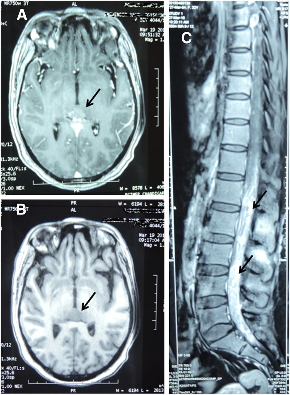
(A and B) On T1-weighted axial CT image, lesion in pineal gland region was isointense before contrast administration (A) and enhanced afterward (B). (C) Sagittal images of dorsolumbar spine after contrast administration showed intensely enhancing sheetlike soft tissue in epidural space.
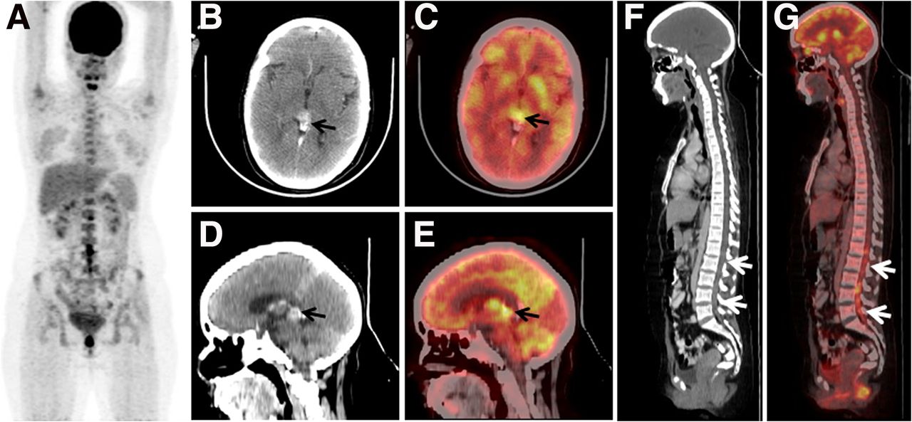
(A) 18F-FDG PET/CT maximum-intensity-projection image revealed abnormal uptake in lumbar spine region. (B–E) Transaxial (top) and sagittal (bottom) CT (left) and PET/CT (right) images of head revealed avidly enhancing nodular lesion (∼1.4 × 1.3 cm) in pineal gland (arrows). (F and G) Sagittal CT (left) and PET/CT (right) images revealed increased uptake in ill-defined densities in spinal canal extending from L1 to L5 (arrows).
DISCUSSION
Tumors of the pineal region account for fewer than 1% of all intracranial tumors, with most being pure germinomas. GCTs commonly affect children and young adults (1). Patients with suspected intracranial GCTs are imaged with unenhanced and enhanced MRI of the head and enhanced sagittal MRI of the spine. Detailed analysis of the images may help in predicting the histologic diagnosis. The levels of α-fetoprotein and β-human chorionic gonadotropin in serum and cerebrospinal fluid are also measured in patients with intracranial GCTs; however, surgical biopsy results may or may not be available in these patients (2). In patients with intracranial germinoma, there has been infrequent observation of spinal and extracranial metastases, which might be missed on regional imaging modalities. Large and multifocal intracranial diseases are independent risk factors for spinal recurrence, and spinal radiotherapy and chemotherapy have been shown to be effective salvage treatment (3). 18F-FDG PET/CT may, in a single session, help in demonstrating not only the primary lesion but also the extent of disease. Most GCTs are hypermetabolic and show increased 18F-FDG uptake, excepting mature teratomas, which show very little or no uptake (4). In one reported case—a 14-y-old boy with a pineal GCT of mixed histopathology—no 18F-FDG uptake was observed in the tumor (5).
CONCLUSION
This case shows the utility of 18F-FDG PET/CT in demonstrating and better characterizing tracer uptake in pineal GCTs and spinal metastases.
DISCLOSURE
No potential conflict of interest relevant to this article was reported.
Footnotes
Published online Mar. 9, 2017.
- Received for publication December 4, 2016.
- Accepted for publication February 3, 2017.


