


Article Figures & Data
Figures
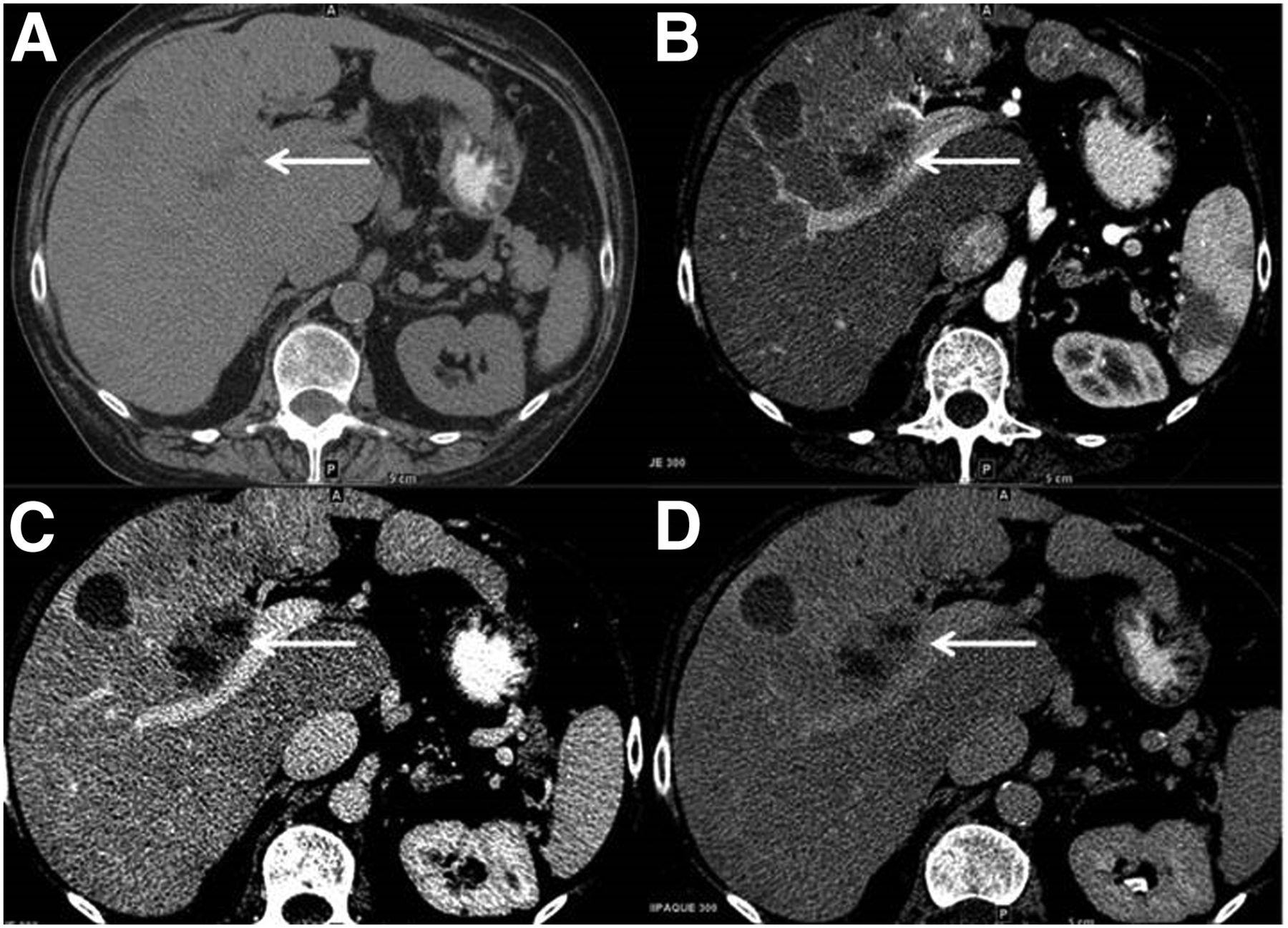
- FIGURE 1.
Multiphasic contrast CT study shows multifocal liver lesions with partial necrosis. (A) A 3.1-cm mildly heterogeneous segment-5 lesion seen on noncontrast axial CT demonstrates minimal arterial enhancement seen in late arterial phase (B) with washout and subtle capsule seen in the portal-venous (C) and delayed phases (D).
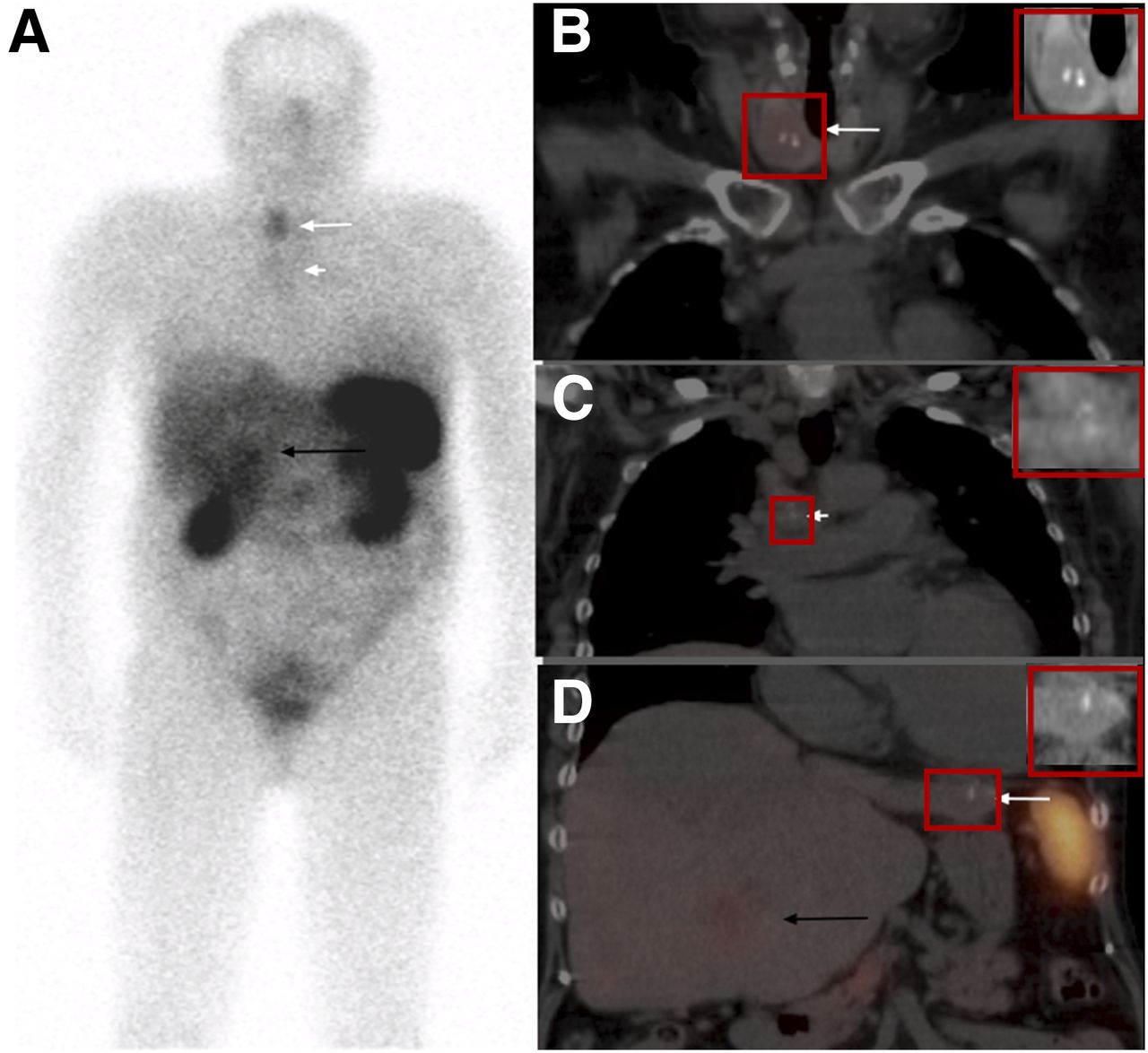
- FIGURE 2.
111In-pentetreotide scan identifies focal uptake in thyroid nodule. (A) Whole-body images (anterior view) show uptake in right thyroid nodule (long white arrow), mild uptake in mediastinal lymph nodes (short white arrow), and heterogeneous liver uptake more prominent in segment-5 lesion (black arrow). (B–D) Fused coronal SPECT/CT images show focal thyroid uptake corresponding to a heterogeneous low attenuation thyroid nodule with dense, coarse calcifications within (arrow in B), mild uptake in mediastinal lymph nodes also containing dense calcifications (arrow in C), and heterogeneous liver uptake due to multiple liver lesions more prominent within one of the lesions in segment 5 (black arrow in D). Small calcification in left lobe of the liver lesion is also seen (white arrow on image D).
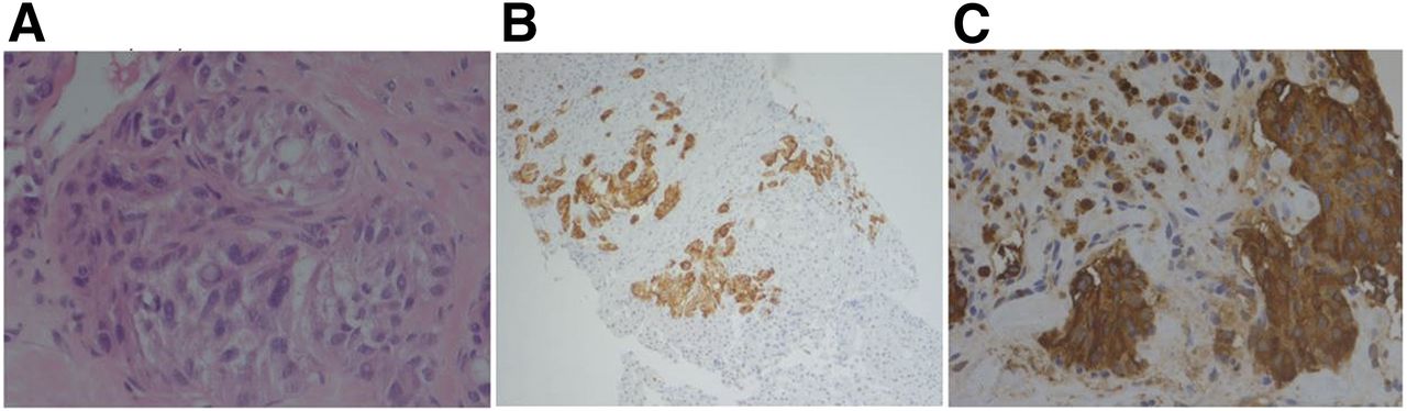
- FIGURE 3.
Histopathologic examination of core liver biopsy (A) shows liver parenchyma largely replaced by neoplastic cells infiltrating in nests and single cells with epithelioid to spindled morphology. Cytologic features include variation in nuclear size and shape with stippled chromatin, intranuclear inclusions, relatively low mitotic rate, and necrosis. Immunostains were positive for chromogranin (B) and calcitonin (C) secretion, consistent with metastatic medullary thyroid carcinoma. Nonneoplastic liver biopsy (not shown) was remarkable for fibrosis and nodularity suggestive of cirrhosis and negative for increased iron. Cytologic impression for fine-needle aspiration of right thyroid nodule also showed medullary thyroid cancer.


Jump to section
Related Articles
Cited By...
- No citing articles found.