


Article Figures & Data
Figures
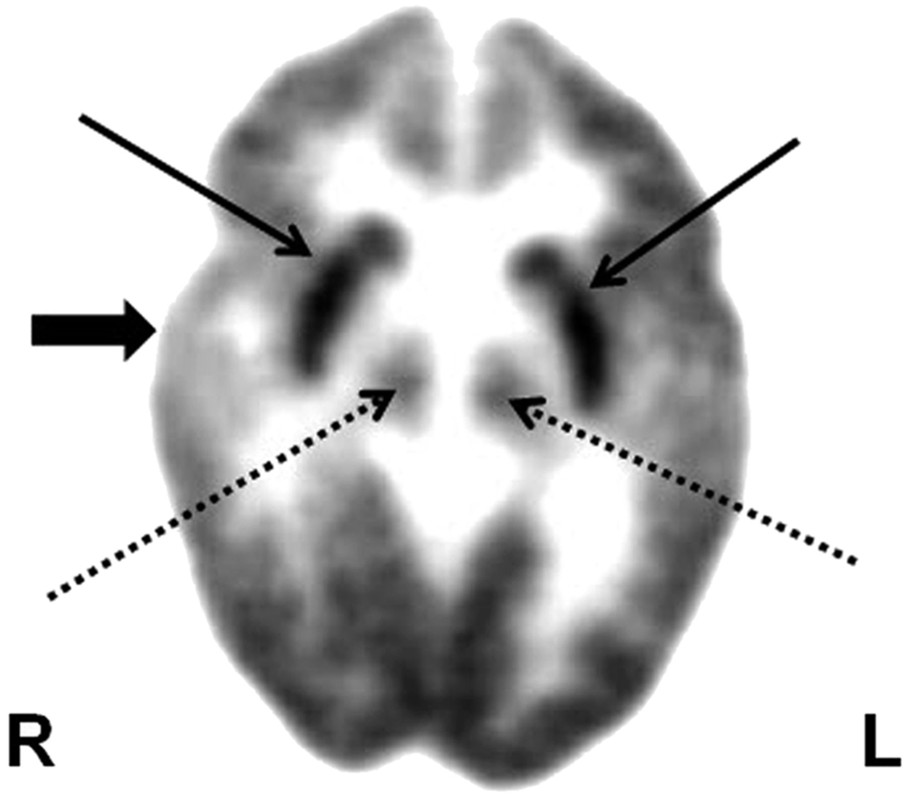
- FIGURE 1.
18F-FDG PET shows focal hypometabolism in right temporal cortex (thick arrow) in child with epileptic spasms and normal MR imaging findings. This child may be considered for focal cortical resection for possible seizure control and cognitive improvement. Bilateral hypermetabolism in basal ganglia (thin solid arrows) and brain stem nuclei (thin dashed arrows) likely implicates their role in spasm generation.
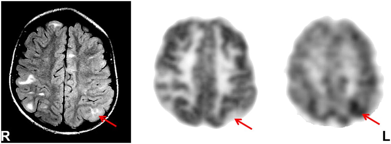
- FIGURE 2.
Fluid-attenuated inversion recovery (FLAIR) MR imaging (left), 18F-FDG PET (middle), and 11C-AMT PET (right) in tuberous sclerosis patient with multiple brain tubers (enhancing lesions on FLAIR MR imaging), intractable epilepsy, and nonlocalizing scalp EEG. Although 18F-FDG PET showed hypometabolism in all tubers and overlying cortices, interictal 11C-AMT PET revealed increased 11C-AMT uptake in left parietal tuber (arrow) only, which likely is epileptogenic, considering the almost 100% specificity of this test in detecting epileptogenic tubers. Most of the increased 11C-AMT uptake is at edge (anteromedial in this case) of tuber, with remainder showing less 11C-AMT uptake.
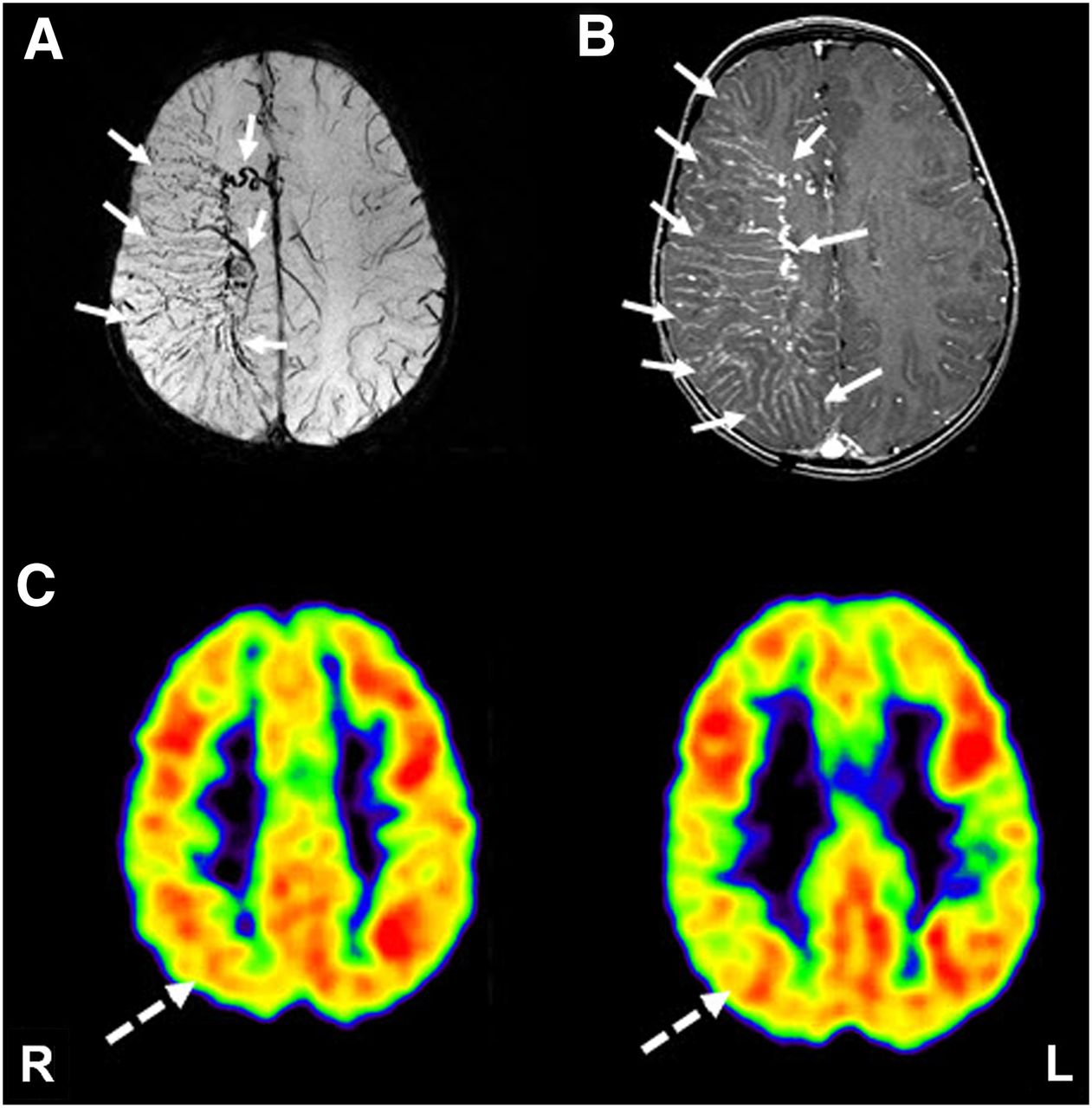
- FIGURE 3.
Susceptibility-weighted MR imaging (A), gadolinium-enhanced T1-weighted MR imaging (B), and 18F-FDG PET imaging (C) in child with SWS. Although both susceptibility-weighted and contrast-enhanced MR imaging showed extensive deep venous abnormalities involving frontoparietal cortex (solid arrows), 18F-FDG PET showed smaller area of hypometabolism, involving parietal cortex only (dashed arrow), suggesting preserved neuronal function despite extensive structural abnormalities.
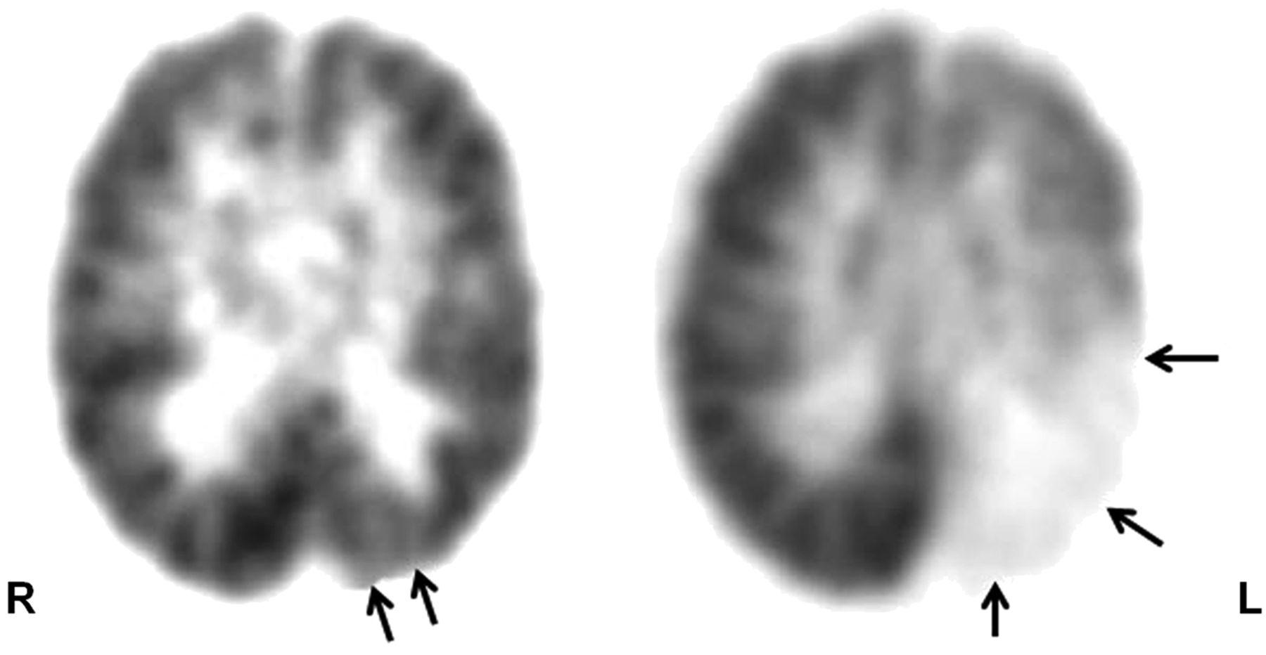
- FIGURE 4.
Serial 18F-FDG PET scans, performed at 18 mo (left) and 6 y (right) old, respectively, show progression of cortical hypometabolism (arrows) in both extent and severity, indicating degenerative changes in brain tissue associated with angioma in child with SWS and meningeal hemangioma of left posterior quadrant. This rapid progression represents demise of abnormal brain tissue and is akin to auto-resection; this child showed improvement in both seizure status and cognitive function.
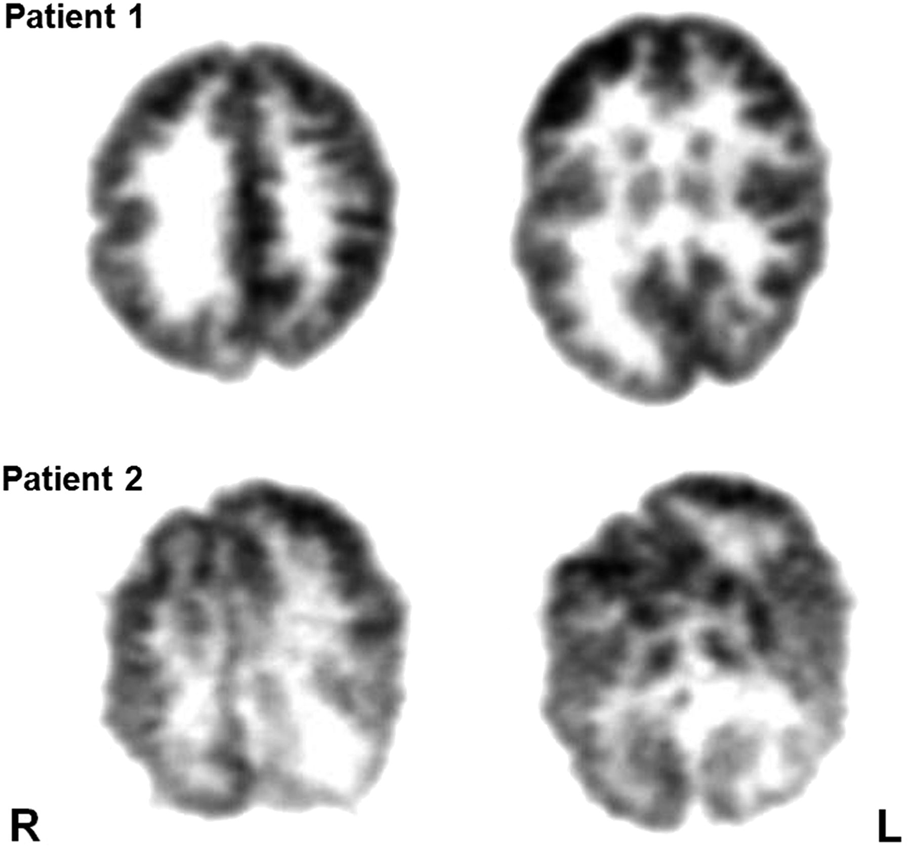
- FIGURE 5.
18F-FDG PET scans of 2 patients with intractable epilepsy due to hemimegalencephaly, right-sided in patient 1 and left-sided in patient 2. Although contralateral hemisphere looks relatively normal in first patient, contralateral hemisphere in second patient looks abnormal, indicating functional impairment of this hemisphere also. Hemispherectomy will have much better outcome in first patient, but seizure and neurocognitive outcome will be poor in second patient.
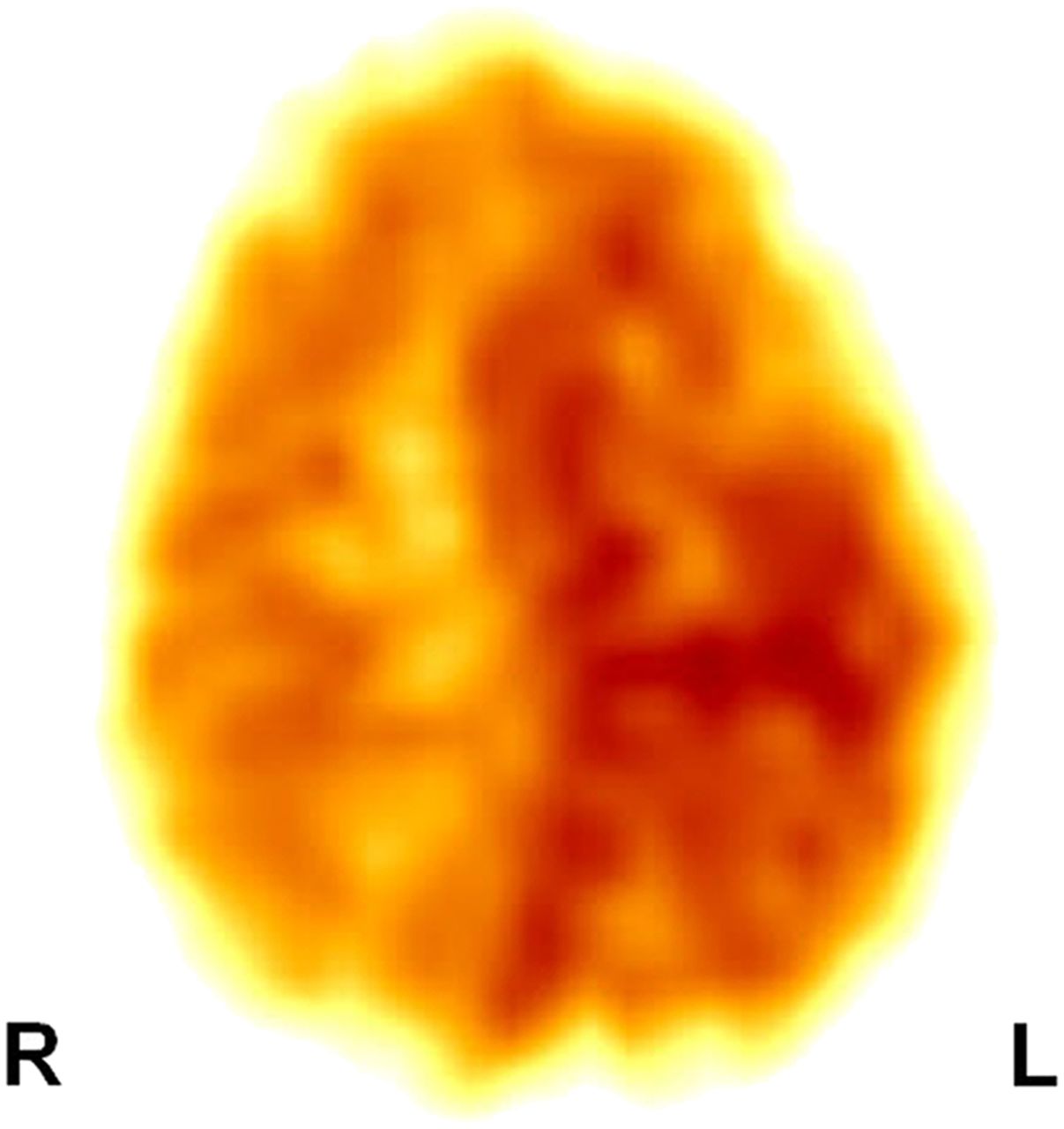
- FIGURE 6.
11C-(R)-PK11195 PET scan showing increased binding in right hemisphere, indicating neuroinflammation mediated by activated microglia, in child with suspected Rasmussen encephalitis.
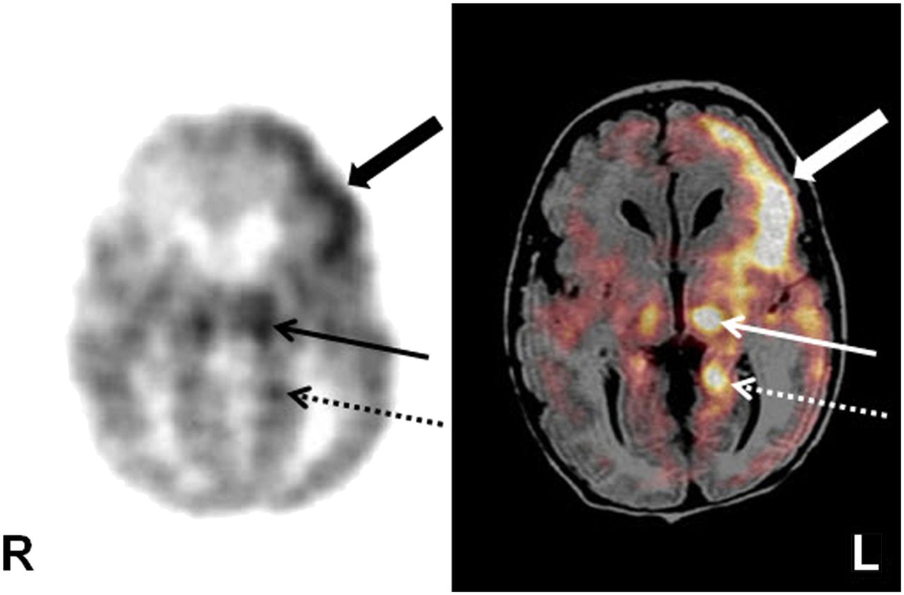
- FIGURE 7.
18F-FDG PET scan alone (left) and coregistered with MR scan (right) in newborn with neonatal seizure shows hypermetabolism in left frontal cortex (thick arrow), as child was seizing during 18F-FDG uptake period. Left thalamus (thin solid arrow) and parahippocampal region (thin dashed arrow) are also hypermetabolic, likely because of their involvement in seizure propagation, without any obvious structural abnormalities seen on MR imaging.


Jump to section
Related Articles
Cited By...
- No citing articles found.