


Article Figures & Data
Figures
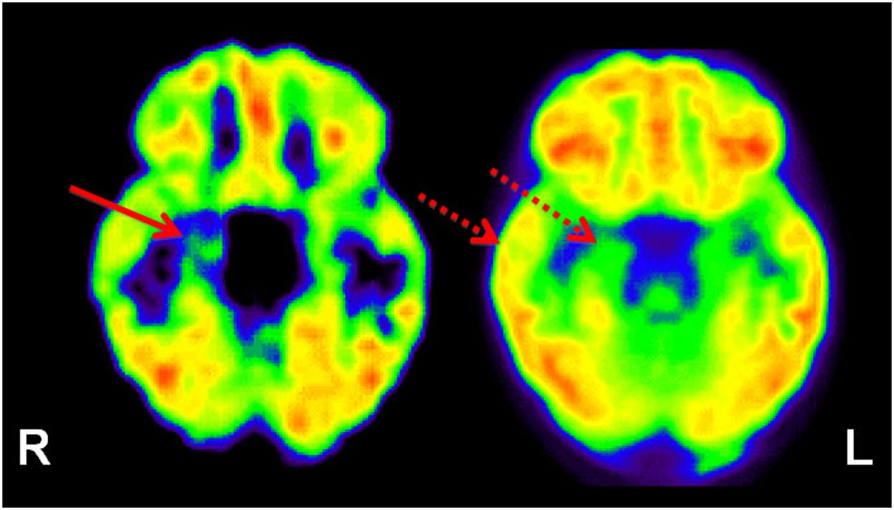
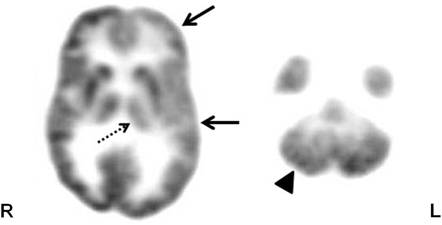
- FIGURE 1.
Axial 18F-FDG PET scan in patient with intractable epilepsy, showing hypometabolism in left frontotemporal cortex (solid arrows). Also seen is hypometabolism in ipsilateral thalamus (broken arrow) and contralateral cerebellum (arrowhead), likely representing thalamic and cerebellar diaschisis, respectively.
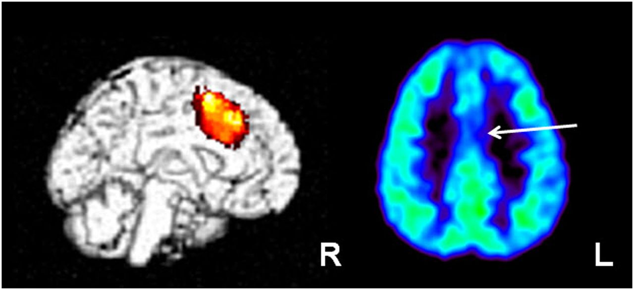
- FIGURE 2.
SPM analysis of 18F-FDG PET scan in child with intractable epilepsy, normal findings on MR imaging, and generalized electroencephalogram changes. Initial reading showed normal findings on 18F-FDG PET; however, SPM analysis revealed area of hypometabolism in left medial frontal/cingulate cortex (left image), which appeared to be suggestive/hypometabolic in rereview of 18F-FDG PET scan (arrow; right image). Child underwent 2-stage epilepsy surgery with intracranial subdural interhemispheric electrode placement, which showed ictal discharges from this region. These medial discharges perhaps resulted in generalized (nonlateralized) electroencephalogram changes. Area was resected and child is seizure-free after surgery.
- FIGURE 3.
11C-flumazenil PET (left image) showing focal abnormality (decreased tracer binding) involving right hippocampus (solid arrow) in patient with medial temporal lobe epilepsy. In comparison, 18F-FDG PET (right image) shows widespread 18F-FDG hypometabolism in right temporal lobe, including neocortex (broken arrows).
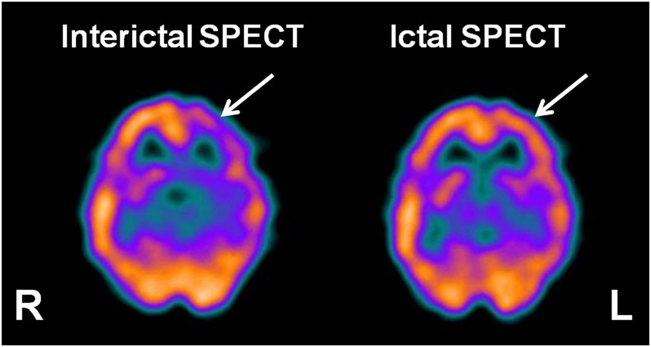
- FIGURE 4.
Interictal 99mTc-ethyl cysteinate dimer SPECT (left image) showing the usual hypoperfusion in presumed epileptogenic focus (arrow) in left frontal cortex region, which becomes hyperperfused during ictal SPECT (right image).
- FIGURE 5.
Axial 18F-FDG PET scan showing subcortical band heterotopia (arrows) in right temporal–occipital white matter in child with intractable epilepsy and normal MR imaging findings. Heterotopic band has higher glucose uptake than adjacent white matter but lower uptake than cortex.
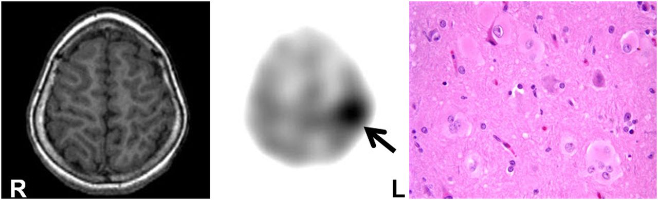
- FIGURE 6.
11C-α-methyl-l-tryptophan PET scan (middle image) showing increased tracer uptake in left parietal lobe (arrow) in child with intractable epilepsy and normal findings on MR imaging (left image). Postsurgical histopathology (right image) revealed type IIB cortical dysplasia with balloon cells (×40, hematoxylin and eosin). Child remains seizure-free after epilepsy surgery.
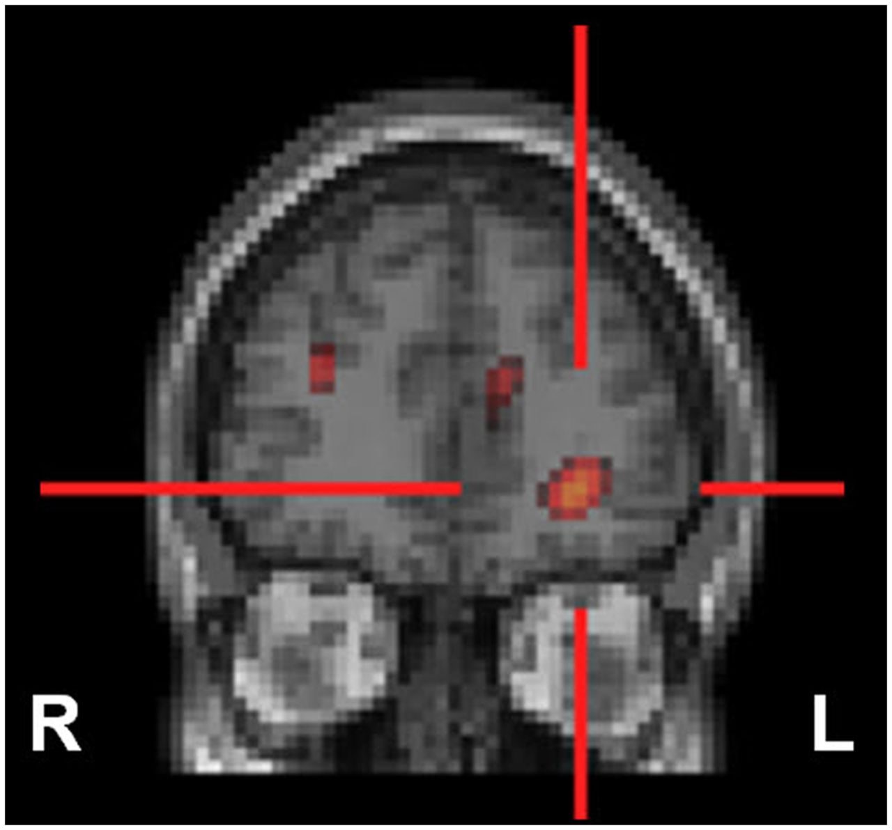
- FIGURE 7.
SISCOM may result in better delineation of epileptogenic focus, which may sometimes be missed even on ictal SPECT. In child with intractable complex partial epilepsy with normal findings on MR imaging and nonlocalizing scalp electroencephalography, SISCOM revealed focus of hyperperfusion in left inferior frontal cortex (red cross), likely representing seizure focus.


Tables
- TABLE 1
Targeted Pathways, Used Radiotracers, and Their Usual Uptake Pattern in Epileptogenic Region
Target Used radiotracers Uptake pattern in epileptogenic region Blood perfusion 15O-H2O, 99mTc-ethyl cysteinate dimer, 99mTc-hexamethyl propyleneamine oxime Interictal decrease; ictal increase Metabolic pathways Glucose metabolism 18F-FDG Interictal decrease*; ictal increase Serotonin/kynurenine metabolism 11C-α-methyl-l-tryptophan Interictal increase Dopamine synthesis 18F-l-DOPA† Interictal decrease Monoamine oxidase 11C-deuterium-deprenyl‡ Interictal increase Receptors Benzodiazepine 11C-flumazenil§ Opiate 11C-carfentanil∥, 18F-cyclofoxy¶, 11C-diprenorphine#, 11C-methylnaltrindole** Interictal increase; interictal decrease 5-hydroxytryptamine 18F-FCWAY††, 11C-WAY††, 18F-MPPF†† Interictal decrease Dopamine 18F-fallypride‡‡, 11C-SCH23390§§ Interictal decrease Peripheral benzodiazepine or translocator protein 11C-PK11195∥∥, 11C-PBR28∥∥ Interictal increase Histamine 11C-doxepin¶¶ Interictal increase N-methyl-d-aspartic acid 11C-ketamine## Interictal decrease Acetylcholine 18F-FA85380*** Interictal decrease FCWAY = trans-4-fluoro-N-2-[4-(2-methoxyphenyl) piperazin-1-yl] ethyl-N-(2-pyridyl) cyclohexanecarboxamide; MPPF = 4-18F-fluoro-N-(2-[4-(2-methoxyphenyl)-l-piperazinyl]ethyl)-N-(2-pyridinyl)benzamide; l-DOPA = l-3,4-dihydroxyphenylalanine (levodopa); PK11195 = 1-(2-chlorophenyl)-N-methyl-N-(1-methylpropyl)-3-isoquinoline carboxamide; PBR28 = N-acetyl-N-(2-[11C-methoxybenzyl)-2-phenoxy-5-pyridinamine.
↵* Can show increased uptake if continuous spiking (interictal) or epileptiform discharges (ictal).
↵† Acts on dopamine receptors.
↵‡ Monoamine oxidase-B inhibitor.
↵§ γ-aminobutyric acid A benzodiazepine receptor.
↵∥ μ-receptor agonist.
↵¶ μ and κ antagonist.
↵# μ-, δ- and κ-receptor antagonist.
↵** δ-receptor antagonist.
↵†† 5HT1A antagonist.
↵‡‡ D2/D3 antagonist.
↵§§ D1 antagonist.
↵∥∥ Peripheral benzodiazepine receptor or translocator protein antagonist.
↵¶¶ 1H receptor antagonist.
#NMDA antagonist.
↵*** α4/β2 nAChR agonist.


