
Main menu

  • Home
  • Content
    • Current
      • JNMT Supplement
    • Ahead of print
    • Past Issues
    • Continuing Education
    • JNMT Podcast
    • SNMMI Annual Meeting Abstracts
  • Subscriptions
    • Subscribers
    • Rates
    • Journal Claims
    • Institutional and Non-member
  • Authors
    • Submit to JNMT
    • Information for Authors
    • Assignment of Copyright
    • AQARA Requirements
  • Info
    • Reviewers
    • Permissions
    • Advertisers
    • Corporate & Special Sales
  • About
    • About Us
    • Editorial Board
    • Contact Information
  • More
    • Alerts
    • Feedback
    • Help
    • SNMMI Journals
  • SNMMI
    • JNMT
    • JNM
    • SNMMI Journals
    • SNMMI

User menu

  • Subscribe
  • My alerts
  • Log in
  • Log out
  • My Cart

Search

  • Advanced search
Journal of Nuclear Medicine Technology
  • SNMMI
    • JNMT
    • JNM
    • SNMMI Journals
    • SNMMI
  • Subscribe
  • My alerts
  • Log in
  • Log out
  • My Cart
Journal of Nuclear Medicine Technology

Advanced Search

  • Home
  • Content
    • Current
    • Ahead of print
    • Past Issues
    • Continuing Education
    • JNMT Podcast
    • SNMMI Annual Meeting Abstracts
  • Subscriptions
    • Subscribers
    • Rates
    • Journal Claims
    • Institutional and Non-member
  • Authors
    • Submit to JNMT
    • Information for Authors
    • Assignment of Copyright
    • AQARA Requirements
  • Info
    • Reviewers
    • Permissions
    • Advertisers
    • Corporate & Special Sales
  • About
    • About Us
    • Editorial Board
    • Contact Information
  • More
    • Alerts
    • Feedback
    • Help
    • SNMMI Journals
  • Watch or Listen to JNMT Podcast
  • Visit SNMMI on Facebook
  • Join SNMMI on LinkedIn
  • Follow SNMMI on Twitter
  • Subscribe to JNMT RSS feeds
Review ArticleTeaching Case Studies

Ectopic Parathyroid Adenoma in the Carotid Sheath

Christopher D. Sanders, Jared D. Kirkland and Ely A. Wolin
Journal of Nuclear Medicine Technology September 2016, 44 (3) 201-202; DOI: https://doi.org/10.2967/jnmt.115.170993
Christopher D. Sanders
David Grant USAF Medical Center, Travis Air Force Base, Fairfield, California
  • Find this author on Google Scholar
  • Find this author on PubMed
  • Search for this author on this site
Jared D. Kirkland
David Grant USAF Medical Center, Travis Air Force Base, Fairfield, California
  • Find this author on Google Scholar
  • Find this author on PubMed
  • Search for this author on this site
Ely A. Wolin
David Grant USAF Medical Center, Travis Air Force Base, Fairfield, California
  • Find this author on Google Scholar
  • Find this author on PubMed
  • Search for this author on this site
  • Article
  • Figures & Data
  • Info & Metrics
  • PDF
Loading

Abstract

Primary hyperparathyroidism is predominantly caused by a single parathyroid adenoma. Knowledge of normal and ectopic locations of parathyroid glands is crucial to help guide surgeons who plan targeted unilateral parathyroidectomy to reduce surgical time and risk. We describe a female patient with clinical primary hyperparathyroidism who underwent a failed initial parathyroidectomy, with subsequent imaging localizing an ectopic parathyroid adenoma in the carotid sheath.

  • hyperparathyroidism
  • ectopic parathyroid adenoma
  • 99mTc-sestamibi

Given that ectopic parathyroid glands are not uncommon in the setting of primary hyperparathyroidism, preoperative localization is critical to the current minimally invasive approaches toward surgical excision. Knowledge of ectopic locations and appropriate imaging guidelines is imperative.

CASE REPORT

Our patient was clinically diagnosed with primary hyperparathyroidism after a workup for incidental hypercalcemia. Dual-phase 99mTc-sestamibi scintigraphy with early SPECT/CT demonstrated uptake posterior and lateral to the superior left thyroid (Figs. 1 and 2). The findings of neck ultrasound were unremarkable.

FIGURE 1.
  • Download figure
  • Open in new tab
  • Download powerpoint
FIGURE 1.

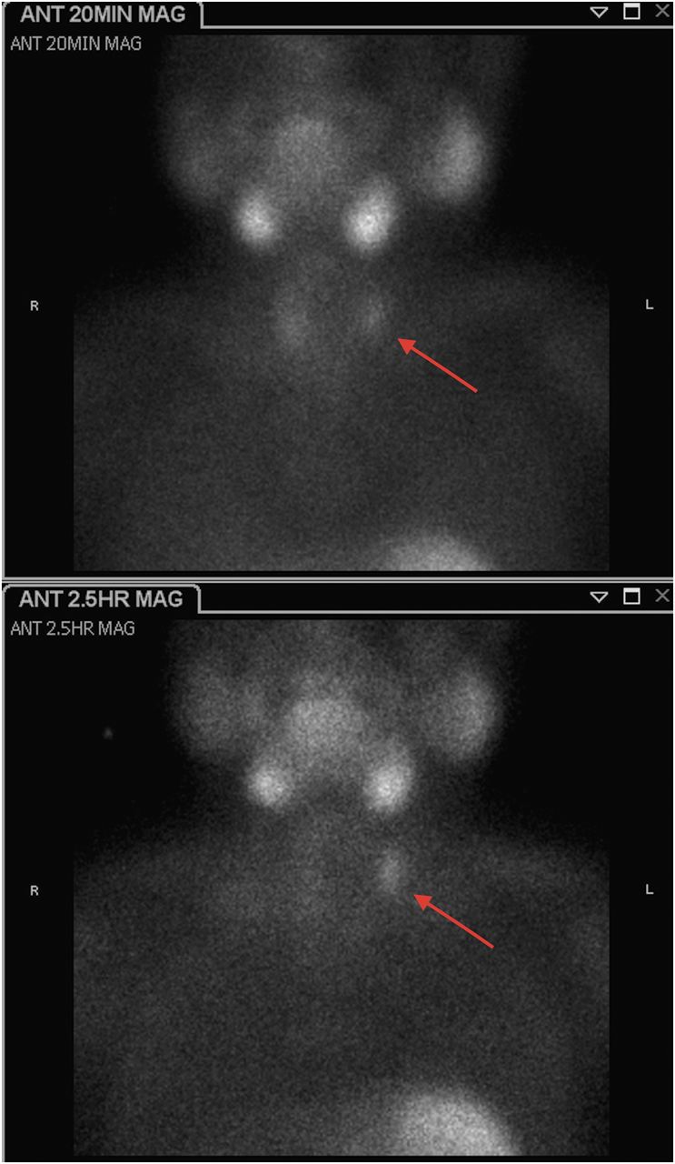
Anterior 20-min (top) and 2.5-h (bottom) 99mTc-sestamibi images demonstrate persistent focal uptake in left neck. MAG = magnified.

FIGURE 2.
  • Download figure
  • Open in new tab
  • Download powerpoint
FIGURE 2.

SPECT/CT data localize uptake posterior and lateral to superior left thyroid gland.

Excision of the left superior parathyroid gland led to no change in intraoperative parathyroid levels. The surgeon proceeded with left hemithyroidectomy and bilateral explorative dissection, with no abnormal tissue found on pathologic examination.

Postoperative MRI did not prospectively identify a parathyroid adenoma. Repeated dual-phase parathyroid scintigraphy confirmed persistence of the adenoma. Repeated SPECT coregistered with dedicated neck CT localized the adenoma to the left carotid sheath (Figs. 3 and 4).

FIGURE 3.
  • Download figure
  • Open in new tab
  • Download powerpoint
FIGURE 3.

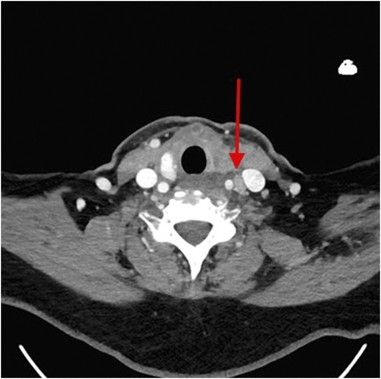
Contrast-enhanced CT demonstrates soft-tissue lesion in left carotid sheath.

FIGURE 4.
  • Download figure
  • Open in new tab
  • Download powerpoint
FIGURE 4.

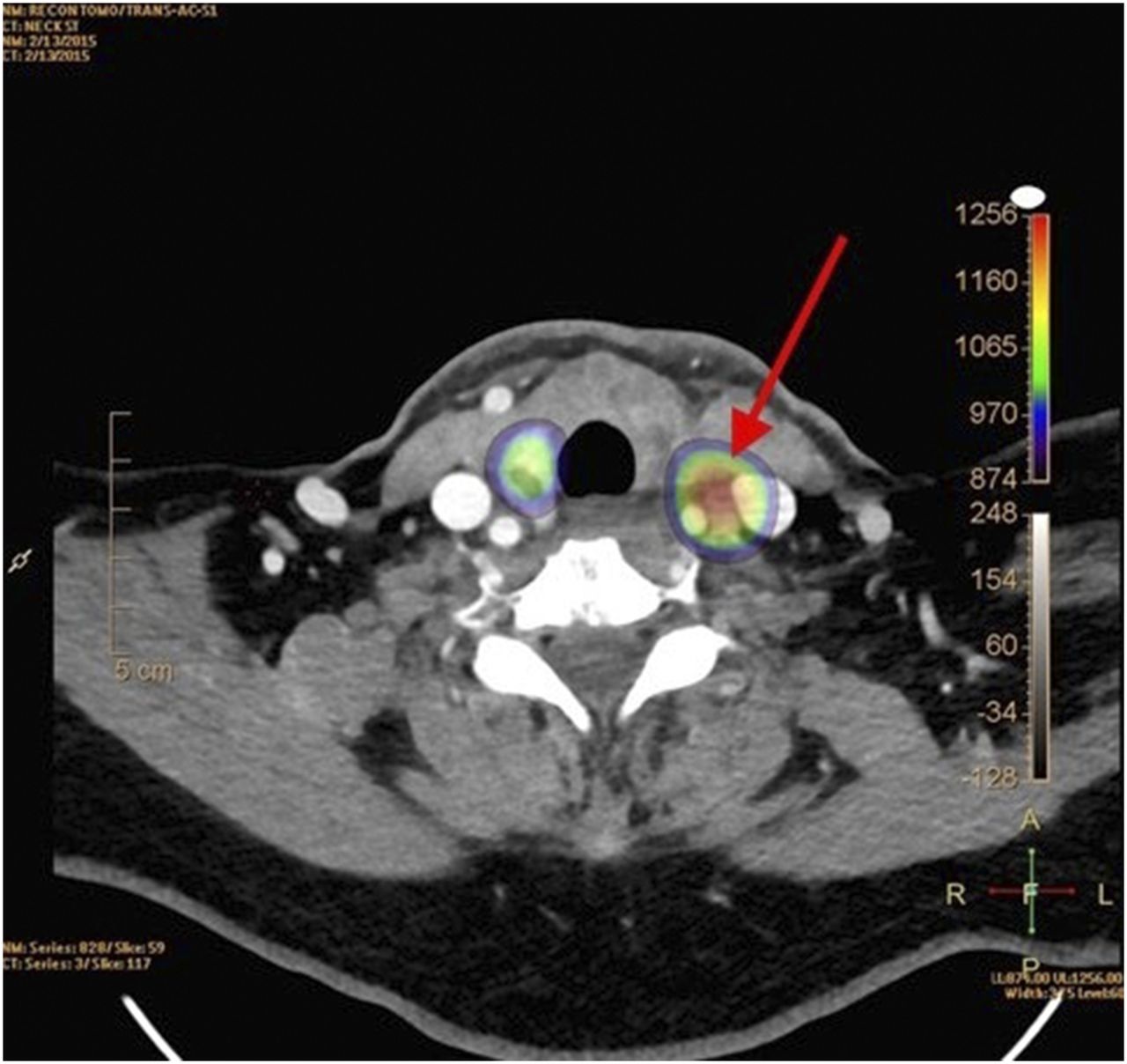
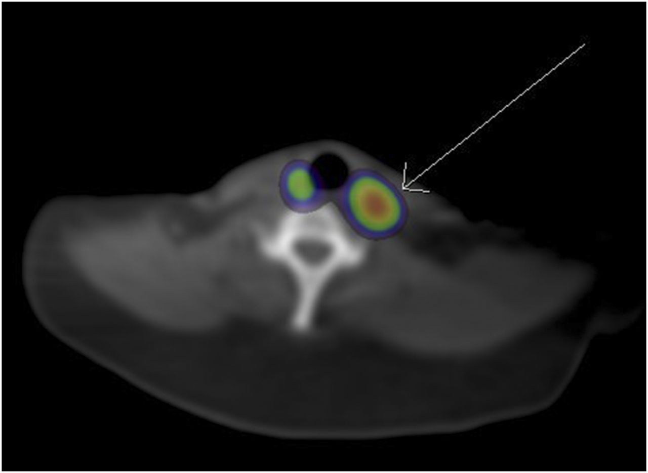
Fused SPECT/high-resolution CT localizes uptake to soft-tissue lesion in left carotid sheath.

Repeated surgery removed a pathology-proven parathyroid adenoma from the left carotid sheath.

DISCUSSION

Primary hyperparathyroidism is a clinical diagnosis based primarily on laboratory results, usually in the fifth to seventh decades of life (1). Hypercalcemia, the primary clinical abnormality, can present with numerous symptoms, including bone or abdominal pain, nephrolithiasis, and psychiatric imbalances (1). Etiologies include a single parathyroid adenoma, multiglandular hyperplasia, multiple adenomas, and, rarely, parathyroid carcinomas (1). Surgical excision of abnormal parathyroid tissue is usually curative. Historically, bilateral cervical dissection has been performed with 95% success (1). Preoperative imaging localization, however, allows for focused surgeries, reducing procedure length and risk (2).

Usually, 2 superior and 2 inferior parathyroid glands are immediately posterior to the thyroid. Ectopic parathyroid glands exist in numerous locations between mouth and diaphragm, with a frequency of 6%–16% (3). Ectopia in the carotid sheath, as occurred in our patient, is reported in less than 1% of cases (2).

Our institution performs a dual-phase 99mTc-sestamibi protocol, with planar images obtained at 20 and 150 min, plus SPECT/CT at 20 min for improved localization (4). The initial examination was limited by the reduced spatial resolution of the coregistered low-dose cone-beam CT scan; subsequent coregistration with high-resolution CT proved invaluable. Four-dimensional CT, along with ultrasound and MRI, can also improve preoperative localization (5).

CONCLUSION

Because focused unilateral parathyroidectomy is curative in most cases of primary hyperparathyroidism, with less surgical time and risk than nontargeted surgery, preoperative localization is crucial. Frequently, the clinical question focuses on whether there is an adenoma—a question readily answered with planar scintigraphy alone. However, the better anatomic localization and spatial resolution provided by advanced imaging is extremely helpful. The initial surgical failure in this case may have been prevented if the initial CT scan had been of higher resolution or if ectopia had been suspected because of the location of the parathyroid adenoma—posterior and lateral to the thyroid.

DISCLOSURE

No potential conflict of interest relevant to this article was reported. The views expressed in this paper are those of the authors and do not reflect the official position of the U.S. Government, the Department of Defense, or the Department of the Air Force.

Footnotes

  • Published online Feb. 4, 2016.

REFERENCES

  1. 1.↵
    1. Johnson NA,
    2. Tublin ME,
    3. Ogilvie JB
    . Parathyroid imaging: technique and role in the preoperative evaluation of primary hyperparathyroidism. AJR. 2007;188:1706–1715.
    OpenUrlCrossRefPubMed
  2. 2.↵
    1. Eslamy HK,
    2. Ziessman HA
    . Parathyroid scintigraphy in patients with primary hyperparathyroidism: 99mTc sestamibi SPECT and SPECT/CT. Radiographics. 2008;28:1461–1476.
    OpenUrlCrossRefPubMed
  3. 3.↵
    1. Roy M,
    2. Mazeh H,
    3. Chen H,
    4. Sippel RS
    . Incidence and localization of ectopic parathyroid adenomas in previously unexplored patients. World J Surg. 2013;37:102–106.
    OpenUrlCrossRefPubMed
  4. 4.↵
    1. Greenspan BS,
    2. Dillehay G,
    3. Intenzo C,
    4. et al
    . SNM practice guideline for parathyroid scintigraphy 4.0. J Nucl Med Technol. 2012;40:111–118.
    OpenUrlFREE Full Text
  5. 5.↵
    1. Smith JR,
    2. Oates E
    . Radionuclide imaging of the parathyroid glands: patterns, pearls, and pitfalls. Radiographics. 2004;24:1101–1115.
    OpenUrlCrossRefPubMed
  • Received for publication December 8, 2015.
  • Accepted for publication January 27, 2016.
PreviousNext
Back to top

In this issue

Journal of Nuclear Medicine Technology: 44 (3)
Journal of Nuclear Medicine Technology
Vol. 44, Issue 3
September 1, 2016
  • Table of Contents
  • About the Cover
  • Index by author
Print
Download PDF
Article Alerts
Sign In to Email Alerts with your Email Address
Email Article

Thank you for your interest in spreading the word on Journal of Nuclear Medicine Technology.

NOTE: We only request your email address so that the person you are recommending the page to knows that you wanted them to see it, and that it is not junk mail. We do not capture any email address.

Enter multiple addresses on separate lines or separate them with commas.
Ectopic Parathyroid Adenoma in the Carotid Sheath
(Your Name) has sent you a message from Journal of Nuclear Medicine Technology
(Your Name) thought you would like to see the Journal of Nuclear Medicine Technology web site.
Citation Tools
Ectopic Parathyroid Adenoma in the Carotid Sheath
Christopher D. Sanders, Jared D. Kirkland, Ely A. Wolin
Journal of Nuclear Medicine Technology Sep 2016, 44 (3) 201-202; DOI: 10.2967/jnmt.115.170993

Citation Manager Formats

  • BibTeX
  • Bookends
  • EasyBib
  • EndNote (tagged)
  • EndNote 8 (xml)
  • Medlars
  • Mendeley
  • Papers
  • RefWorks Tagged
  • Ref Manager
  • RIS
  • Zotero
Share
Ectopic Parathyroid Adenoma in the Carotid Sheath
Christopher D. Sanders, Jared D. Kirkland, Ely A. Wolin
Journal of Nuclear Medicine Technology Sep 2016, 44 (3) 201-202; DOI: 10.2967/jnmt.115.170993
Twitter logo Facebook logo LinkedIn logo Mendeley logo
  • Tweet Widget
  • Facebook Like
  • Google Plus One
Bookmark this article

Jump to section

  • Article
    • Abstract
    • CASE REPORT
    • DISCUSSION
    • CONCLUSION
    • DISCLOSURE
    • Footnotes
    • REFERENCES
  • Figures & Data
  • Info & Metrics
  • PDF

Related Articles

  • No related articles found.
  • PubMed
  • Google Scholar

Cited By...

  • No citing articles found.
  • Google Scholar

More in this TOC Section

  • High-Sensitivity Troponin Elevation in a Young Woman with Typical Chest Pain: The Heart of the Matter
  • Pulmonary Adenocarcinoma Revealed by Parathyroid Scintigraphy: An Incidental Case to Remember
  • Prominent Right Ventricular Tracer Uptake: A Harbinger of Multivessel Coronary Artery Disease
Show more Teaching Case Studies

Similar Articles

Keywords

  • Hyperparathyroidism
  • Ectopic Parathyroid Adenoma
  • 99mTc-sestamibi
SNMMI

© 2025 SNMMI

Powered by HighWire