


Abstract
Prostate-specific membrane antigen (PSMA) is a type II transmembrane protein. It has been shown to be expressed in various solid malignant neoplasms. We report a case of a prostate cancer patient who underwent 68Ga-PSMA PET/CT imaging. There is a large thyroid nodule in the right thyroid gland, which had intense PSMA accumulation. Follicular thyroid lesions can be seen on 68Ga-PSMA PET/CT imaging.
The expanding use of 18F-FDG PET has led to the identification of increasing numbers of patients with an incidentaloma in the thyroid gland. The risk of malignancy in these thyroid incidentalomas can be 27.8%–74% (1). Prostate-specific membrane antigen (PSMA) is a type II transmembrane protein with high expression in prostate carcinoma cells (2). PSMA ligand 68Ga-HBED-CC (Glu-NH-CO-NH-Lys-(Ahx)-[68Ga (HBED-CC)]) (68Ga-PSMA PET/CT) has been shown to be expressed in various solid malignant neoplasms such as neuroendocrine tumors, renal cell carcinoma, breast cancer, and differentiated thyroid cancer (3).
CASE REPORT
We report a case of a 72-y-old male patient who underwent 68Ga-PSMA PET/CT imaging for prostate cancer evaluation with a Gleason score of 4 + 4. No recurrent or metastatic lesion for prostate cancer was detected. However, a large thyroid nodule in right thyroid gland, which had intense PSMA accumulation on the peripheric side of the nodules, was seen (Fig. 1A; multiple image projection). On CT slices and fusion images, a nearly 4-cm thyroid nodule was seen (Figs. 1B and 1C). Thyroid ultrasound and fine-needle aspiration biopsy was performed, and follicular lesion of undetermined significance was reported. This PSMA-positive thyroid nodule was determined to be a malignant thyroid nodule using ultrasound imaging and clinical data. Our patient did not want to undergo surgery.
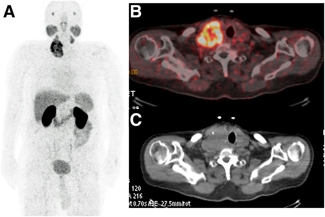
(A) Multiple image projection of 68Ga-PSMA imaging of 72-y-old patient with prostate cancer. Large thyroid nodule in right thyroid gland has intense PSMA accumulation on peripheric side of nodules. (B and C) Fusion and CT images, respectively, of patient. Nearly 4-cm thyroid nodule, which has PSMA accumulation, is seen.
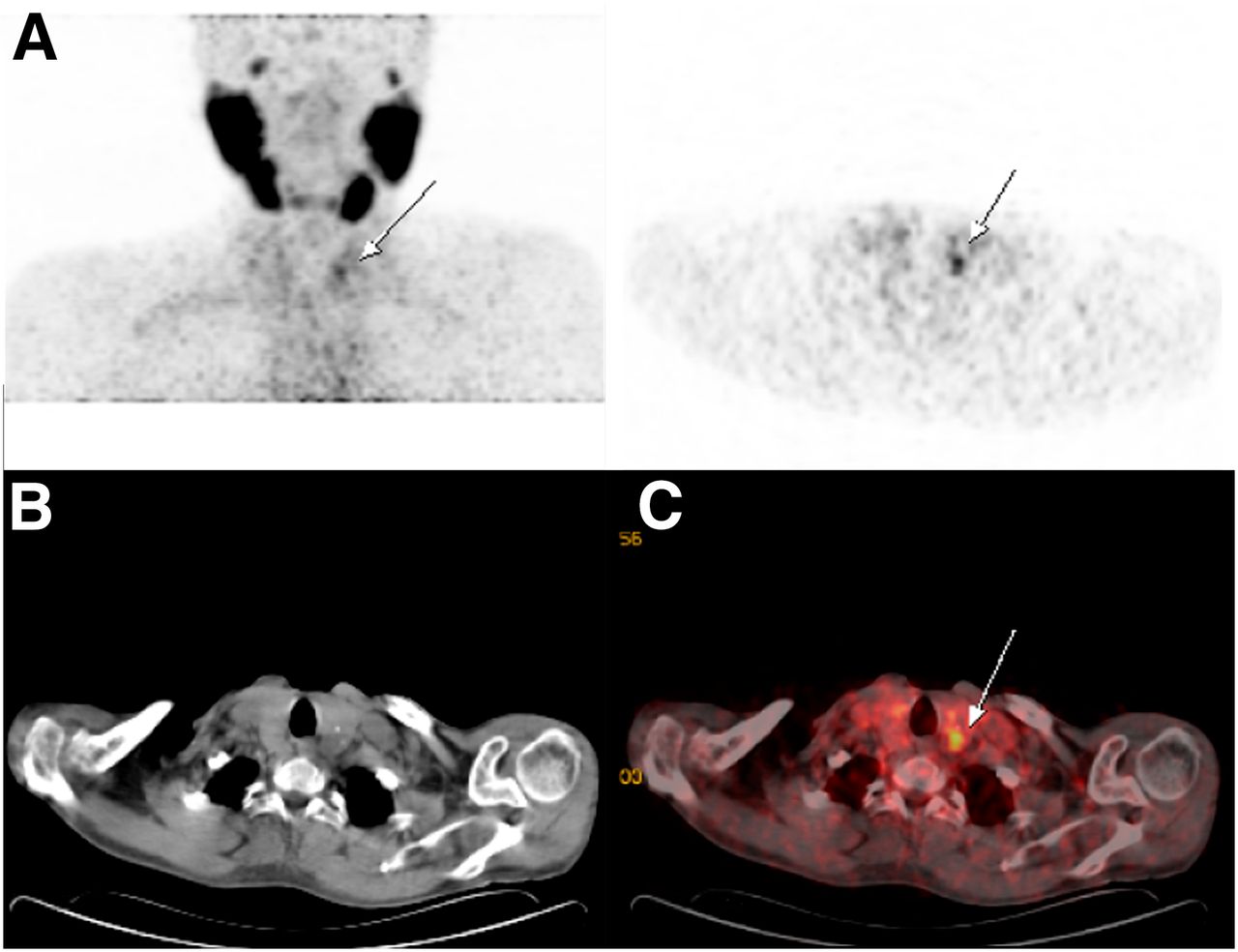
We also performed 68Ga-PSMA PET/CT imaging in a 62-y-old prostate cancer patient with a thyroid nodule. Slightly increased uptake in the left thyroid gland on the 68Ga-PSMA image was seen (Fig. 2A). Our patient underwent surgery, and his pathology result was Hürthle cell angioinvasive follicular thyroid cancer with a 0.4-cm diameter. Papillary thyroid cancer on the right thyroid gland (0.5 cm) was also reported in the pathology results. However, on PSMA imaging there was no increased activity on the right thyroid gland.

68Ga-PSMA PET (A), CT (B), and fusion images (C) of 62-y-old patient with prostate cancer. Slightly increased uptake on nodule in left thyroid gland is seen.
DISCUSSION
68Ga-PSMA can be used for PET/CT-based staging of prostate cancer. PSMA expression can be seen on the cell membranes of endothelial cells of the tumor neovasculature in several other cancers such as renal cell carcinoma, colon carcinoma, neuroendocrine tumors, melanoma, or breast cancer (3,4). It is important to be aware of thyroid incidentalomas in 68Ga-PSMA imaging to avoid scan misinterpretation. Fine-needle aspiration biopsy of PSMA-avid thyroid lesions should be considered to exclude a primary thyroid neoplasm. It is problematic to distinguish benign follicular nodules from follicular carcinomas. 99mTc-methoxyisobutylisonitrile and 18F-FDG PET/CT images were obtained to predict malignant thyroid nodules (5). 68Ga-PSMA imaging might be useful to distinguish follicular thyroid lesions.
CONCLUSION
Follicular thyroid lesions can be seen on 68Ga-PSMA PET/CT imaging. It is important to be aware of thyroid incidentalomas in 68Ga-PSMA imaging to avoid scan misinterpretation.
DISCLOSURE
No potential conflict of interest relevant to this article was reported.
Footnotes
Published online Mar. 10, 2016.
REFERENCES
- Received for publication December 22, 2015.
- Accepted for publication February 5, 2016.


