
Main menu

  • Home
  • Content
    • Current
      • JNMT Supplement
    • Ahead of print
    • Past Issues
    • Continuing Education
    • JNMT Podcast
    • SNMMI Annual Meeting Abstracts
  • Subscriptions
    • Subscribers
    • Rates
    • Journal Claims
    • Institutional and Non-member
  • Authors
    • Submit to JNMT
    • Information for Authors
    • Assignment of Copyright
    • AQARA Requirements
  • Info
    • Reviewers
    • Permissions
    • Advertisers
    • Corporate & Special Sales
  • About
    • About Us
    • Editorial Board
    • Contact Information
  • More
    • Alerts
    • Feedback
    • Help
    • SNMMI Journals
  • SNMMI
    • JNMT
    • JNM
    • SNMMI Journals
    • SNMMI

User menu

  • Subscribe
  • My alerts
  • Log in
  • My Cart

Search

  • Advanced search
Journal of Nuclear Medicine Technology
  • SNMMI
    • JNMT
    • JNM
    • SNMMI Journals
    • SNMMI
  • Subscribe
  • My alerts
  • Log in
  • My Cart
Journal of Nuclear Medicine Technology

Advanced Search

  • Home
  • Content
    • Current
    • Ahead of print
    • Past Issues
    • Continuing Education
    • JNMT Podcast
    • SNMMI Annual Meeting Abstracts
  • Subscriptions
    • Subscribers
    • Rates
    • Journal Claims
    • Institutional and Non-member
  • Authors
    • Submit to JNMT
    • Information for Authors
    • Assignment of Copyright
    • AQARA Requirements
  • Info
    • Reviewers
    • Permissions
    • Advertisers
    • Corporate & Special Sales
  • About
    • About Us
    • Editorial Board
    • Contact Information
  • More
    • Alerts
    • Feedback
    • Help
    • SNMMI Journals
  • Watch or Listen to JNMT Podcast
  • Visit SNMMI on Facebook
  • Join SNMMI on LinkedIn
  • Follow SNMMI on Twitter
  • Subscribe to JNMT RSS feeds
Research ArticleImaging

Determining the Minimal Required Radioactivity of 18F-FDG for Reliable Semiquantification in PET/CT Imaging: A Phantom Study

Ming-Kai Chen, David H. Menard and David W. Cheng
Journal of Nuclear Medicine Technology March 2016, 44 (1) 26-30; DOI: https://doi.org/10.2967/jnmt.115.165258
Ming-Kai Chen
1Radiology and Biomedical Imaging, Yale University School of Medicine, New Haven, Connecticut
  • Find this author on Google Scholar
  • Find this author on PubMed
  • Search for this author on this site
David H. Menard III
2Yale–New Haven Hospital, New Haven, Connecticut; and
  • Find this author on Google Scholar
  • Find this author on PubMed
  • Search for this author on this site
David W. Cheng
3Sidra Medical and Research Center, Doha, Qatar
  • Find this author on Google Scholar
  • Find this author on PubMed
  • Search for this author on this site
  • Article
  • Figures & Data
  • Info & Metrics
  • PDF
Loading

Article Figures & Data

Figures

  • FIGURE 1.
    • Download figure
    • Open in new tab
    • Download powerpoint
    FIGURE 1.

    (A) Standard Jaszczak ECT phantom with solid spheres replaced by hollow spheres. (B) Hollow spheres with diameters of 3.4, 2.1, 1.5, 1.2, 1.0, and 0.5 cm. (C) Image of spheres filled with 18F-FDG (165 kBq/mL). (D) Transaxial reconstructed images at 0, 4, 6, 8, 10, 12, 13.5, 14.5, 15.5, 16.5, 17.5, and 18.5 h of decay (displayed in 3-min reconstruction).
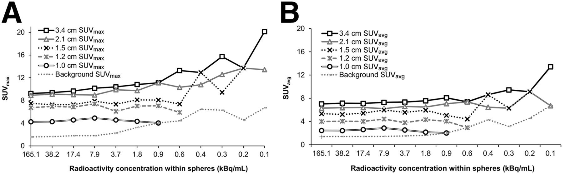
  • FIGURE 2.
    • Download figure
    • Open in new tab
    • Download powerpoint
    FIGURE 2.

    Impact of radioactivity concentration on SUVmax (A) and SUVavg (B) in spheres of various sizes using 10-min acquisition. Radioactivity concentrations on x-axis correspond to acquisition time points of approximately 0, 4, 6, 8, 10, 12, 13.5, 14.5, 15.5, 16.5, 17.5, and 18.5 h.
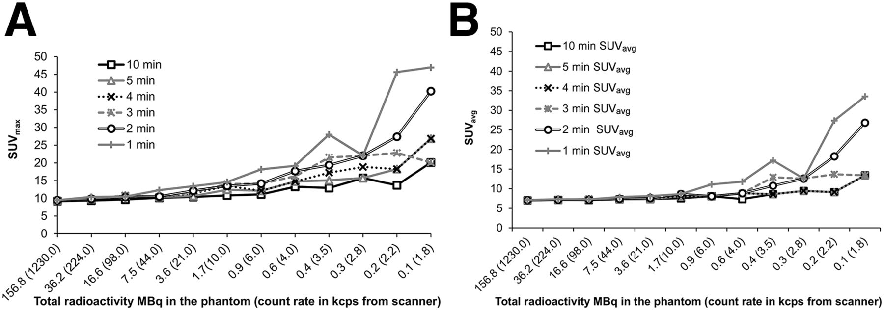
  • FIGURE 3.
    • Download figure
    • Open in new tab
    • Download powerpoint
    FIGURE 3.

    Impact of total radioactivity on SUVmax (A) and SUVavg (B) in 3.4-cm sphere at various acquisition times. SUVmax is significantly overestimated at earlier acquisitions (47 at 1-min acquisition compared with 9–10 at later acquisitions). Likewise, SUVavg is overestimated at earlier acquisitions (33.6 at 1-min acquisition compared with 7–8 at later acquisitions). Counting rates are not corrected for random counts or dead time.
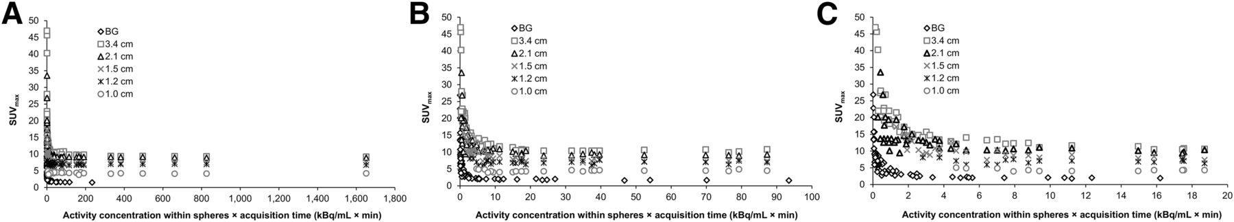
  • FIGURE 4.
    • Download figure
    • Open in new tab
    • Download powerpoint
    FIGURE 4.

    Use of radioactivity concentration–acquisition time product to determine reproducibility of SUVmax in spheres of various sizes. SUVmax is overestimated within a low range of concentration–time product. Data for typical (A) and expanded (B and C) x-axes are shown.

PreviousNext
Back to top

In this issue

Journal of Nuclear Medicine Technology: 44 (1)
Journal of Nuclear Medicine Technology
Vol. 44, Issue 1
March 1, 2016
  • Table of Contents
  • About the Cover
  • Index by author
Print
Download PDF
Article Alerts
Sign In to Email Alerts with your Email Address
Email Article

Thank you for your interest in spreading the word on Journal of Nuclear Medicine Technology.

NOTE: We only request your email address so that the person you are recommending the page to knows that you wanted them to see it, and that it is not junk mail. We do not capture any email address.

Enter multiple addresses on separate lines or separate them with commas.
Determining the Minimal Required Radioactivity of 18F-FDG for Reliable Semiquantification in PET/CT Imaging: A Phantom Study
(Your Name) has sent you a message from Journal of Nuclear Medicine Technology
(Your Name) thought you would like to see the Journal of Nuclear Medicine Technology web site.
Citation Tools
Determining the Minimal Required Radioactivity of 18F-FDG for Reliable Semiquantification in PET/CT Imaging: A Phantom Study
Ming-Kai Chen, David H. Menard, David W. Cheng
Journal of Nuclear Medicine Technology Mar 2016, 44 (1) 26-30; DOI: 10.2967/jnmt.115.165258

Citation Manager Formats

  • BibTeX
  • Bookends
  • EasyBib
  • EndNote (tagged)
  • EndNote 8 (xml)
  • Medlars
  • Mendeley
  • Papers
  • RefWorks Tagged
  • Ref Manager
  • RIS
  • Zotero
Share
Determining the Minimal Required Radioactivity of 18F-FDG for Reliable Semiquantification in PET/CT Imaging: A Phantom Study
Ming-Kai Chen, David H. Menard, David W. Cheng
Journal of Nuclear Medicine Technology Mar 2016, 44 (1) 26-30; DOI: 10.2967/jnmt.115.165258
Twitter logo Facebook logo LinkedIn logo Mendeley logo
  • Tweet Widget
  • Facebook Like
  • Google Plus One
Bookmark this article

Jump to section

  • Article
    • Abstract
    • MATERIALS AND METHODS
    • RESULTS
    • DISCUSSION
    • CONCLUSION
    • DISCLOSURE
    • Acknowledgments
    • Footnotes
    • REFERENCES
  • Figures & Data
  • Info & Metrics
  • PDF

Related Articles

  • No related articles found.
  • PubMed
  • Google Scholar

Cited By...

  • Enhancing PET/CT Assessment with Porous 3D-Printed Grids - A Pilot Study
  • Determining the Minimal Required Ultra-Low-Dose CT Dose Level for Reliable Attenuation Correction of 18F-FDG PET/CT: A Phantom Study
  • Google Scholar

More in this TOC Section

  • Early 10-Minute Postinjection [18F]F-FAPI-42 uEXPLORER Total-Body PET/CT Scanning Protocol for Staging Lung Cancer Using HYPER Iterative Reconstruction
  • Single- Versus Dual-Time-Point Imaging for Transthyretin Cardiac Amyloid Using 99mTc-Pyrophosphate
  • Does Arthrography Improve Accuracy of SPECT/CT for Diagnosis of Aseptic Loosening in Patients with Painful Knee Arthroplasty: A Systematic Review and Metaanalysis
Show more Imaging

Similar Articles

Keywords

  • PET
  • phantom
  • dosimetry
  • Dose reduction
  • acquisition time
  • standard uptake value
SNMMI

© 2025 SNMMI

Powered by HighWire