


Article Figures & Data
Figures
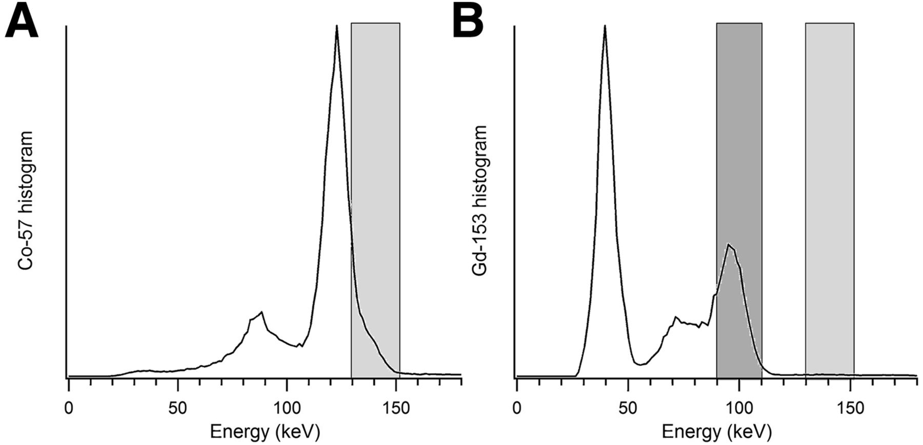
- FIGURE 1.
Energy spectra acquired for 57Co flood source (A) and 153Gd flood source (B). 99mTc emission window (140 keV, 15% width) is shown in light gray. With 57Co flood source, many transmission photons are acquired in emission window, providing body outline in emission image but contributing crosstalk and reduced SNR. With 153Gd flood source, transmission photons are acquired in separate window (100 keV, 20%) shown in dark gray. 153Gd flood source contributes few counts to 99mTc emission window, arising from simultaneous detection of europium x-rays (40-keV peak) along with primary γ-photons (100-keV peak).
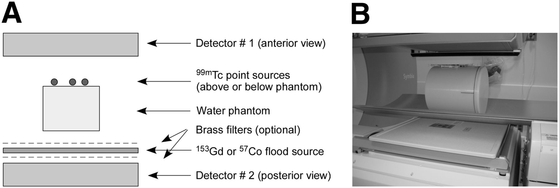
- FIGURE 2.
Schematic (A) and photograph (B) of phantom experiments simulating lymphoscintigraphy studies with flood source shadowing. In photograph, point sources are contained in 1-mL syringes taped to top of water-filled phantom. Images also were acquired with phantom rotated 180°, to position point sources on posterior side. Flood source (white) is visible in photograph (B), shown with 1 brass filter below source and none above source.
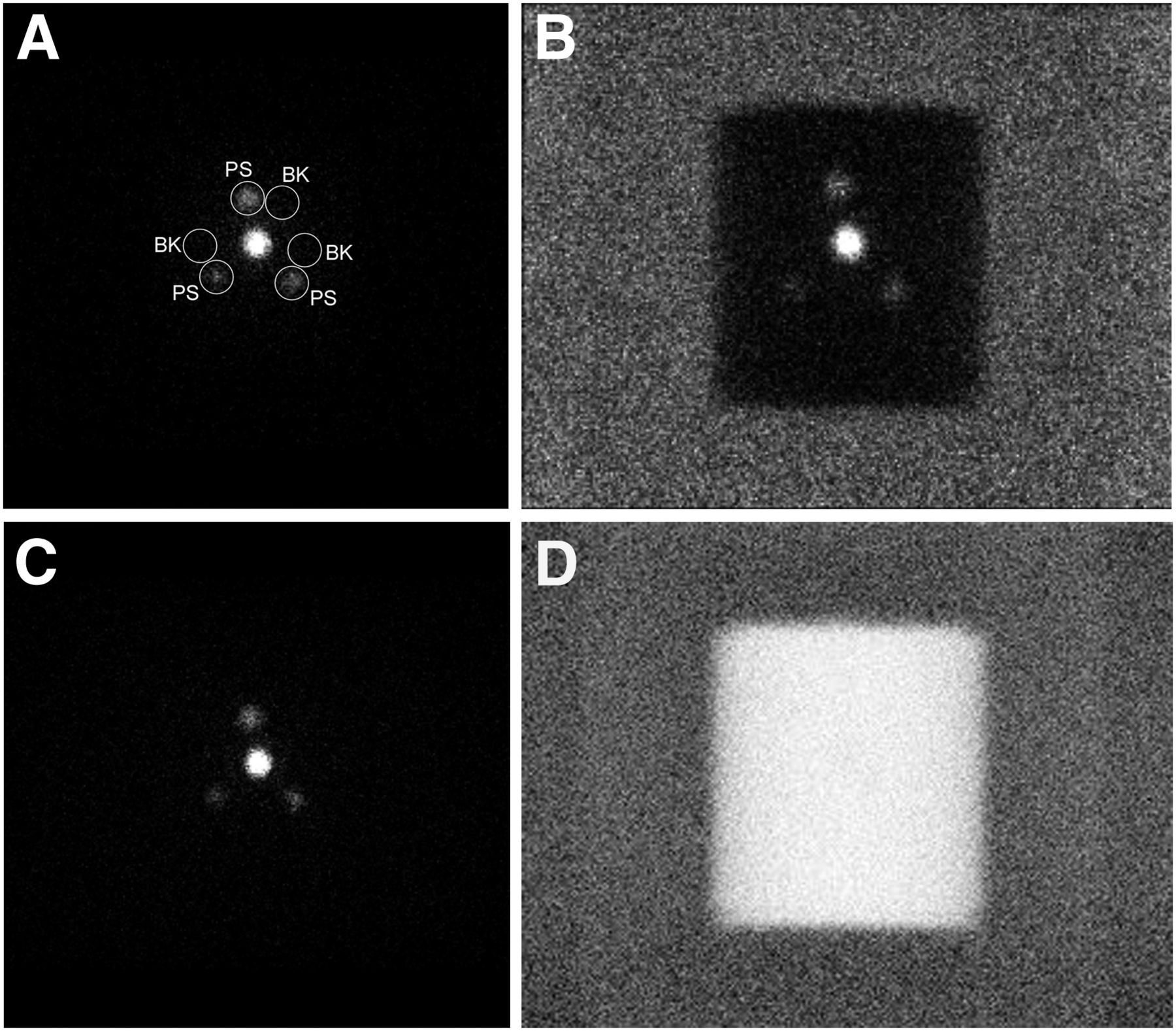
- FIGURE 3.
Anterior-view scintigraphy emission images (140-keV window) are shown in gray scale of water phantom with 99mTc point sources located below phantom: with no flood source (A), with 57Co flood source (B), and with 153Gd flood source (C). In A, circular regions of interest (diameter, 33 mm) for measuring point source counts (labeled PS) and adjacent background counts (labeled BK) are shown. For 153Gd flood source, corresponding transmission image (100-keV window) (D) is shown in inverse gray scale. Point source activities were 3,770 kBq in center, 800 kBq at top, 380 kBq at lower-right, and 220 kBq at lower-left. All emission images are shown with equivalent intensity scaling relative to central point source.
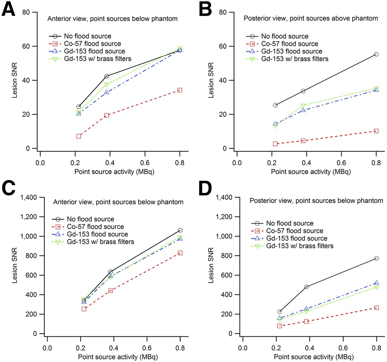
- FIGURE 4.
Graphs of SNR versus activity for 3 peripheral point sources in various phantom and flood configurations: anterior view, point sources below phantom (A); posterior view, point sources above phantom (B); anterior view, point sources above phantom (C); and posterior view, point sources below phantom (D).
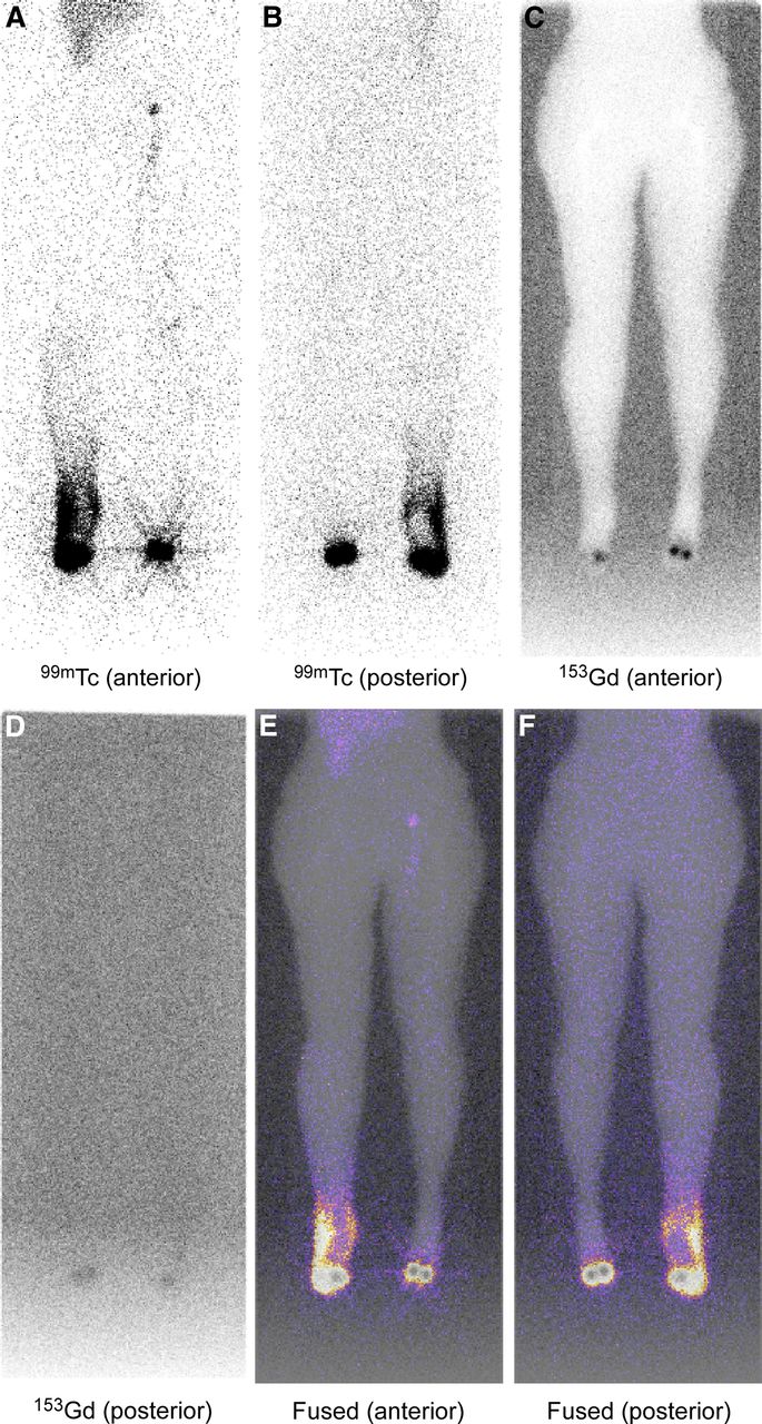
- FIGURE 5.
Example images from whole-body lymphedema scan: anterior and posterior views of 99mTc data, 153Gd data, and fused 99mTc (color) and 153Gd (inverse gray scale) data. Dermal backflow is clearly visible in 99mTc images, extent of which would not have been as apparent if 57Co flood source had been used instead.
- FIGURE 6.
Example fused 99mTc (color) and 153Gd (inverse gray scale) images from breast lymphoscintigraphy scan. 99mTc color window has been enhanced in oblique and lateral views to reveal lymph node with low tracer activity, which was not visible in anterior view and which would have had lower SNR if using 57Co flood source and single energy window.
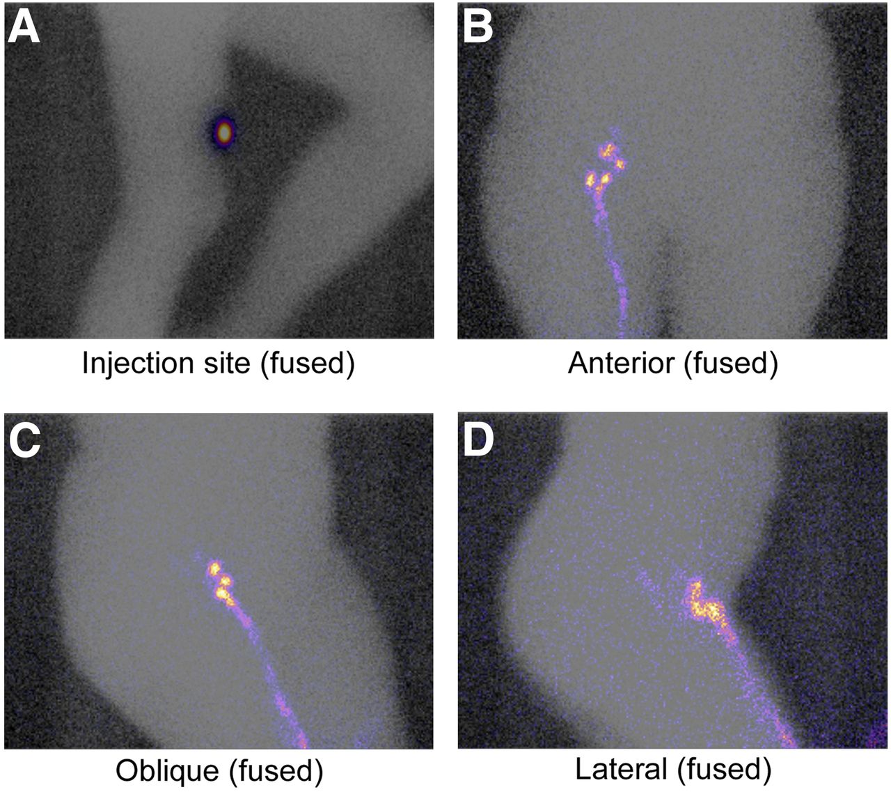
- FIGURE 7.
Example fused 99mTc (color) and 153Gd (inverse gray scale) images from melanoma lymphoscintigraphy scan of injection site and of multiple views of inguinal lymph nodes.


