
Main menu

  • Home
  • Content
    • Current
      • JNMT Supplement
    • Ahead of print
    • Past Issues
    • Continuing Education
    • JNMT Podcast
    • SNMMI Annual Meeting Abstracts
  • Subscriptions
    • Subscribers
    • Rates
    • Journal Claims
    • Institutional and Non-member
  • Authors
    • Submit to JNMT
    • Information for Authors
    • Assignment of Copyright
    • AQARA Requirements
  • Info
    • Reviewers
    • Permissions
    • Advertisers
    • Corporate & Special Sales
  • About
    • About Us
    • Editorial Board
    • Contact Information
  • More
    • Alerts
    • Feedback
    • Help
    • SNMMI Journals
  • SNMMI
    • JNMT
    • JNM
    • SNMMI Journals
    • SNMMI

User menu

  • Subscribe
  • My alerts
  • Log in
  • My Cart

Search

  • Advanced search
Journal of Nuclear Medicine Technology
  • SNMMI
    • JNMT
    • JNM
    • SNMMI Journals
    • SNMMI
  • Subscribe
  • My alerts
  • Log in
  • My Cart
Journal of Nuclear Medicine Technology

Advanced Search

  • Home
  • Content
    • Current
    • Ahead of print
    • Past Issues
    • Continuing Education
    • JNMT Podcast
    • SNMMI Annual Meeting Abstracts
  • Subscriptions
    • Subscribers
    • Rates
    • Journal Claims
    • Institutional and Non-member
  • Authors
    • Submit to JNMT
    • Information for Authors
    • Assignment of Copyright
    • AQARA Requirements
  • Info
    • Reviewers
    • Permissions
    • Advertisers
    • Corporate & Special Sales
  • About
    • About Us
    • Editorial Board
    • Contact Information
  • More
    • Alerts
    • Feedback
    • Help
    • SNMMI Journals
  • Watch or Listen to JNMT Podcast
  • Visit SNMMI on Facebook
  • Join SNMMI on LinkedIn
  • Follow SNMMI on Twitter
  • Subscribe to JNMT RSS feeds
Review ArticleTeaching Case Studies

Interesting Layering of Excreted 18F-FDG in the Urinary Bladder in Patients with Urinary Tract Infection and Distended Bladder

Guohua Shen, Wenjie Zhang, Zhiyun Jia and Houfu Deng
Journal of Nuclear Medicine Technology September 2015, 43 (3) 234-235; DOI: https://doi.org/10.2967/jnmt.114.148031
Guohua Shen
Department of Nuclear Medicine, West China Hospital, Sichuan University, Chengdu, Sichuan, People’s Republic of China
  • Find this author on Google Scholar
  • Find this author on PubMed
  • Search for this author on this site
Wenjie Zhang
Department of Nuclear Medicine, West China Hospital, Sichuan University, Chengdu, Sichuan, People’s Republic of China
  • Find this author on Google Scholar
  • Find this author on PubMed
  • Search for this author on this site
Zhiyun Jia
Department of Nuclear Medicine, West China Hospital, Sichuan University, Chengdu, Sichuan, People’s Republic of China
  • Find this author on Google Scholar
  • Find this author on PubMed
  • Search for this author on this site
Houfu Deng
Department of Nuclear Medicine, West China Hospital, Sichuan University, Chengdu, Sichuan, People’s Republic of China
  • Find this author on Google Scholar
  • Find this author on PubMed
  • Search for this author on this site
  • Article
  • Figures & Data
  • Info & Metrics
  • PDF
Loading

Abstract

Settling of 18F-FDG in the bladder is often noted on whole-body PET/CT images, but this phenomenon has never received any careful attention and the mechanism has been unclear. The 2 patients described in this report, one with a T1 pathologic fracture and another with widespread bone and lymph node metastases from an unknown primary tumor, underwent PET/CT. Both had urinary tract infection and a distended bladder during scanning. The interesting layering of 18F-FDG in the urinary bladder was observed in both patients. The presence of this phenomenon demands careful evaluation of the urine by the clinician, and the mechanism is hypothesized to be slow 18F-FDG excretion in patients with a distended urinary bladder, resulting in delayed mixing with urine. In addition, urinary tract infection may be a potential cause. Images showing this interesting layering should be interpreted with care.

  • 18F-FDG layering
  • positron emission tomography/computed tomography
  • urinary bladder

Accumulation of 18F-FDG in the urinary bladder usually makes evaluation of bladder lesions on PET/CT difficult. At our institution, urinary bladder activity is minimized by asking the patient to drink at least 500 mL of plain water and to void before image acquisition. However, settling of 18F-FDG in the urinary bladder is occasionally observed and can impose an interpretation challenge, obscuring or simulating pathologic findings (1). A recent study about intravenous contrast PET/CT stated that the higher specific gravity of the contrast material displaces the excreted 18F-FDG, resulting in anterior layering of the radiotracer (2). We report here 2 patients with bladder layering of excreted 18F-FDG on PET/CT without intravenous contrast material. We hypothesize that the phenomenon is due mainly to delayed mixing of 18F-FDG excretion with nonradioactive urine in a distended bladder and urinary tract infection. However, the mechanism needs to be further investigated in a more comprehensive study.

CASE REPORT

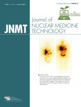
The first patient was a 63-y-old woman with neck and back pain and lower-limb weakness. She had undergone PET/CT examination, and the images showed a pathologic fracture in the first thoracic vertebra. There was interesting layering of excreted 18F-FDG in the urinary bladder, showing 18F-FDG–loaded urine at the bottom of a distended bladder and clear urine at the top (Fig. 1A). The combination of radiologic images and clinical examination revealed no bladder abnormalities but indicated a urinary tract infection. The second patient, who was 67 y old and male with fever of unknown origin, was diagnosed with widespread bone and lymph node metastases from an unknown primary tumor. In addition, he had symptoms of lung infection. A clear demarcation between the urine without 18F-FDG at the top of the bladder and 18F-FDG–loaded urine at the bottom was also seen (Fig. 1B). We noted that the urinary bladder was full during scanning. Both patients had clear symptoms of urinary tract infection. The urine was cloudy and had elevated urinary epithelial cell counts, red blood cell counts, white blood cell counts, and total protein content. Urine analysis was also positive for bacteria and fungus. The relevant features are summarized in Table 1.

FIGURE 1.
  • Download figure
  • Open in new tab
  • Download powerpoint
FIGURE 1.

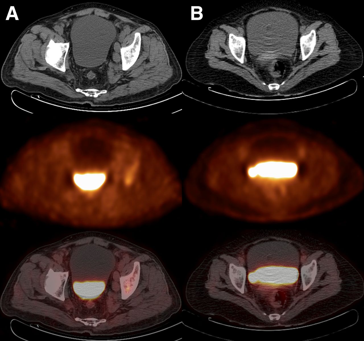
CT (top), PET (middle), and PET/CT (bottom) images showing layering of 18F-FDG in bladder. (A) A 63-y-old woman with T1 pathologic fracture and urinary tract infection. (B) A 67-y-old man with widespread bone and lymph node metastases from unknown primary tumor, also with urinary tract infection.

View this table:
  • View inline
  • View popup
TABLE 1

Clinical and Pathologic Details of the 2 Patients

DISCUSSION

Several examples of layering within the urinary bladder have been reported (3–5). Carrington et al. described a characteristic appearance on CT, with urine that contains contrast medium and has a high specific gravity being layered posteriorly in the dependent portion of the bladder, whereas lower-specific-gravity, nonopacified urine is layered at the uppermost part of the bladder (3). Another study showed that anterior layering of excreted 18F-FDG in the bladder is commonly observed on PET/CT scans obtained with intravenous iodinated contrast material and is due to displacement of 18F-FDG by excreted iodinated contrast material with higher specific gravity (5).

However, the 2 patients in our study did not undergo CT or MR imaging examinations before the PET/CT evaluation, and the urine did not contain contrast medium. Both had a urinary tract infection, and this might have caused the layering phenomenon. The metabolically active cellular components of the urine, such as inflammatory cells, bacteria, and fungi, take up excreted 18F-FDG in the urine for their metabolism, and these cells settle to the bottom of the bladder because of gravity during PET scanning, when a patient lies supine for some time (4). Some patients do not empty the bladder before scanning. The bladder already contains a considerable amount of urine with metabolically active cells and subsequently receives 18F-FDG–loaded urine during scanning. We found that the urinary bladders of our 2 patients were full during scanning. In a distended bladder, delayed mixing of 18F-FDG excretion with nonradioactive urine causes the layering. Moreover, PET/CT images of patients with an empty urinary bladder may help in further interpreting this phenomenon, as settling seems to be better visualized in a full bladder.

To the best of our understanding, there was no standard explanation for settling of 18F-FDG in the urinary bladder that may have alerted the clinicians to check more carefully for lesions on or near the bladder. We may infer that urinary tract infection and a distended bladder are capable of inducing urine layering; however, a 100% cause-and-effect relationship could not be established because of the limited number of cases and the limited information. A definitive explanation needs to be sought with a larger prospective study involving systematic analysis of urine, bladder volume, bladder pressure, and specific gravity in patients with layering, compared with those without layering.

CONCLUSION

These 2 cases illustrate the interesting layering of 18F-FDG in the bladder, the causes of which might be urinary tract infection and a distended bladder; however, the mechanism needs to be further investigated in a more comprehensive study. Moreover, if settling of 18F-FDG in the urinary bladder is seen on imaging, this finding should be reported with the aim of alerting clinicians to look at the urine more carefully. All shades of clinical presentations are possible.

DISCLOSURE

This study was supported by the National Natural Science Foundation (grants 81271532, 81171456, and 30900378) and by Fundamental Research Funds for the Central Universities (project 2015SCU04B09). No other potential conflict of interest relevant to this article was reported.

Footnotes

  • Published online Dec. 23, 2014.

REFERENCES

  1. 1.↵
    1. Subhas N,
    2. Patel PV,
    3. Pannu HK,
    4. et al
    . Imaging of pelvic malignancies with in-line FDG PET-CT: case examples and common pitfalls of FDG PET. Radiographics. 2005;25:1031–1043.
    OpenUrlCrossRefPubMed
  2. 2.↵
    1. Purcell DD,
    2. Coakley FV,
    3. Franc BL,
    4. et al
    . Anterior layering of excreted 18F-FDG in the bladder on PET/CT: frequency and cause. AJR. 2007;189:W96–W99.
    OpenUrlCrossRefPubMed
  3. 3.↵
    1. Carrington BM,
    2. Johnson RJ
    . Case report: inverted contrast medium–urine level in the bladder on computed tomography. Clin Radiol. 1992;46:216–217.
    OpenUrlCrossRefPubMed
  4. 4.↵
    1. Kumar K
    . Settlement of [18F]-FDG in the urinary bladder: a new sign. Nucl Med Commun. 2009;30:37–40.
    OpenUrlCrossRefPubMed
  5. 5.↵
    1. Samii VF
    . Inverted contrast medium-urine layering in the canine urinary bladder on computed tomography. Vet Radiol Ultrasound. 2005;46:502–505.
    OpenUrlCrossRefPubMed
  • Received for publication August 31, 2014.
  • Accepted for publication October 27, 2014.
PreviousNext
Back to top

In this issue

Journal of Nuclear Medicine Technology: 43 (3)
Journal of Nuclear Medicine Technology
Vol. 43, Issue 3
September 1, 2015
  • Table of Contents
  • About the Cover
  • Index by author
Print
Download PDF
Article Alerts
Sign In to Email Alerts with your Email Address
Email Article

Thank you for your interest in spreading the word on Journal of Nuclear Medicine Technology.

NOTE: We only request your email address so that the person you are recommending the page to knows that you wanted them to see it, and that it is not junk mail. We do not capture any email address.

Enter multiple addresses on separate lines or separate them with commas.
Interesting Layering of Excreted 18F-FDG in the Urinary Bladder in Patients with Urinary Tract Infection and Distended Bladder
(Your Name) has sent you a message from Journal of Nuclear Medicine Technology
(Your Name) thought you would like to see the Journal of Nuclear Medicine Technology web site.
Citation Tools
Interesting Layering of Excreted 18F-FDG in the Urinary Bladder in Patients with Urinary Tract Infection and Distended Bladder
Guohua Shen, Wenjie Zhang, Zhiyun Jia, Houfu Deng
Journal of Nuclear Medicine Technology Sep 2015, 43 (3) 234-235; DOI: 10.2967/jnmt.114.148031

Citation Manager Formats

  • BibTeX
  • Bookends
  • EasyBib
  • EndNote (tagged)
  • EndNote 8 (xml)
  • Medlars
  • Mendeley
  • Papers
  • RefWorks Tagged
  • Ref Manager
  • RIS
  • Zotero
Share
Interesting Layering of Excreted 18F-FDG in the Urinary Bladder in Patients with Urinary Tract Infection and Distended Bladder
Guohua Shen, Wenjie Zhang, Zhiyun Jia, Houfu Deng
Journal of Nuclear Medicine Technology Sep 2015, 43 (3) 234-235; DOI: 10.2967/jnmt.114.148031
Twitter logo Facebook logo LinkedIn logo Mendeley logo
  • Tweet Widget
  • Facebook Like
  • Google Plus One
Bookmark this article

Jump to section

  • Article
    • Abstract
    • CASE REPORT
    • DISCUSSION
    • CONCLUSION
    • DISCLOSURE
    • Footnotes
    • REFERENCES
  • Figures & Data
  • Info & Metrics
  • PDF

Related Articles

  • No related articles found.
  • PubMed
  • Google Scholar

Cited By...

  • No citing articles found.
  • Google Scholar

More in this TOC Section

  • High-Sensitivity Troponin Elevation in a Young Woman with Typical Chest Pain: The Heart of the Matter
  • Pulmonary Adenocarcinoma Revealed by Parathyroid Scintigraphy: An Incidental Case to Remember
  • Prominent Right Ventricular Tracer Uptake: A Harbinger of Multivessel Coronary Artery Disease
Show more Teaching Case Studies

Similar Articles

Keywords

  • 18F-FDG layering
  • positron emission tomography/computed tomography
  • urinary bladder
SNMMI

© 2025 SNMMI

Powered by HighWire