


Article Figures & Data
Figures
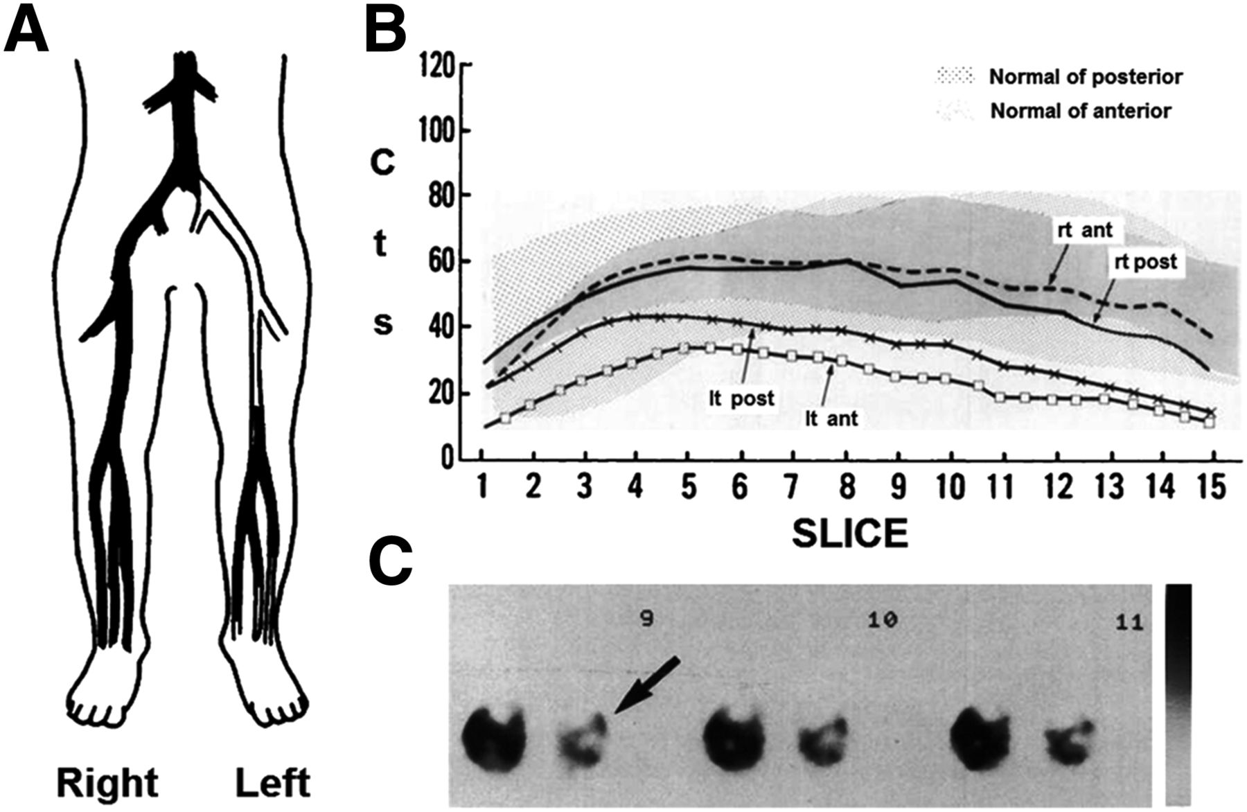
- FIGURE 1.
Arteriography (A) demonstrates unilateral PVD, which is confirmed by abnormal 201Tl SPECT stress perfusion profile curves of anterior and posterior tibial muscle components of the left leg (B) as well as visual inspection of 201Tl SPECT transverse images (C; perfusion defect noted by arrow). (Reprinted from (24).)
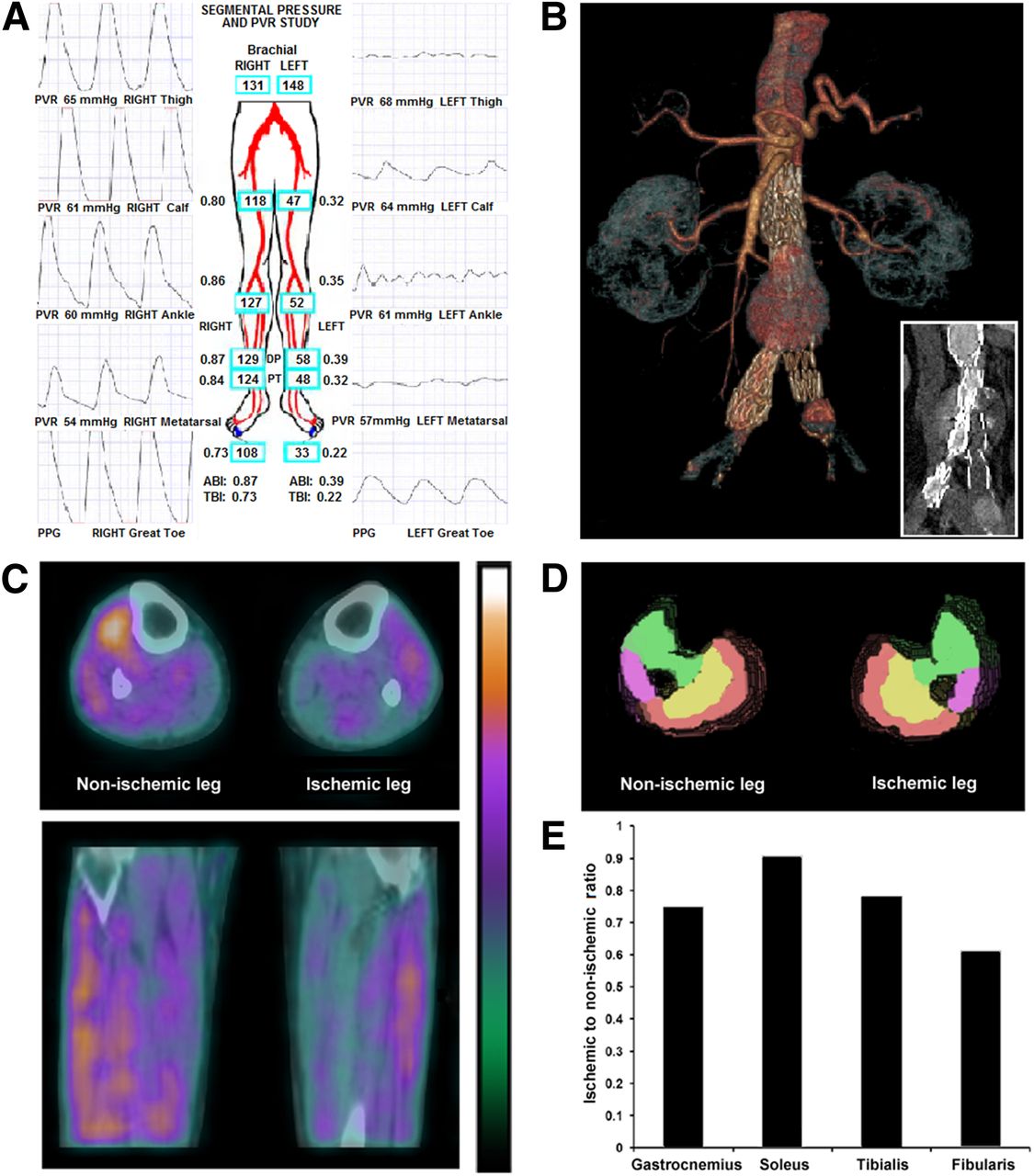
- FIGURE 2.
Multimodality evaluation with ankle–brachial indices (A), CT angiography (B), and hybrid 99mTc-tetrofosmin SPECT/CT (C) reveals impaired lower-extremity pressures and tissue perfusion in PVD patient with previously implanted aortoiliac stents (B). Segmentation of muscle groups into 3-dimensional regions of interest by CT attenuation images (D) confirmed differences in regional tissue perfusion between legs (E). Red = gastrocnemius; yellow = soleus; green = tibialis; purple = fibularis; ABI = ankle–brachial index; PPG = photoplethysmograph; PVR = pulse volume recording; TBI = toe–brachial index.
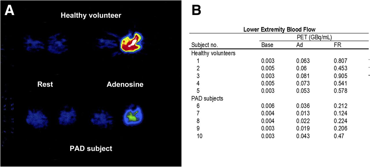
- FIGURE 3.
Lower-extremity PET H215O imaging of healthy subject and PVD patient during selective adenosine infusion into left leg (A). Baseline and adenosine stress blood flow was assessed, and flow reserve was expressed as ratio of adenosine flow to baseline flow (B). Flow reserve was significantly lower in PVD patients than in healthy subjects. (Reprinted from (32).).
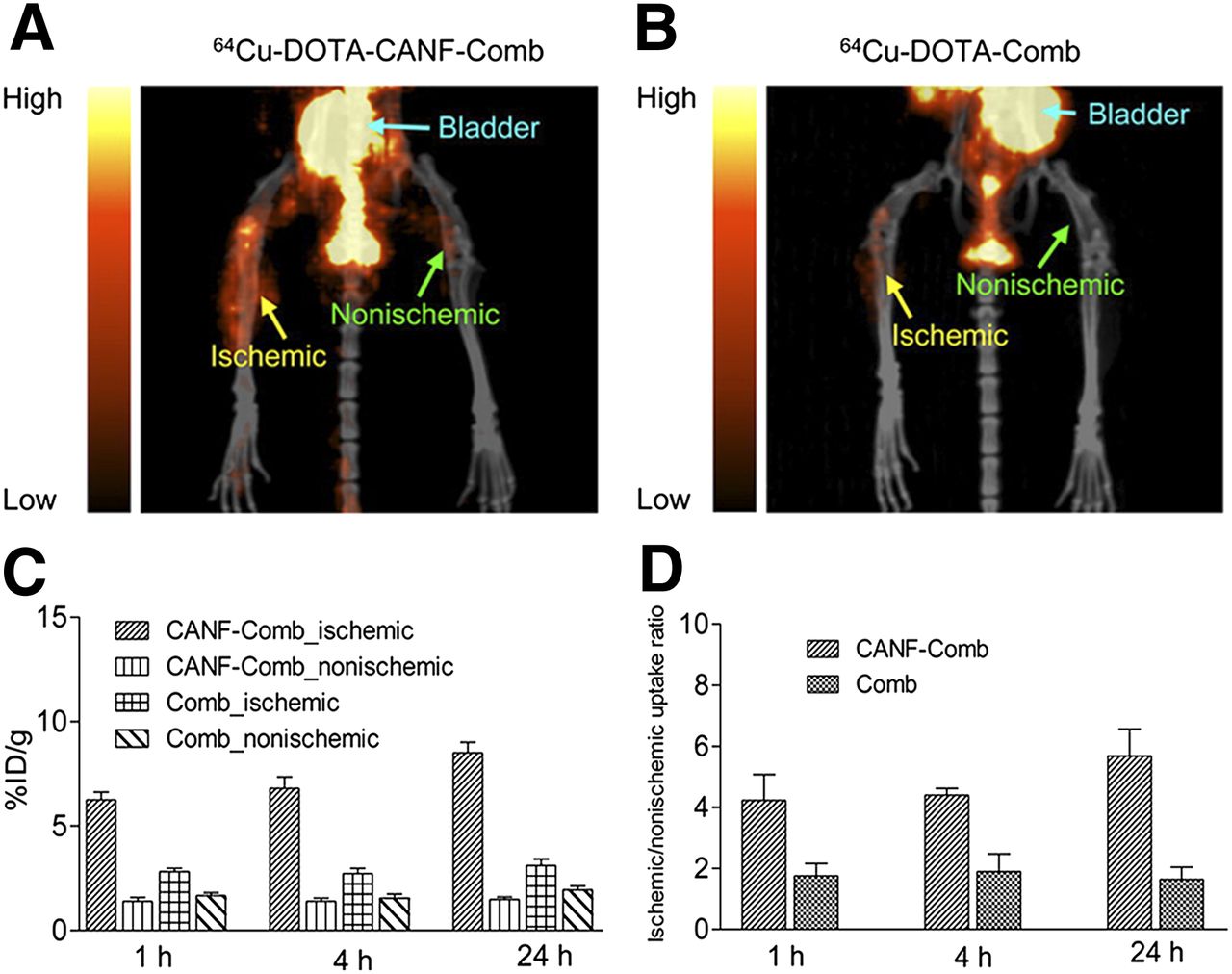
- FIGURE 4.
PET/CT imaging with 64Cu-DOTA–C-type atrial natriuretic factor–comb (A) and 64Cu-DOTA–comb (B) in mouse model of hind limb ischemia, 7 d after femoral artery occlusion. 64Cu-DOTA–C-type atrial natriuretic factor–comb uptake was significantly higher than uptake of 64Cu-DOTA–comb in ischemic limb when percentage injected tracer dose per gram of tissue (C) or ischemic-to-nonischemic leg ratios (D) were examined. (Reprinted from (52).) CANF = C-type atrial natriuretic factor.
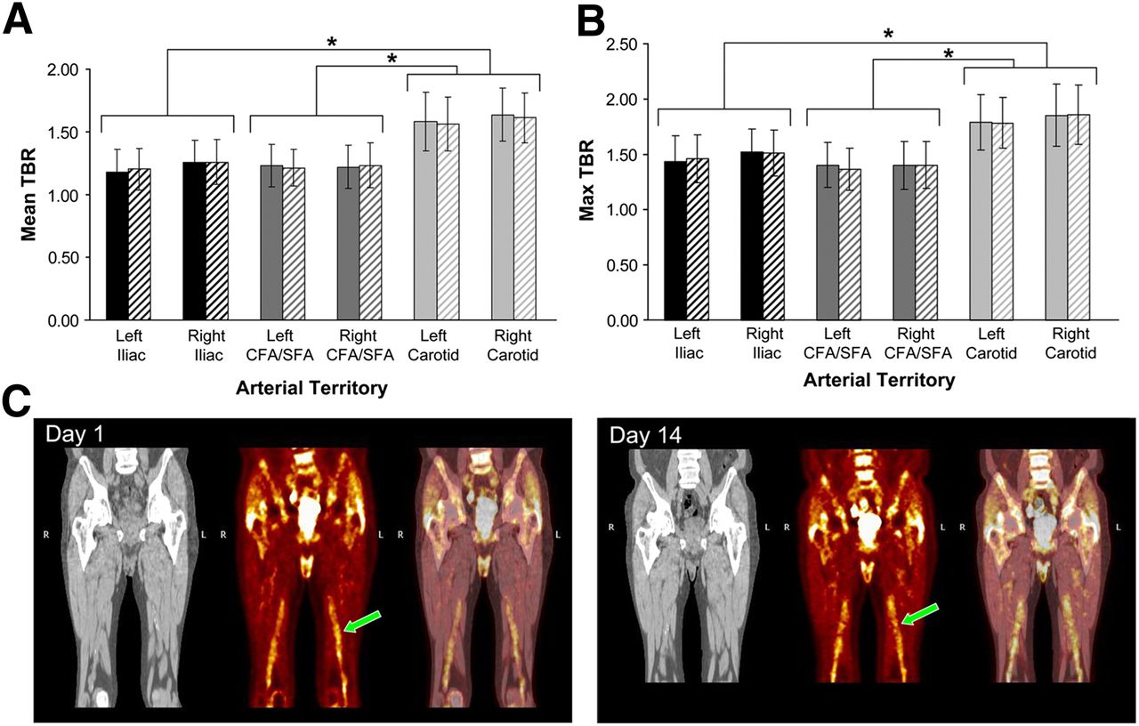
- FIGURE 5.
Analysis of mean (A) and maximum (B) 18F-FDG tumor-to-background ratios within multiple arterial regions on day 1 (solid bars) and 14 d later (hatched bars). No significant differences were observed within arterial regions across time. Carotid artery uptake was significantly higher than 18F-FDG uptake within lower-extremity arteries on days 1 and 14. 18F-FDG PET (C, middle) fused with CT imaging (C, right) revealed similar uptake in femoral arteries on days 1 and 14 (noted by arrows). CFA = common femoral artery; SFA = superficial femoral artery; TBR = tumor-to-background ratio. *P < 0.001. (Reprinted from (58).)


Tables
Modality Perfusion/blood flow Angiogenesis Atherosclerosis SPECT 201Tl (7,21,24–26) 99mTc-NC100692 (47,48) 99mTc-sestamibi (27,29,31) 111In-VEGF121 (45) 99mTc-pyrophosphate (76) 125I-c(RGD(I)yV) (77) 99mTc-tetrofosmin PET 15O-water (32,33,36,40) 76Br-nanoprobe (50) 18F-FDG (58–62) C15O2 (35) 68Ga-NOTA-RGD (49) 18F-sodium fluoride (66) 15O2 (35,36) 64Cu-DOTA-CANF-comb (52) 11C-acetate (67) 13N-ammonia (34) 64Cu-DOTA-VEGF121 (46) 64Cu-DOTA-CANF (65) CANF = C-type atrial natriuretic factor.


