
Main menu

  • Home
  • Content
    • Current
      • JNMT Supplement
    • Ahead of print
    • Past Issues
    • Continuing Education
    • JNMT Podcast
    • SNMMI Annual Meeting Abstracts
  • Subscriptions
    • Subscribers
    • Rates
    • Journal Claims
    • Institutional and Non-member
  • Authors
    • Submit to JNMT
    • Information for Authors
    • Assignment of Copyright
    • AQARA Requirements
  • Info
    • Reviewers
    • Permissions
    • Advertisers
    • Corporate & Special Sales
  • About
    • About Us
    • Editorial Board
    • Contact Information
  • More
    • Alerts
    • Feedback
    • Help
    • SNMMI Journals
  • SNMMI
    • JNMT
    • JNM
    • SNMMI Journals
    • SNMMI

User menu

  • Subscribe
  • My alerts
  • Log in
  • My Cart

Search

  • Advanced search
Journal of Nuclear Medicine Technology
  • SNMMI
    • JNMT
    • JNM
    • SNMMI Journals
    • SNMMI
  • Subscribe
  • My alerts
  • Log in
  • My Cart
Journal of Nuclear Medicine Technology

Advanced Search

  • Home
  • Content
    • Current
    • Ahead of print
    • Past Issues
    • Continuing Education
    • JNMT Podcast
    • SNMMI Annual Meeting Abstracts
  • Subscriptions
    • Subscribers
    • Rates
    • Journal Claims
    • Institutional and Non-member
  • Authors
    • Submit to JNMT
    • Information for Authors
    • Assignment of Copyright
    • AQARA Requirements
  • Info
    • Reviewers
    • Permissions
    • Advertisers
    • Corporate & Special Sales
  • About
    • About Us
    • Editorial Board
    • Contact Information
  • More
    • Alerts
    • Feedback
    • Help
    • SNMMI Journals
  • Watch or Listen to JNMT Podcast
  • Visit SNMMI on Facebook
  • Join SNMMI on LinkedIn
  • Follow SNMMI on Twitter
  • Subscribe to JNMT RSS feeds
Review ArticleContinuing Education

Technical Considerations in Brain Amyloid PET Imaging with 18F-Florbetapir

LisaAnn Trembath, Maureen Newell and Michael D. Devous
Journal of Nuclear Medicine Technology September 2015, 43 (3) 175-184; DOI: https://doi.org/10.2967/jnmt.115.156679
LisaAnn Trembath
Avid Radiopharmaceuticals, Inc., Philadelphia, Pennsyvania
CNMT, MSM, CCRA, FSNMMI-TS
  • Find this author on Google Scholar
  • Find this author on PubMed
  • Search for this author on this site
Maureen Newell
Avid Radiopharmaceuticals, Inc., Philadelphia, Pennsyvania
CNMT, RT(N)
  • Find this author on Google Scholar
  • Find this author on PubMed
  • Search for this author on this site
Michael D. Devous Sr.
Avid Radiopharmaceuticals, Inc., Philadelphia, Pennsyvania
PhD
  • Find this author on Google Scholar
  • Find this author on PubMed
  • Search for this author on this site
  • Article
  • Figures & Data
  • Info & Metrics
  • PDF
Loading

Article Figures & Data

Figures

  • Tables
  • FIGURE 1.
    • Download figure
    • Open in new tab
    • Download powerpoint
    FIGURE 1.

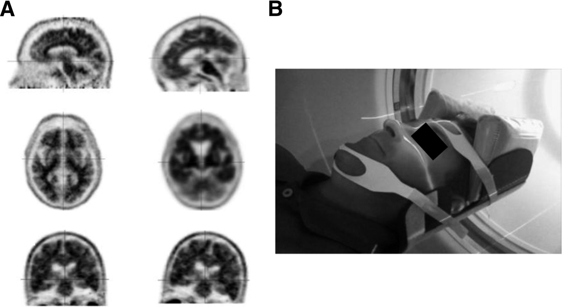
    (A) Top row of images demonstrates proper canthomeatal positioning on left and head pitched too far forward on right. In middle row are corresponding axial projections, with standard appearance on left and nonstandard appearance on right, with frontal lobes prominent and occipital lobes just becoming visible. Bottom two images demonstrate proper coronal positioning (left), and right image shows prominent roll to left. (Courtesy of Avid Radiopharmaceuticals.) (B) Volunteer properly positioned in head holder (GE Healthcare) using laser system, cushions, chin strap, and forehead strap. (Courtesy of Desert Advanced [RadNet Imaging Centers], Palm Springs, California.)

  • FIGURE 2.
    • Download figure
    • Open in new tab
    • Download powerpoint
    FIGURE 2.

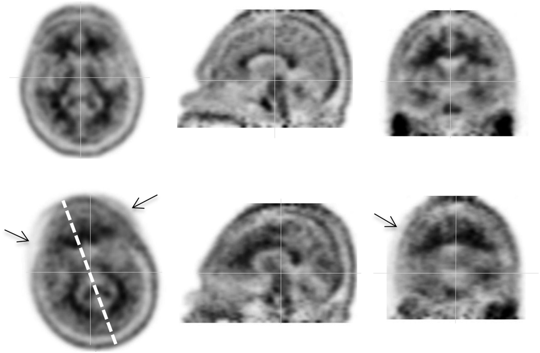
    Ten-minute dynamic PET acquisition, with frame 1 at top and frame 2 at bottom. Frame 2 is representative of subject motion, most notably yaw and roll. Dashed line indicates degree of motion between frames. Arrows indicate image blurring. (Courtesy of Avid Radiopharmaceuticals.)

  • FIGURE 3.
    • Download figure
    • Open in new tab
    • Download powerpoint
    FIGURE 3.

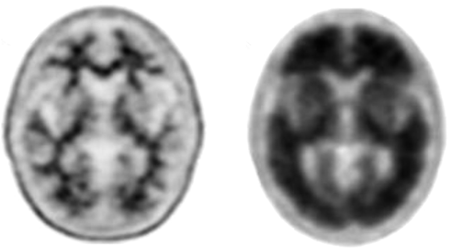
    Representative amyloid-negative (left) and amyloid-positive (right) transaxial slices from 18F-florbetapir PET scans. (Courtesy of Avid Radiopharmaceuticals.)

  • FIGURE 4.
    • Download figure
    • Open in new tab
    • Download powerpoint
    FIGURE 4.

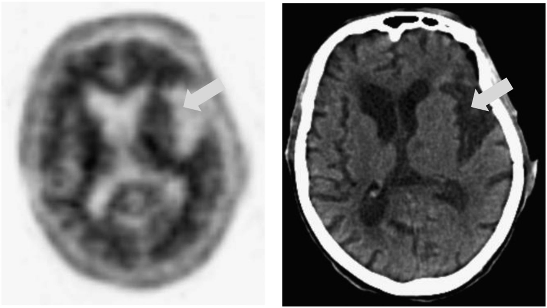
    18F-florbetapir PET slice (left) and corresponding CT slice (right), with arrows indicating location of atrophy. (Courtesy of Avid Radiopharmaceuticals.)

  • FIGURE 5.
    • Download figure
    • Open in new tab
    • Download powerpoint
    FIGURE 5.

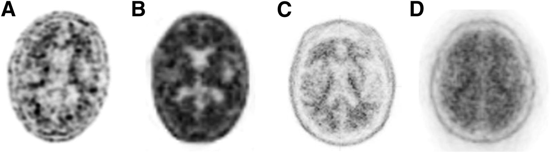
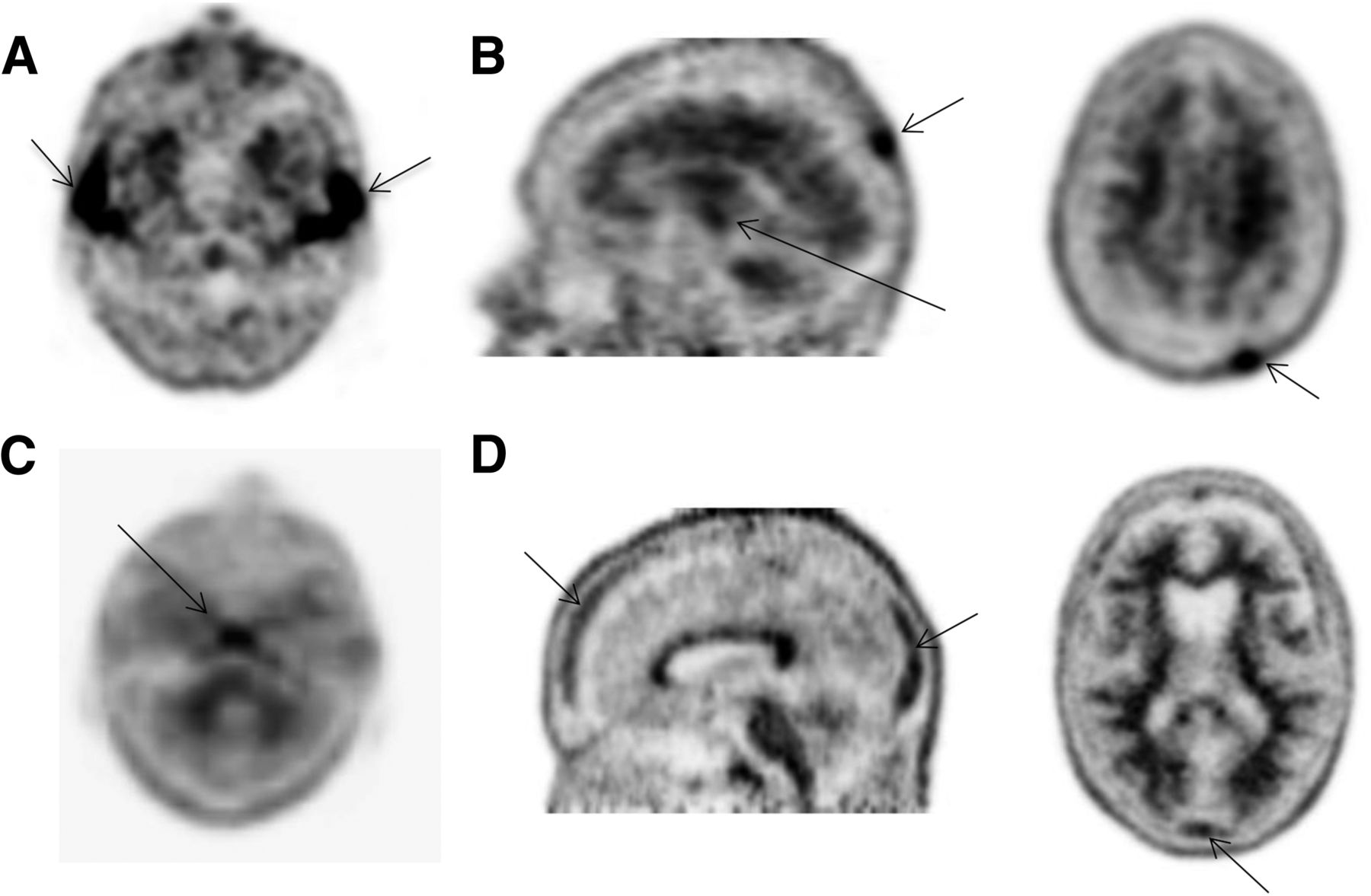
    18F-florbetapir images with poor resolution due to dose infiltration (A), imaging beyond recommended time window (B), inadequate smoothing (2 mm FWHM) (C), and scanner issue during acquisition (D). (Courtesy of Avid Radiopharmaceuticals.)

  • FIGURE 6.
    • Download figure
    • Open in new tab
    • Download powerpoint
    FIGURE 6.

    (A) Normal salivary gland uptake on 18F-florbetapir PET image. (B) Nonspecific bony uptake in skull and clivus bone confirmed by CT. (C) Nonspecific uptake in clivus bone. (D) Nonspecific uptake in skull. (Courtesy of Avid Radiopharmaceuticals.)

  • FIGURE 7.
    • Download figure
    • Open in new tab
    • Download powerpoint
    FIGURE 7.

    (A) Streak artifact on CT image (left) and PET/CT image (right) due to incomplete warm-up of CT tube. (B) Image resulting from detector block failure. (C) Image resulting from calibration error due to out-of-date normalization (left), and reprocessed image with new normalization (right). (D) Out-of-date normalization seen as streaks on non–attenuation-corrected images. (Courtesy of Avid Radiopharmaceuticals.)

Tables

  • Figures
    • View popup
    TABLE 1

    18F-Florbetapir–Estimated Absorbed Radiation Dose (4)

    Organ or tissueμGy/MBq
    Adrenal14
    Bone
     Osteogenic cells28
     Red marrow14
    Brain10
    Breasts6
    Gallbladder wall143
    Gastrointestinal tract
     Lower large intestine wall28
     Small intestine66
     Stomach wall12
     Upper intestine wall74
    Heart wall13
    Kidneys14
    Liver64
    Lungs9
    Muscle9
    Ovaries18
    Pancreas14
    Skin6
    Spleen9
    Testes7
    Thymus7
    Thyroid7
    Urinary bladder wall27
    Uterus16
    Total body12
    Effective dose*19*
    • ↵* μSv/MBq.

PreviousNext
Back to top

In this issue

Journal of Nuclear Medicine Technology: 43 (3)
Journal of Nuclear Medicine Technology
Vol. 43, Issue 3
September 1, 2015
  • Table of Contents
  • About the Cover
  • Index by author
Print
Download PDF
Article Alerts
Sign In to Email Alerts with your Email Address
Email Article

Thank you for your interest in spreading the word on Journal of Nuclear Medicine Technology.

NOTE: We only request your email address so that the person you are recommending the page to knows that you wanted them to see it, and that it is not junk mail. We do not capture any email address.

Enter multiple addresses on separate lines or separate them with commas.
Technical Considerations in Brain Amyloid PET Imaging with 18F-Florbetapir
(Your Name) has sent you a message from Journal of Nuclear Medicine Technology
(Your Name) thought you would like to see the Journal of Nuclear Medicine Technology web site.
Citation Tools
Technical Considerations in Brain Amyloid PET Imaging with 18F-Florbetapir
LisaAnn Trembath, Maureen Newell, Michael D. Devous
Journal of Nuclear Medicine Technology Sep 2015, 43 (3) 175-184; DOI: 10.2967/jnmt.115.156679

Citation Manager Formats

  • BibTeX
  • Bookends
  • EasyBib
  • EndNote (tagged)
  • EndNote 8 (xml)
  • Medlars
  • Mendeley
  • Papers
  • RefWorks Tagged
  • Ref Manager
  • RIS
  • Zotero
Share
Technical Considerations in Brain Amyloid PET Imaging with 18F-Florbetapir
LisaAnn Trembath, Maureen Newell, Michael D. Devous
Journal of Nuclear Medicine Technology Sep 2015, 43 (3) 175-184; DOI: 10.2967/jnmt.115.156679
Twitter logo Facebook logo LinkedIn logo Mendeley logo
  • Tweet Widget
  • Facebook Like
  • Google Plus One
Bookmark this article

Jump to section

  • Article
    • Abstract
    • INDICATIONS AND CONTRAINDICATIONS
    • ADVERSE REACTIONS
    • MECHANISM OF ACTION AND BIODISTRIBUTION
    • RADIATION DOSIMETRY
    • PATIENT PREPARATION
    • DOSING AND ADMINISTRATION
    • POSITIONING THE PATIENT
    • SCANNING PARAMETERS
    • PROCESSING PARAMETERS
    • DISPLAY AND INTERPRETATION
    • ARTIFACTS, PITFALLS, AND NORMAL VARIANTS
    • CONCLUSION
    • DISCLOSURE
    • Footnotes
    • REFERENCES
  • Figures & Data
  • Info & Metrics
  • PDF

Related Articles

  • No related articles found.
  • PubMed
  • Google Scholar

Cited By...

  • PET-derived amyloid patterns in gray and white matter across the Alzheimers disease: A high-model-order ICA
  • Amyloid Imaging Update: How the Amyloid Landscape Is Changing in Light of the Recent Food and Drug Administration Approval of Antiamyloid Therapeutics
  • Google Scholar

More in this TOC Section

  • Illuminating the Hidden: Standardizing Cardiac MIBG Imaging for Sympathetic Dysfunction
  • PET/CT Case Series: Unmasking the Mystery of Cardiac Sarcoidosis
  • Delivery Methods of Radiopharmaceuticals: Exploring Global Strategies to Minimize Occupational Radiation Exposure
Show more Continuing Education

Similar Articles

Keywords

  • florbetapir
  • PET
  • Brain Imaging
  • amyloid
  • Technical
SNMMI

© 2025 SNMMI

Powered by HighWire