


Article Figures & Data
Figures
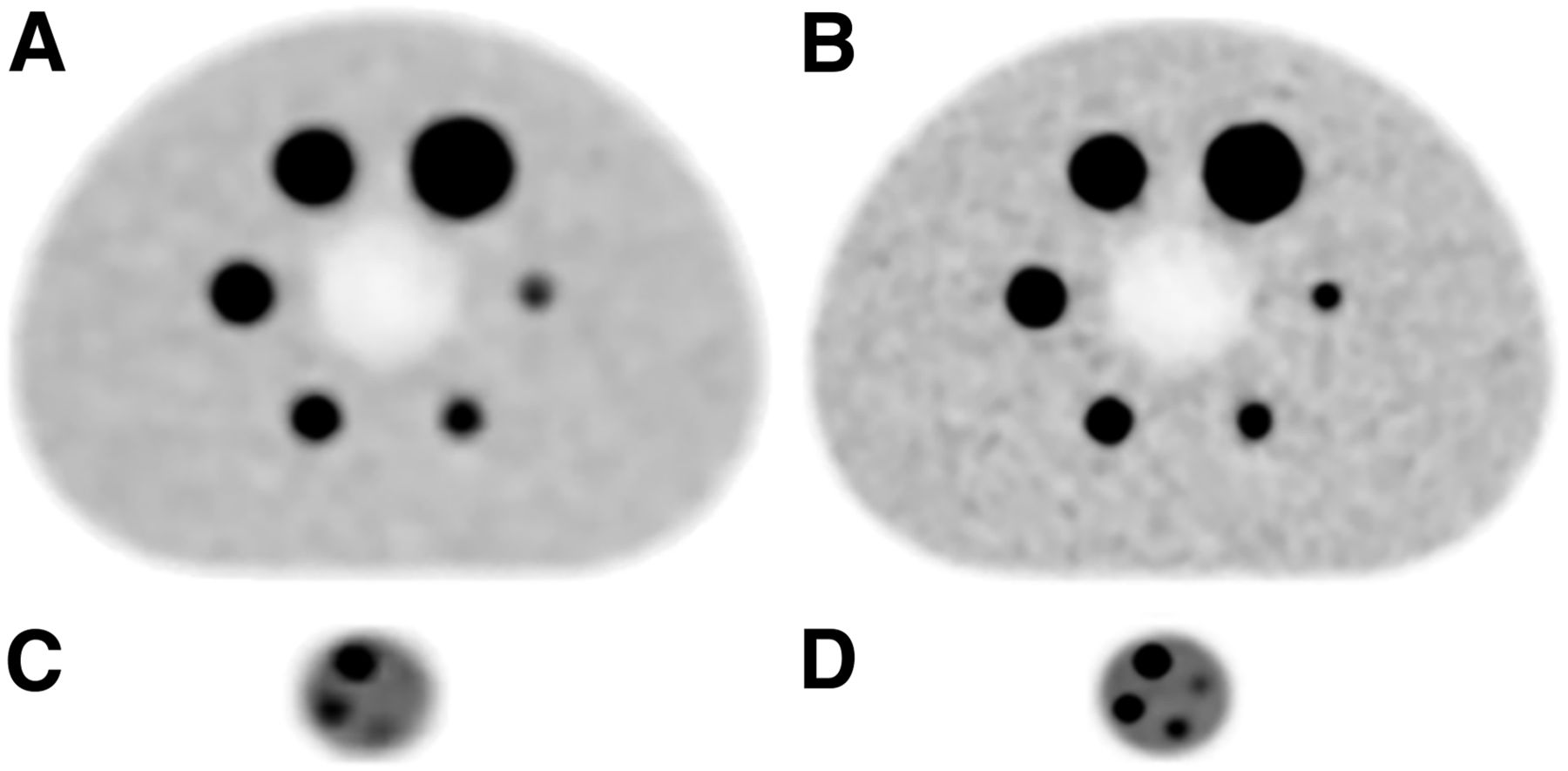
- FIGURE 1.
18F-FDG PET images of NEMA phantom (A and B) and microphantom (C and D) using standard-voxel reconstruction (A and C) and small-voxel reconstructions (B and D). Sphere sizes for NEMA phantom were 10, 13, 17, 22, 28, and 37 mm, inner diameter, and sphere sizes for microphantom were 4, 5, 6, and 8 mm, inner diameter. For all spheres with diameter of 13 mm or less, contrast is clearly increased using small-voxel reconstruction. Moreover, smallest microphantom sphere cannot be distinguished from background on standard-voxel reconstruction (C), yet it can be detected on small-voxel reconstruction (D).
- FIGURE 2.
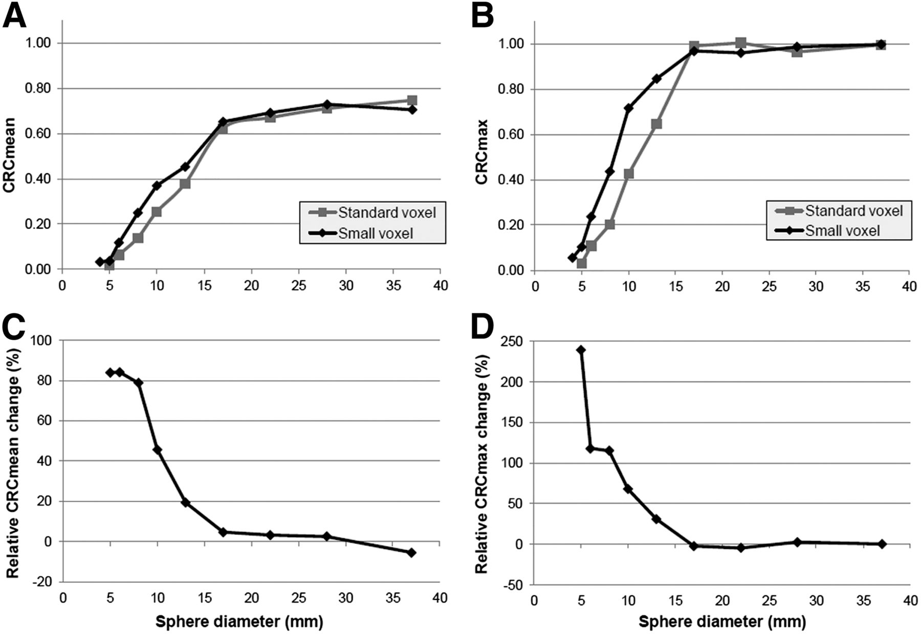
CRCmean (A) and CRCmax (B) for phantom spheres using standard- and small-voxel reconstructions, with relative changes (%) for both parameters presented in plot C and D. For small spheres (≤13 mm), we found increases for CRCmean and CRCmax using small-voxel reconstruction, with highest relative increases for 5- and 6-mm small spheres. As 4-mm small spheres could not be distinguished from background on standard-voxel reconstruction, no relative CRC changes were determined.
- FIGURE 3.
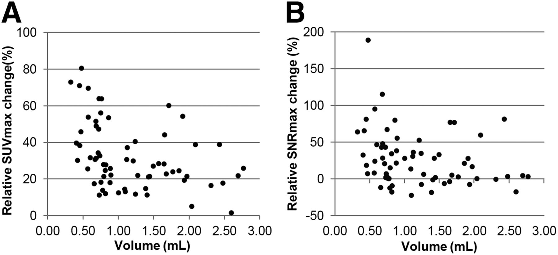
Relative changes in SUVmax (A) and SNRmax (B) for all 66 included lesions using small-voxel reconstruction instead of standard-voxel reconstruction. Average changes in SUVmax and SNRmax across all lesions were 32% and 27%, respectively. For lesions smaller than 0.75 mL, we found average SUVmax and SNRmax increases of 44% and 46%, respectively.
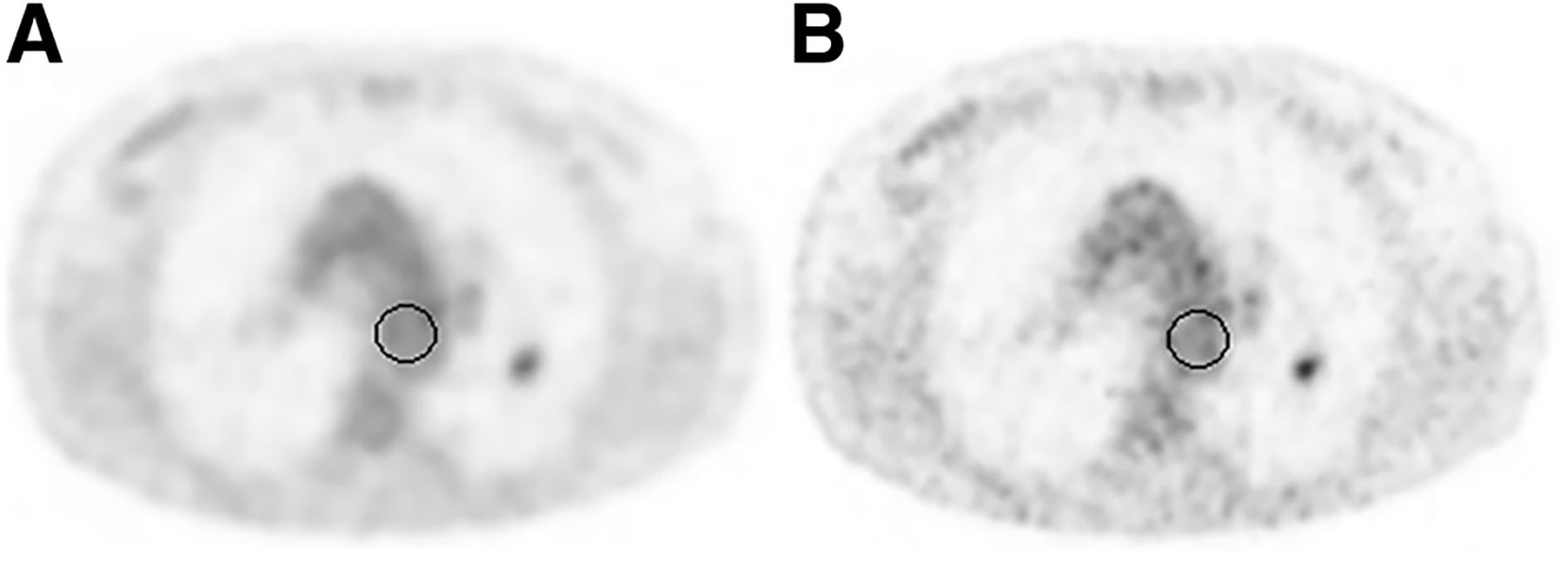
- FIGURE 4.
Transverse 18F-FDG PET images using standard-voxel reconstruction (A) and small-voxel reconstruction (B). Lesion in left lung (volume, 0.68 mL) with SUVmax of 2.6 using standard-voxel reconstruction increased with 54% to 4.0 using small-voxel reconstruction. SNRmax increased with 115% (from 3.1 to 6.6). ROIs used for background measurements are illustrated by black circles.
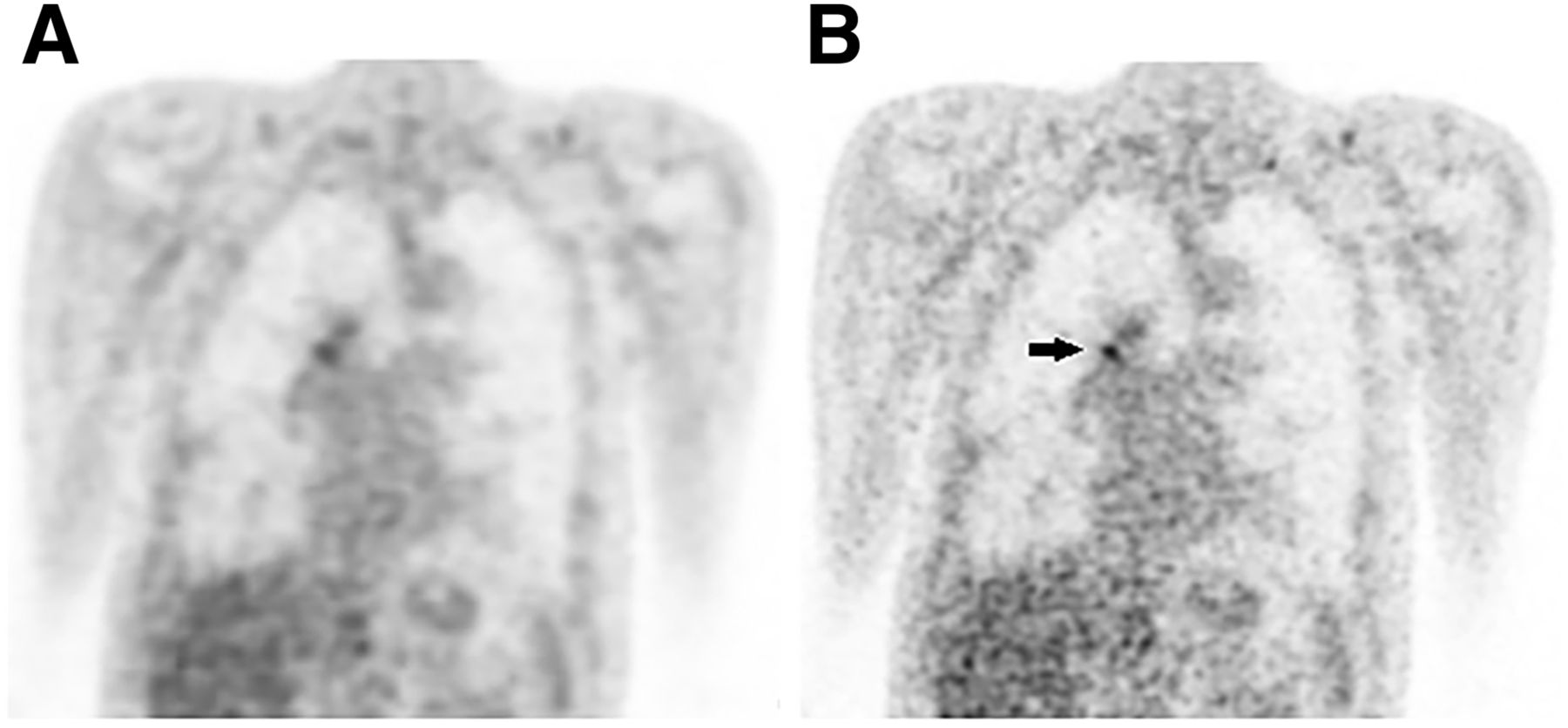
- FIGURE 5.
Coronal 18F-FDG PET images with standard-voxel reconstruction (A) and small-voxel reconstruction (B). SUVmax of lesion in right hilar region (volume, 0.50 mL) with SUVmax of 3.0 using standard-voxel reconstruction increased with 46% to 4.4 on small-voxel reconstruction (black arrow). SNRmax increased with 77% (from 9.8 to 13.3).


Tables
- TABLE 1
CRCmean, CRCmax, SNRmean, and SNRmax for 10 Phantom Spheres for Both Voxel Reconstructions, Including Relative Changes (%)
Microphantom sphere diameter (mm) NEMA phantom sphere diameter (mm) Parameter 4 5 6 8 10 13 17 22 28 37 CRCmean Standard N/A 0.02 0.06 0.14 0.25 0.38 0.62 0.67 0.71 0.75 Small 0.03 0.04 0.12 0.25 0.37 0.45 0.65 0.69 0.73 0.71 % N/A 84% 84% 79% 46% 19% 5% 3% 3% −6% CRCmax Standard N/A 0.03 0.11 0.20 0.43 0.65 0.99 1.01 0.96 1.00 Small 0.06 0.10 0.24 0.44 0.72 0.85 0.97 0.96 0.99 1.00 % N/A 239% 118% 115% 68% 31% −2% −4% 2% 0% SNRmean Standard N/A 5 16 56 42 63 104 112 118 124 Small 9 10 30 64 42 52 74 79 83 80 % N/A 87% 85% 79% 0% −18% −28% −29% −30% −35% SNRmax Standard N/A 8 28 52 71 108 165 167 161 166 Small 14 27 61 112 82 97 110 110 113 114 % N/A 242% 119% 116% 15% −10% −33% −35% −30% −31% Standard = standard-voxel reconstruction (4 × 4 × 4 mm); N/A = not applicable; small = small-voxel reconstruction (2 × 2 × 2 mm).


Jump to section
Related Articles
Cited By...
- Performance of Digital PET Compared with High-Resolution Conventional PET in Patients with Cancer
- High Spatial Resolution Digital Positron Emission Tomography Images With Dedicated Source-to-background Algorithm for Radiotherapy Planning
- Assessing 18F-FDG Uptake in the Sentinel Lymph Node in Breast Cancer
- Time-of-Flight Information Improved the Detectability of Subcentimeter Spheres Using a Clinical PET/CT Scanner
- Low-dose High-resolution 18F-FDG-PET/CT Using Time-of-flight and Point-spread Function Reconstructions: A Role in the Detection of Breast Carcinoma Axillary Lymph Node Metastases