


Abstract
Sentinel node lymphoscintigraphy using colloidal particles has become common practice at many institutions. The ideal particle size for colloids such as filtered 99mTc-sulfur colloid (99mTc-FSC) in sentinel node studies is 15–100 nm. It is reported that the use of a reduced heating time during the reconstitution process results in an increased number of smaller particles (<30 nm). However, it is unclear whether these smaller particles (>15 nm) would be of benefit in sentinel node studies. This study sought to better define particle size by using electron microscopy, as well as to evaluate the radiochemical purity (RCP) of 99mTc-FSC at various time points after filtration. Methods: One group of 99mTc-sulfur colloid (99mTc-SC) preparations was reconstituted using the standard heating time of 5 min, and another group was prepared using a reduced heating time of 3 min. The 99mTc-SC preparations were passed through a 0.2-μm filter, and retained filter activity was measured. RCP values were collected at 0, 1, 3, and 6 h after filtration, and the particle sizes were measured at 0 and 6 h after filtration. Results: Average RCP values (± SD) for 99mTc-FSC with 5-min heating were 98.4% ± 3.0% and 98.3% ± 1.8% for 0 h and 6 h, respectively (n = 6). Average RCP values for 99mTc-FSC with 3-min heating were 98.4% ± 4.1% and 96.9% ± 3.1% for 0 h and 6 h, respectively (n = 6). Electron microscopy data showed that median particle sizes for the 3-min heating at 0 and 6 h were 24 and 35 nm, respectively. Median particle sizes for the 5-min heating at 0 and 6 h were 29 and 27 nm, respectively. The proportion of particles within the ideal range for sentinel node lymphoscintigraphy was similar between the heating methods (91.1% for 3-min heating at 0 h and 88.8% for 5-min heating at 0 h, P = 0.1851). Conclusion: Our results indicate that although there are slight significant differences in RCP value, particle size, and particle number for 99mTc-FSC prepared using either a standard or a reduced heating time, both methods produce particles within the optimum range for sentinel node studies.
The radiopharmaceutical 99mTc-labeled sulfur colloid (99mTc-SC) is currently used in several nuclear medicine studies. Using a microfiltration process, smaller particles of filtered 99mTc-SC (99mTc-FSC) can be used to assess potential lymphatic blockages as well as detect the sentinel node of tumor drainage for patients with breast cancer or melanoma (1).
The sentinel node is the first node in the lymph node basin to be encountered by lymphatic fluid draining from a tumor (2). Various studies show that the status of the sentinel node draining the primary tumor can accurately predict the condition of lymph nodes farther along the lymphatic channels. Biopsy of the sentinel node has therefore become a standard component for initial staging in breast cancer and melanoma (3). Although other cancers such as colorectal, gastric, esophageal, head and neck, thyroid, lung, gynecologic, and urologic cancers can be assessed using this method, they often have a reduced accuracy due to more complex lymphatic drainage patterns (3).
99mTc-FSC used in conjunction with isosulfan blue dye is a proven and successful method for identifying sentinel nodes and has a low false-negative rate (4). This method for sentinel node lymphoscintigraphy entails an intradermal injection of 99mTc-FSC around the site of the primary lesion. The 99mTc-FSC then progresses along the lymphatic channels and is phagocytosed by macrophages in the sentinel node (5). The radiolabeled colloid can then be imaged using various types of γ-camera imaging. Although planar imaging is sufficient in many practices, SPECT and SPECT/CT imaging can detect significantly more sentinel nodes and can better delineate the locations of the sentinel nodes (6). Finally, the sentinel node is often then located with a hand-held γ-detecting intraoperative probe to guide surgeons in lymph node dissections. Additionally, blue dye is often injected during surgery to more accurately determine the location of the lymph node (1).
The speed at which the radiolabeled colloid travels and the degree of nodal uptake is highly dependent on the size of the particles; unfiltered 99mTc-SC, prepared according to the package insert, has an average particle size of 61–445 nm (7). Although this size range is suitable for some studies, many of these particles would be too large for adequate lymph node uptake in lymphoscintigraphy. Larger particles (>100 nm) get trapped in the interstitial space instead of migrating to the sentinel node, and too small a particle (less than a few nanometers) will diffuse out of the interstitial space into venous capillaries (8). The optimal radiocolloid size for sentinel node lymphoscintigraphy is reported to be around 15–100 nm (9). This size allows for quick lymphatic drainage for imaging purposes as well as sufficient retention in the lymph node.
Current practice uses a 0.22-μm microfiltration method to obtain particles closer to the optimum range. Eshima et al. previously showed that reduced heating time during the reconstitution process of 99mTc-SC can produce a larger portion of smaller particles (10). However, this finding was determined using a microfiltration method to size the particles, and the method did not characterize particles smaller than 30 nm. It is crucial to characterize the particles within this range (i.e., 15–100 nm) so as to establish whether the reduced heating time is beneficial in producing more optimally sized particles (>15 nm) for lymphoscintigraphy or particles too small (<15 nm) to be of benefit.
Therefore, the objectives of this study were to prepare 99mTc-SC with a reduced heating time and use electron microscopy to determine whether there is an increase in optimally sized particles while still maintaining acceptable radiochemical purity (RCP) until 6 h.
MATERIALS AND METHODS
99mTc-FSC Preparation
Two groups of 99mTc-SC were prepared (cold kits from Pharmalucence, Inc.) using 2 different heating times. The first group of 99mTc-SC was prepared according to the package insert using 11.1–16.65 GBq (300–450 mCi) of sodium 99mTc-pertechnetate with less than a 12-h ingrowth (Ultra-TechneKow FM; Mallinckrodt Pharmaceuticals) (11). This preparation was considered the baseline. The second group of preparations was reconstituted in the same manner as the baseline with the exception of a reduced heating time of 3 min instead of the recommended 5-min heating time. In total, 12 samples were prepared; 6 used the 5-min recommended heating time and 6 used the reduced heating time (3 min). All preparations were passed through a sterile and nonpyrogenic 29-mm-diameter membrane filter of 0.22-μm pore size (Millex VV; Millipore Corp.). Residual filter and syringe activity was measured for each filtration.
RCP Testing
RCP values were determined at 0, 1, 3, and 6 h after filtration using the approved method given in the Mayo Clinic Nuclear Pharmacy Procedure Manual (12). A drop of 99mTc-FSC was placed on the bottom of a strip of Whatman 31ET CHR chromatography paper (GE Healthcare U.K. Limited/Amersham Place). The strip was placed in a test tube of 85% methanol, taking care to keep the 99mTc-FSC spot above the solvent line. A rubber stopper was placed on top of the tube to maintain equilibrium. Once the solvent front reached the top of the strip, the strip was then removed from the tube and cut in half (12). Both the top and bottom portions of the strip were measured separately in a dose calibrator, and the activity was recorded. According to the United States Pharmacopeia, 92% bound and above has satisfactory RCP (13).
Electron Microscope Grid Preparation and Evaluation
Samples were taken from each preparation at 0 and 6 h after filtration, and particle sizes of 99mTc-FSC were analyzed using electron microscopy. A large drop of 99mTc-FSC was mounted on a plastic-coated copper grid and allowed to dry. Distilled water was pipetted onto the grid and removed several times with chromatography paper to eliminate any water-soluble salts. The remaining 99mTc-FSC on the grid was then allowed to dry before being examined under the electron microscope. Grids were viewed with an electron microscope (Tecnai 12 transmission electron microscope; FEI), and images were captured from various areas of the grid at ×59,000 zoom. A distance scale was produced when the images were captured; this scale was applied to determine the size of the particles using the Java-based image processing program Image J.
Data Analysis
The data obtained from the RCP testing were summarized as mean ± SD and analyzed using an unpaired t test. Samples within the ideal range for sentinel node lymphoscintigraphy were summarized as a percentage with corresponding 95% confidence interval (CI) and compared between heating time and collection times using the χ2 test. The distribution of particle sizes for each heating time and sampling time were summarized as a median with corresponding 25th and 75th percentiles due to the skewness of the data. The nonparametric Kolmogorov–Smirnov (KS) test was used to compare the distribution of particle sizes between subgroups defined by the amount of heating time (3 min vs. 5 min) and sampling time (0 h vs. 6 h). The KS test was used because it is sensitive to both location and scale shifts between distributions of data. The average overall density was obtained by combining data from replicate samples into a single sample and estimating the corresponding density of the combined data.
RESULTS
99mTc-FSC Preparation
Filter activity as well as initial and final syringe activity were documented for each filtration to estimate particles larger than 220 nm (Table 1).
Retained 0.22-μm Filtered Activity of 99mTc-SC Prepared with 3- or 5-Minute Heating at 0 Hours
RCP Testing
The range of RCP values for the 5-min heating from 0 to 6 h was 95.2%–99.8%. The range of RCP values for the 3-min heating from 0 to 6 h was 93.7%–99.8%. The RCP value for the 3-min heating decreased slightly over time but was still well above the 92% limit (Fig. 1). There was no statistical difference in RCP between the 5- and 3-min heating times at 0 h (P = 0.99) or between the 5- and 3-min heating times at 6 h (P = 0.12).
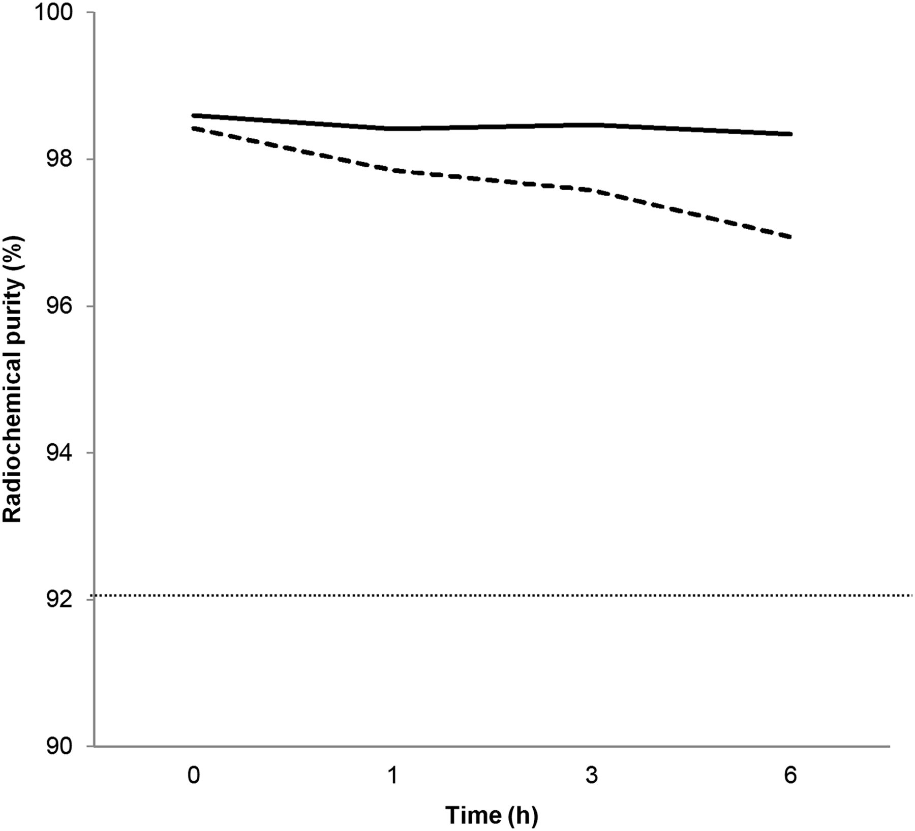
RCP values of 99mTc-FSC over 6 h. Solid line represents 5-min heating. Dashed line represents 3-min heating. Dotted line represents RCP acceptance level as per United States Pharmacopeia (92%).
Electron Microscope Evaluation
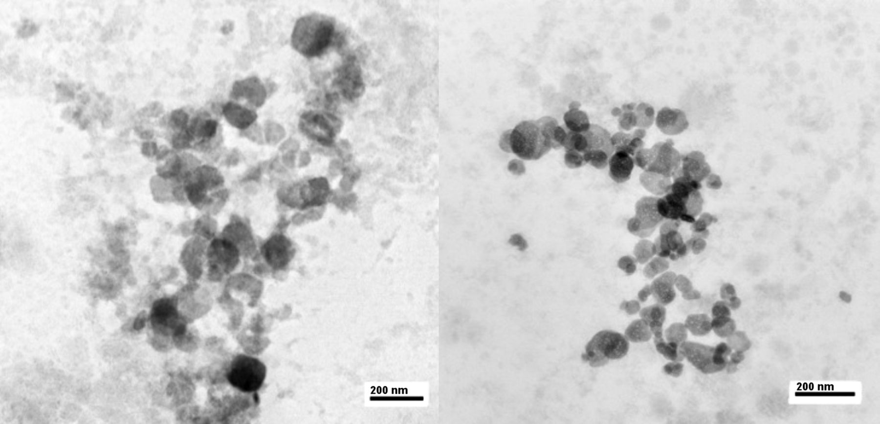
Images of 99mTc-FSC sample preparations at 0 h after the 5- and 3-min heating times are shown in Figure 2. The particles of 99mTc-FSC prepared with 3-min heating at this point in time (i.e., 0 h after filtration) looked smaller than the particles prepared with 5-min heating (Fig. 2).

Electron micrographs of 99mTc-FSC particles at ×59,000 magnification. (Left) 99mTc-FSC particles with 5-min heating at 0 h. (Right) 99mTc-FSC particles with 3-min heating at 0 h.
Particle Size Distribution for Standard and Reduced Heating Times
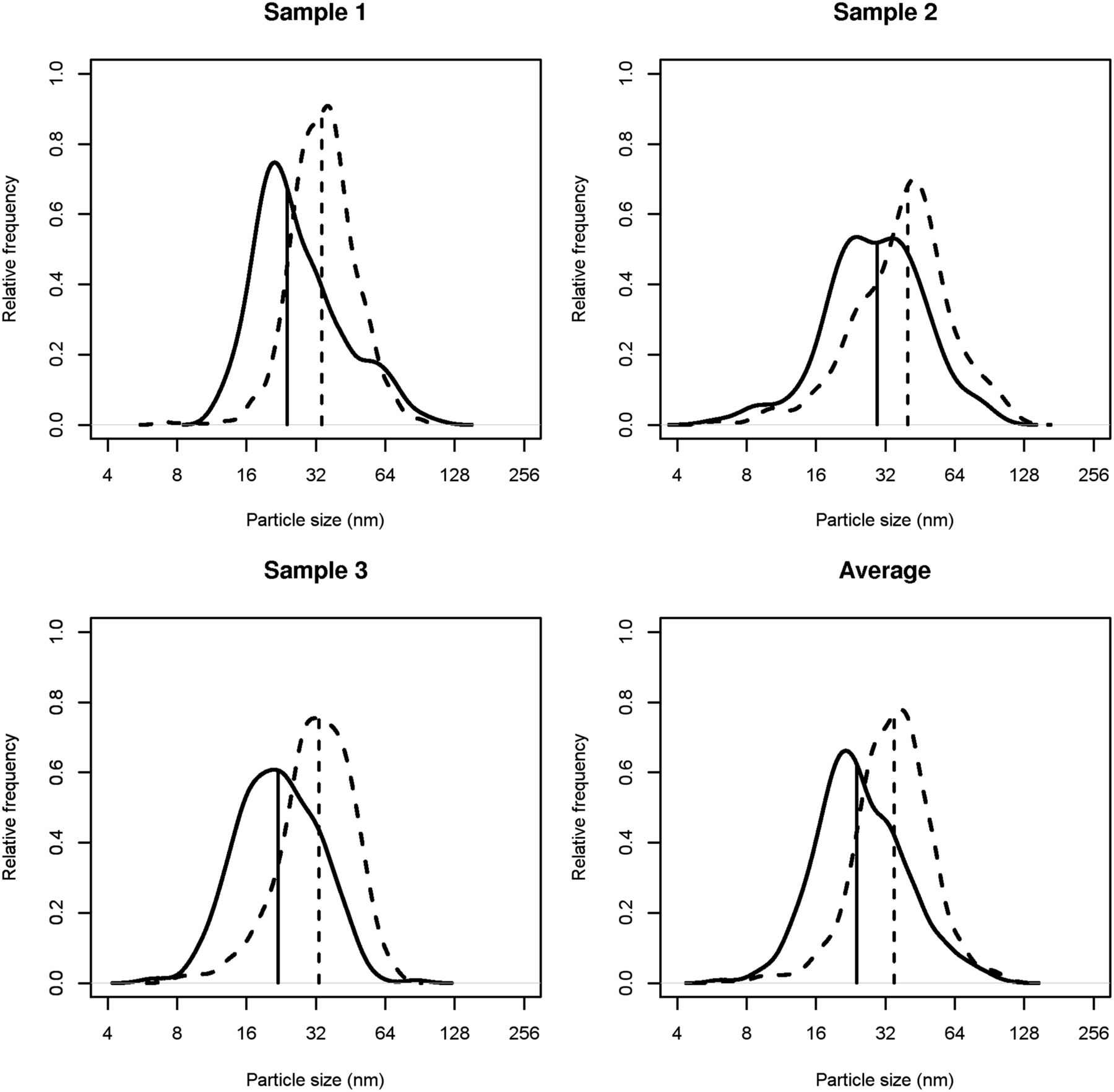
According to the KS test, more particle sizes were smaller for the 3-min heating at 0 h than for the 5-min heating at 0 h, as the density curves are shifted downward and the median values are smaller (KS test, P < 0.0001) (Table 2; Figs. 3 and 4). Additionally, there appeared to be a slight decrease in particle sizes from the 0- and 6-h reads for the 5-min heating (KS test, P = 0.0151) (Table 2; Figs. 3 and 4). Lastly, there seemed to be a tendency for the 3-min heating at 6 h to have larger particle sizes than those seen for the 5-min heating (KS test, P < 0.0001) (Table 2; Figs. 3 and 4).
Particle Size Distribution of 99mTc-FSC

Relative frequency of particle size for samples with 3-min heating. Along x-axis are particle sizes (nm), and y-axis represents relative frequency (%) that particle sizes (within small range) occur in sample. Solid lines represent 0-h readings, and dashed lines represent 6-h readings. Vertical lines indicate median particle size for sample.
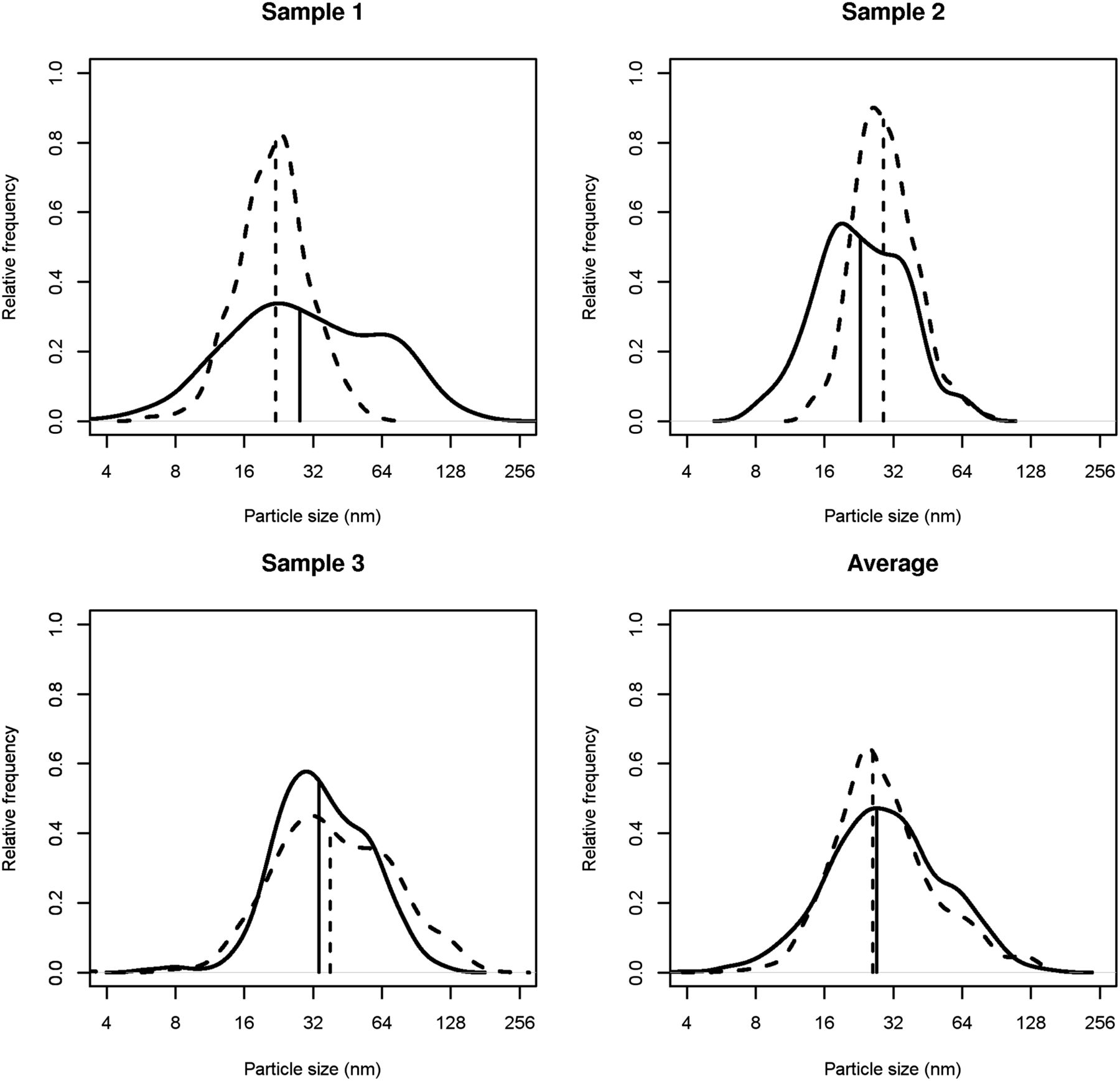
Relative frequency of particle size for samples with 5-min heating. Along x-axis are particle sizes (nm), and y-axis represents relative frequency (%) that particle sizes (within small range) occur in sample. Solid lines represent 0-h readings, and dashed lines represent 6-h readings. Vertical lines indicate median particle size for sample.
Although the samples contained statistically different particles sizes, they still followed a relatively similar distribution when their 25th percentile, median, and 75th percentile were examined (Table 2). Each sample had a large number of particles within the ideal size range (15–100 nm) (Table 3). The 3-min heating at 6 h had the highest percentage of particles counted within the ideal range (97.5%; 95% CI: 96.1%, 98.3%). After that, the order from most to least particles within the ideal range was the 5-min heating at 6 h (91.3%; 95% CI: 89.2%, 93%), the 3-min heating at 0 h (91.0%; 95% CI: 88.7%, 93%), and the 5-min heating at 0 h (88.8%; 95% CI: 86.1%, 91%) (Table 3).
Relative Frequency of 99mTc-FSC Particles Within Specified Size Ranges
DISCUSSION
The comparable retained filter activities for the 3- and 5-min heating times suggest that there is no significant difference in the number of particles being filtered with either heating time (P = 0.62) (Table 1). RCP was measured from 0 to 6 h because of an effort to increase the shelf life of 99mTc-FSC at our facility. In addition to the RCP, it was thought important that the particle size stay relatively stable until 6 h so as not to affect lymphatic drainage and sentinel node retention. As stated in the Results section, there was no statistical difference in RCP between the 3- and 5-min heating samples at 0 h or between the 3- and 5-min heating times at 6 h, of which all were still above the 92% limit set by United States Pharmacopeia (Fig. 1). This finding is consistent with the reaction of 99mTc-SC during the reconstitution process, for which more unbound 99mTc remains with the 3-min heating as there is less time to react with the sulfur to form particles (14).
Figure 3 can be described as follows: the relative frequency curves are very much like histograms of the data in which the histograms are smoothed over all possible bin sizes. The peak of the relative frequency gives an idea of where the highest frequency of particle sizes occurs. The vertical lines indicate the median particle size for a sample and may or may not be the same as the peak. If the data are symmetric, then the median particle size will occur at the peak frequency. The 3-min heating at 0 h versus the 3-min heating at 6 h gives a sense of stability of the preparation over time, and according to the analysis this particle size gets significantly larger (Table 2). Along with that, there appears to be a tendency for the 3-min heating at 6 h to have significantly larger particle sizes than those seen for the 5-min heating at 6 h (Table 2). However, our results for the 5-min heating group showed that the particle size at 6 h was slightly smaller than the particle size at 0 h (Table 2). Although this difference is statistically significant, the actual size differences in both situations are very small. The 3-min heating at 0 h versus the 5-min heating at 0 h gives a sense of the effect of heating time on initial particle size distributions, and initially the 3-min heating gave smaller particle sizes (Table 2; Figs. 3 and 4). The smaller particle size for the 3-min heating at 0 h than for the 5-min heating at 0 h fits well with our hypothesis that a reduced heating time may reduce the particle sizes. The reduced heating time is supposed to produce smaller particles, and such is exactly the case for the 3-min heating at 0 h in our study. This group had the smallest particles among the 4 groups (Table 2; Figs. 3 and 4). However, the reduced heating time might not provide an optimal condition to sufficiently terminate the sulfur-coating process, as the particles appeared to grow larger over time; the actual mechanism of this process is unknown to us.
There were many benefits to using an electron microscope versus filter analysis. Because of pore clogging, there are often many particles remaining on the filter, even if they are not larger than the pore size. The biggest benefit of the electron microscope was the ability to have genuine visualization of the particles rather than the indirect method that filtration relies on.
Although electron microscopy analysis was able to determine the exact size of the SC particles, it was limited in its ability to represent the entire sample. Although we had sufficient sampling, it would have been beneficial to have filtrations using multiple pore sizes to reaffirm that the small sample drop was representative of the entire kit. Additionally, the relative density of 99mTc-FSC proved to be a challenge because of the nature of electron microscopy, which favors dense particles; 99mTc-FSC particles consist mostly of sulfur, with a density close to 2 g/cm3. Another limiting factor was the extra debris formed within the preparation. This made some grids particularly difficult to analyze and the particles difficult to count because of the inability, at times, to distinguish debris from 99mTc-FSC.
Although we reported the optimal size particles to be 15–100 nm (9), this differs from the 100–200 nm reported by some (15). We speculate that this difference may be from the intended procedural use for the sentinel node biopsy. Although 15- to 100-nm particles are useful for immediate imaging because of quick lymphatic drainage, the larger 100- to 200-nm range may be retained better for delayed imaging and next-day biopsy.
A future experiment would combine the microfiltration analysis to determine the size of particles of the entire solution. Filtrations would be done at 0 and 6 h to show that particles are not clumping over time. The sample could then be analyzed using electron microscopy to visualize the particles and determine whether the data correlate with the microfiltration method.
CONCLUSION
On the basis of the results, 99mTc-FSC using a standard or reduced heating method remains stable until 6 h in terms of RCP and particle size. Although there was a statistically significant difference in particle size according to the KS test from 0 to 6 h for both the 3-min and the 5-min heating times, there was a maximum difference of merely 11 nm. As a result, most of the particles for both heating times still fell within the optimal range for particle size in sentinel node mapping.
DISCLOSURE
This research was funded in part by the Mayo Clinic Department of Radiology Advance Award. No other potential conflict of interest relevant to this article was reported.
Acknowledgments
We thank Jon E. Charlesworth, Electron Microscopy RES CORE Facility, Department of Biochemistry and Molecular Biology, Mayo Clinic, and the electron microscopy faculty for the long hours of procedural assistance given in determining particle size. Additionally, we thank the radiopharmaceutical laboratory technologists for their assistance in gathering RCP data. Finally, we thank Dr. Patrick J. Peller for providing relevant literature, as well as his involvement in coming up with the initial idea for this research.
Footnotes
Published online Aug. 7, 2014.
REFERENCES
- Received for publication July 8, 2014.
- Accepted for publication July 14, 2014.


Jump to section
Related Articles
Cited By...
- No citing articles found.