


Article Figures & Data
Figures
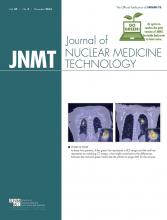
- FIGURE 1.
Gelatin-based liver phantom containing suspended 90Y resin microspheres. (A) Maximum-intensity projection of liver phantom in oblique view. Dotted lines outline liver phantom. (B and C) 90Y PET/CT scan in coronal plane demonstrates heterogeneous distribution of 90Y resin microspheres within 2 tumor inserts, T1 and T2, and throughout nontumorous liver, L. Small air pockets can be seen within and around tumor inserts and liver phantom. (D–F) 90Y PET/CT in transaxial view demonstrates CT artifacts caused by a magnetic stir-bar (SB) at bottom of T1 tumor insert. (A color version of this figure is available as a supplemental file online at http://tech.snmjournals.org/.)
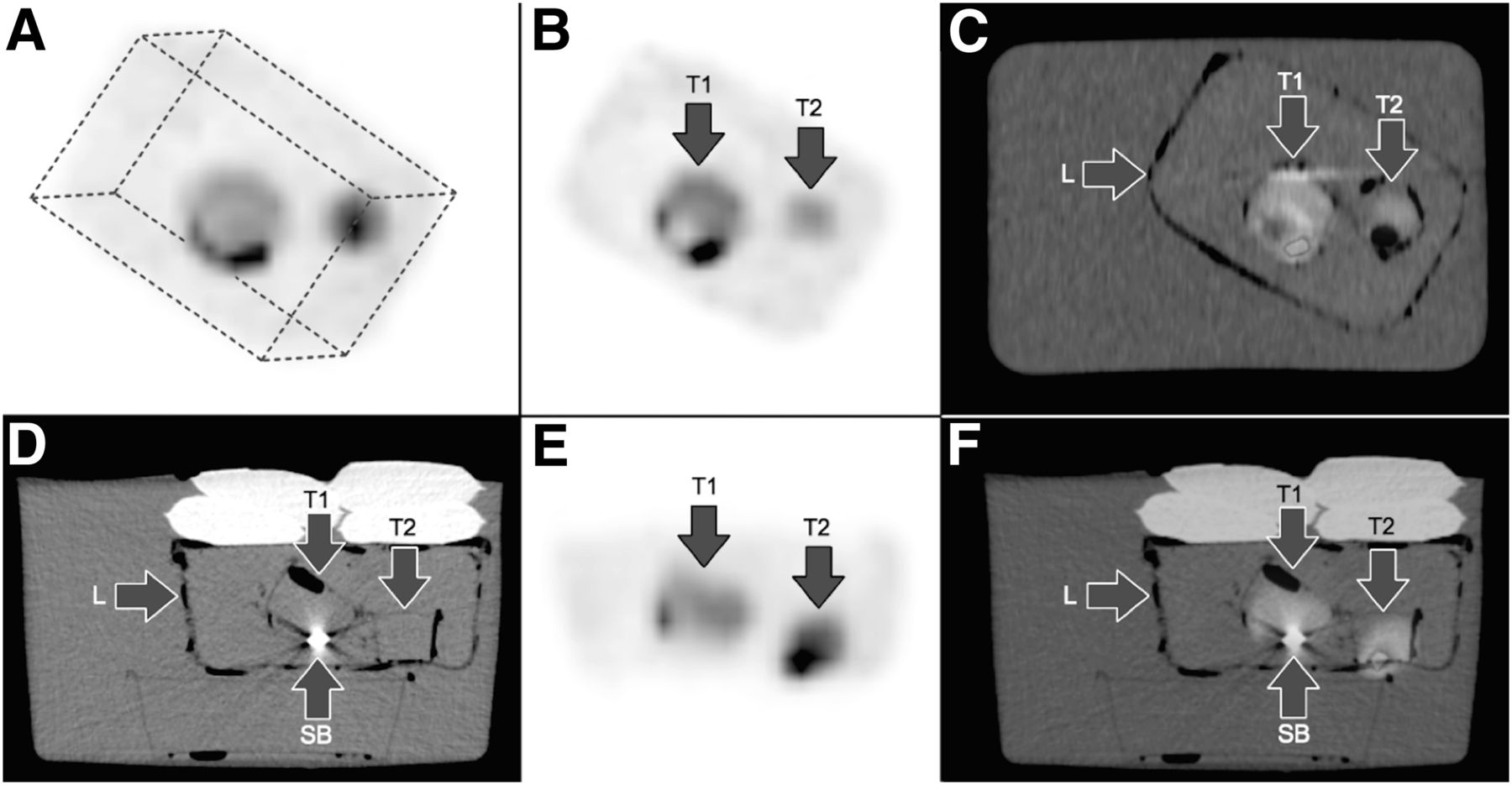
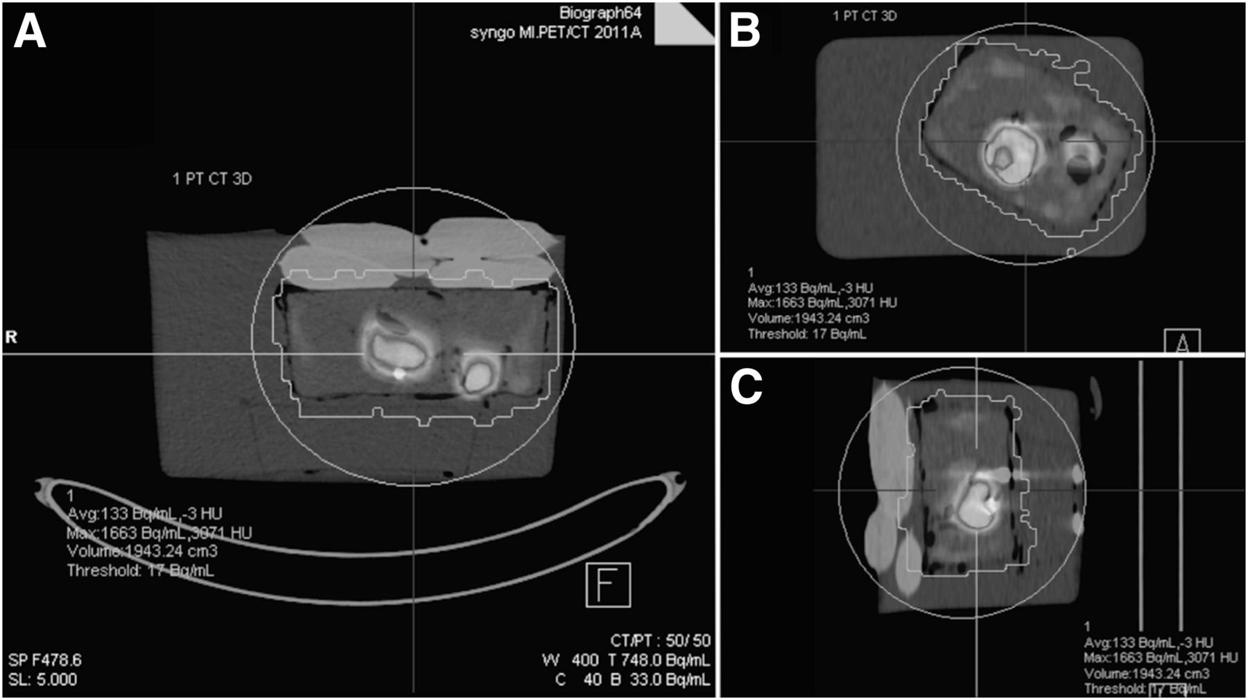
- FIGURE 2.
Example of volume-of-interest analysis of 90Y PET/CT phantom scan generated using 1% volumetric isocontour threshold, which may be used for quantitative analysis. Images are shown in transaxial (A), coronal (B), and sagittal planes (C). (A color version of this figure is available as a supplemental file online at http://tech.snmjournals.org/.)


Additional Files
Color versions of figures
Files in this Data Supplement:


Jump to section
Related Articles
Cited By...
- No citing articles found.