


Article Figures & Data
Figures
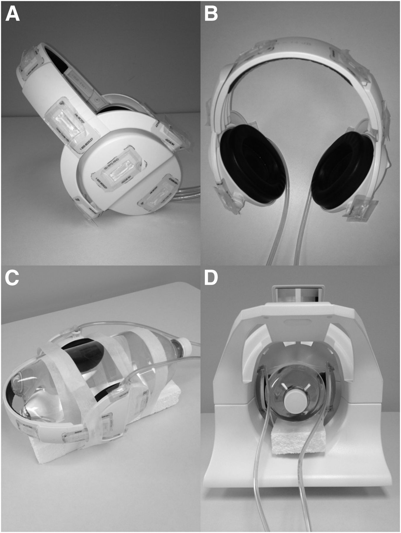
- FIGURE 1.
(A and B) Side view of headphones showing 12 fiducial markers (A) and top view of headphones with fiducials (B). (C and D) Headphones secured to Styrofoam (C) and placed in head coil (D).
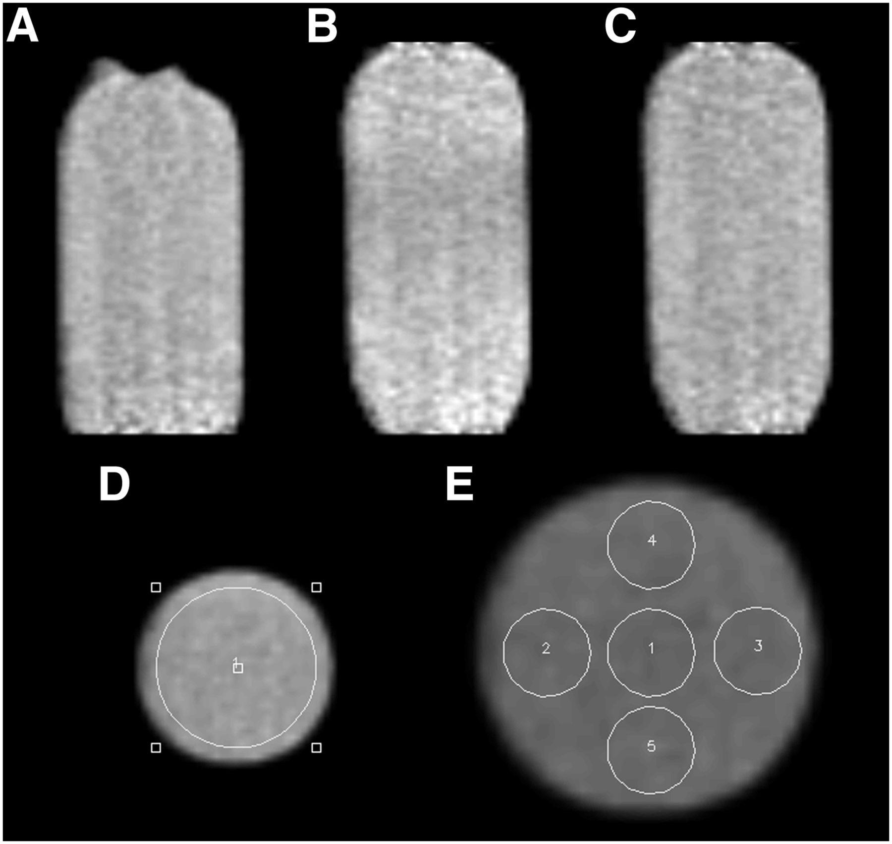
- FIGURE 2.
(A) Three-liter radioactive phantom without headphones demonstrates uniform distribution of activity. (B) Three-liter radioactive phantom demonstrates decrease in activity in center region with headphones on uncorrected. (C) Three-liter phantom using corrected μ maps demonstrates uniform activity concentration with headphones on. (D) Large, circular region of interest replicated across all transverse planes. (E) Five small, circular regions of interest replicated across all transverse planes. 1 = center; 2 = right; 3 = left; 4 = upper; 5 = lower.
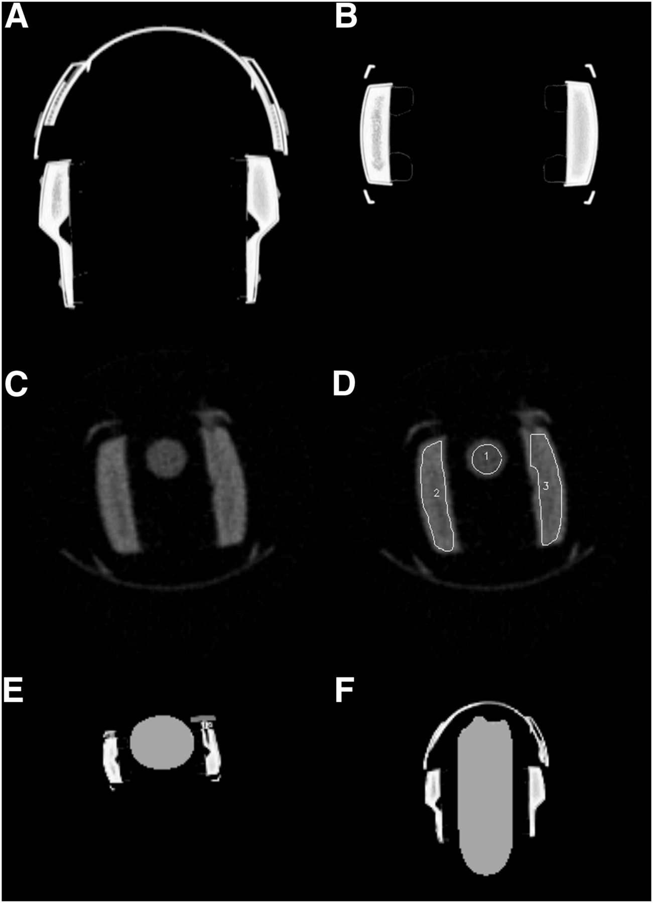
- FIGURE 3.
(A) View of coronal CT template created from high-resolution CT transmission scan. (B) Transaxial CT template. (C) Axial view of 57Co transmission scan. (D) Axial 57Co transmission with representative view of how regions of interest were drawn on headphones and water-filled centrifuge tube. (E) Axial view of modified μ map. (F) Coronal view of modified μ map. 1 = center; 2 = right; 3 = left.
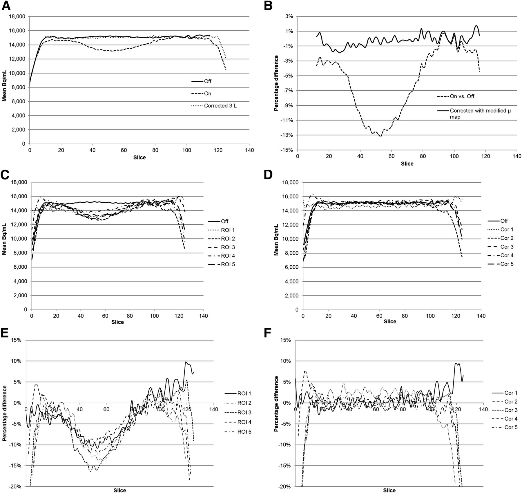
- FIGURE 4.
(A) Difference in activity concentration between headphones off, on, and corrected for each slice of phantom. (B) Percentage difference in counts between headphones off vs. on and off vs. corrected for each slice of phantom. (C) Difference in activity concentration of the 5 regions between headphones off and on for each slice of bottle. (D) Difference in activity concentration of the 5 regions between headphones off and corrected for each slice of bottle. (E) Percentage difference between headphones on compared with off of the 5 smaller regions of interest. (F) Percentage difference in activity concentration using corrected μ map in headphone area (slices 20–100). Cor = corrected. A color version of this figure is available as a supplemental file at http://tech.snmjournals.org.
- FIGURE 5.
Maximum and average percentage difference for off vs. on and off vs. corrected. (A) Large regions of interest (slices 20–100). (B) Small regions of interest (slices 20–100).
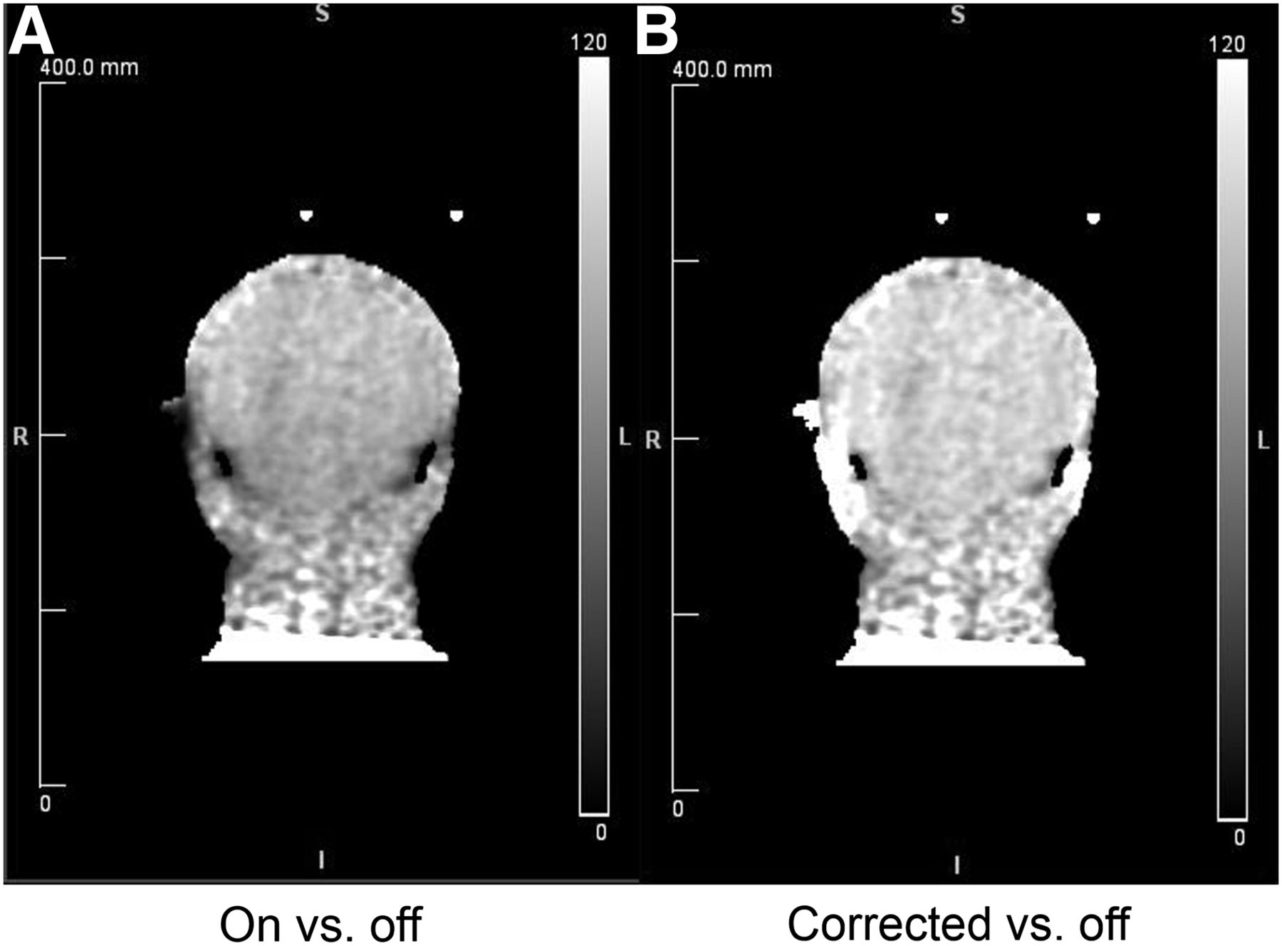
- FIGURE 6.
(A) Coronal image showing ratio of measured activity concentration from PET scan acquired with headphones over scan without headphones. (B) Coronal image showing ratio of activity concentration of patient PET scan with headphone attenuation correction over scan without headphones. A color version of this figure is available as a supplemental file at http://tech.snmjournals.org.


Tables
MR parameters MRAC Dixon AC MRAC UTE AC MPRAGE Acquisition time (min) 00:19 01:40 04:45 Voxel size (mm) 2.6 × 2.6 × 3.12 1.6 × 1.6 × 1.6 1.1 × 1.1 × 1.2 Orientation Coronal Transverse Sagittal Slices per slab 128 192 176 Matrix size 192 × 126 192 × 192 256 × 256 Field of view (mm) 500 300 270 Repetition time (ms) 3.60 11.94 2,100 First and second echo time (ms) 1.23 and 2.46 0.07 and 2.46 2.95 Flip angle 10.0° 10.0° 9.0 Parallel acquisition technique mode GRAPPA 2 (Siemens) None GRAPPA 2 (Siemens) PET parameters Emission scan 5 min Reconstruction iterations 3 Reconstruction subsets 21 Postreconstruction filter Gaussian, 4 mm Number of slices 127 Matrix 256 × 256 Orientation Transverse Voxel size (mm) 1.4 × 1.4 × 2.03 AC = attenuation correction; GRAPPA = generalized autocalibrating partially parallel acquisition; MRAC = MR-based attenuation correction.
Structure or region Difference before correction Difference after correction Cerebellar–cortical −14.4% 0.2% Inferior temporal −14.0% 2.1% Temporal pole −12.6% 2.2% Fusiform −11.5% 1.6% Superior temporal −7.8% 2.8% Medial orbitofrontal −6.7% −0.2% Caudate −3.6% 2.2% Rostral anterior cingulate −2.4% 3.0% Superior frontal 0.3% 3.9% Cuneus 0.3% 2.9% Inferior parietal 0.9% 4.2% Precuneus 1.0% 4.0%
Supplemental Data
Files in this Data Supplement:


Jump to section
Related Articles
Cited By...
- No citing articles found.