


Abstract
For many types of cancer, 18F-FDG PET/CT is commonly used in evaluation and management, including tumor diagnosis, staging, restaging, treatment monitoring, and radiation therapy planning. Meticulous patient preparation including restrictions of diet and activity and management of blood glucose levels in diabetic patients, as well as an awareness of the effect of medications and environmental conditions, plays an important role toward obtaining good-quality images, which are essential for accurate interpretation. Protocol guidelines for performing PET/CT have been proposed by various societies and groups, including the Society of Nuclear Medicine and Molecular Imaging, the European Association of Nuclear Medicine, the American College of Radiology, and the National Cancer Institute. Standardization of the PET/CT procedure is necessary to enable use of metabolic parameters as imaging biomarkers in routine clinical decision making and to ensure reproducibility and allow comparison examinations across different sites. Though several published articles, including various society guidelines, have addressed the relevant patient preparation variables individually, we believe there is need for further clarification. This article summarizes existing data and proposes a standard patient preparation protocol.
In clinical practice, 18F-FDG PET/CT is commonly used in the evaluation and management of many types of cancer, including tumor diagnosis, staging, restaging, treatment monitoring, and radiation therapy planning. Oncologic imaging with PET has recently gained particular importance in the quest to identify new and effective therapies and to understand the role of molecular biomarkers in treating cancer.
According to the 2012 PET Imaging Market Summary Report published by International Marketing Ventures, PET and PET/CT studies were performed at over 2,200 U.S. locations using fixed or mobile scanners and, per a 2011 census, a total of 1,853,700 clinical PET/CT and PET studies were performed in the United States, with oncology, cardiology, neurology, and others constituting 94% (1,736,800), 3% (53,300), 3% (58,000), and less than 1% (5,600), respectively (1).
Meticulous patient preparation plays an important role toward obtaining good-quality images, which are essential for accurate interpretation of the PET/CT studies. Relevant considerations before the study include restrictions of diet and activity and management of blood glucose levels in diabetic patients, as well as an awareness of the effect of medications and environmental conditions. Important protocol guidelines for performing PET and PET/CT have been proposed by various societies and groups, including the Society of Nuclear Medicine and Molecular Imaging (SNMMI), the European Association of Nuclear Medicine (EANM), the American College of Radiology (ACR), the National Cancer Institute (NCI), and the Netherlands Society of Nuclear Medicine (2–6). Quantification of 18F-FDG uptake is a useful tool in addition to qualitative interpretation of images, as the percentage change in tumor standardized uptake value (SUV) is more reproducible than the percentage change in CT tumor size (7). However, SUV is affected by several variables, including those related to patient preparation, blood glucose, uptake period, scan acquisition, image reconstruction, and region-of-interest parameters (8,9).
The published literature has reported considerable variability in the way PET/CT scans are obtained. This variability likely makes it difficult to quantitatively compare studies performed at different centers. An Imaging Response Assessment Team funded by the NCI developed and conducted a 34-question survey among 15 institutions about how clinical PET/CT studies were performed and found considerable variability (10). Protocol variability was also reported by another international study based on a World Wide Web survey of public and private imaging centers performed to assess current PET/CT protocols (11).
Therefore, standardization of the PET/CT procedure is necessary to enable use of metabolic parameters as imaging biomarkers in routine clinical decision making, ensure reproducibility, and allow comparison examinations across different sites. Though several published articles, including various society guidelines, have addressed the relevant patient preparation variables individually, we believe there is need for further clarification. This article summarizes existing data and proposes a standard patient preparation protocol for oncologic 18F-FDG studies that can be easily incorporated into daily clinical practice. The proposed protocol is described in Table 1, and a comparison of our proposed protocol with the recommendations of the SNMMI, EANM, ACR, and NCI is presented in Table 2. Readers are directed to previous publications (12–16) for detailed information on oral and intravenous contrast agents, CT protocols for diagnostic whole-body 18F-FDG PET/CT, and dedicated brain and cardiac 18F-FDG PET imaging, which are beyond the scope of this review.
Proposed Standard Patient Preparation Protocol for 18F-FDG PET and PET/CT
Patient Preparation Recommendations
DIETARY INSTRUCTIONS
Fasting
To minimize dietary glucose–related competitive inhibition of 18F-FDG uptake and reduce serum insulin to near basal levels, we recommend complete fasting for a minimum of 6 h before the scan, including cessation of tube feeding, dextrose-containing intravenous fluids, and parenteral hyperalimentation. During this time, only plain (unflavored) water is permitted, and there should be absolutely no sugar or carbohydrate intake of any kind, including gum, candy, or breath mints. Similar recommendations with subtle differences are found in the various society guidelines. Per the SNMMI, patients should be instructed to fast and not consume liquids except for water for at least 4–6 h before radiotracer injection. Intravenous fluids containing dextrose or parenteral feedings should also be withheld for 4–6 h (2). The EANM suggests that patients not consume any food or sugar for at least 6 h before injection of 18F-FDG. Thus, patients scheduled to undergo the PET study in the morning should not eat after midnight and preferably should have a light meal without alcohol on the evening before the PET study. Those scheduled for an afternoon examination may have a light breakfast before 8:00 am (i.e., up to 2 sandwiches without sugars or sugar-containing sandwich fillings). Parenteral nutrition and intravenous fluids containing glucose should be discontinued at least 4 h before the PET/CT examination (3). The NCI consensus recommendations proposed that patients should fast for a minimum of 4 h before 18F-FDG injection and that if the study is scheduled for the afternoon, a light breakfast with minimal carbohydrate-containing foods is acceptable (4). The ACR recommends a minimum of 4 h of fasting, with no oral or intravenous fluids containing sugar or dextrose (5). Per the survey of the Imaging Response Assessment Team (10), the most reasonable recommendation was fasting for at least 4 h and preferably 6 h. According to the international Web survey (11), the average fasting period before oncologic 18F-FDG PET/CT varied among users: 0–4 h (27%), 5–6 h (51%), 7–8 h (9%), or more than 8 h (13%).
Hydration
Most guidelines recommend good hydration, typically oral, before the study for radiation safety reasons and to ensure a low 18F-FDG concentration in urine. The EANM recommends 1 L of water by mouth in the 2 h before injection and favors another 0.5 L during the uptake period as tolerated (3). The NCI recommends that patients should consume at least 2–3 12-oz (355 mL each) glasses of water during fasting and another 250–500 mL of water after injection and before scanning (4). Our preference is to have patients drink 1–2 L of plain water as tolerated during the 4 h immediately before the PET/CT scan. All patients are asked to void just before imaging.
Specific Diet
We recommend a high-protein, low-carbohydrate diet for 24 h before scanning to minimize dietary glucose–related competitive inhibition of 18F-FDG uptake. A sample menu is described in the proposed standard patient preparation protocol (Table 1). Alcohol and nicotine products are completely avoided for 12 h before the scan. The NCI also recommends a low-carbohydrate diet for 24 h before the study (4). About half of the Imaging Response Assessment Team sites recommended a low-carbohydrate diet to their patients (10). Per the international survey (11), about one third of the responders reported requiring dietary restrictions such as no caffeine, a low-carbohydrate diet, or nothing taken orally.
ACTIVITY RESTRICTION
We recommend that exercises such as jogging, cycling, weightlifting, strenuous housework, yard work, and sexual activity be avoided for a minimum of 24 h (ideally 48 h) before the scan to minimize uptake of radiotracer in skeletal muscles (Fig. 1). Patients are also advised not to chew gum for 24 h before the study to avoid activation of masticatory muscles (17). The EANM suggests that all patients avoid (extreme) exercise for at least 6 h before the PET study (3). Both the NCI and the ACR recommend avoiding strenuous activity for 24 h before injection (4,5).

Effect of exercise on skeletal muscle 18F-FDG uptake. Maximum-intensity-projection image of 18F-FDG PET study demonstrates prominent diffuse 18F-FDG uptake in skeletal and cardiac muscles in patient who performed strenuous exercises in the 2 d before undergoing PET.
MEDICATION
All prescription medications should be taken as directed (insulin and oral hypoglycemics are discussed in greater detail in the next section). The PET center personnel need to recognize that several commonly prescribed medications can elevate serum glucose levels, including glucocorticoids, phenothiazines, lithium, tricyclic antidepressants, phenytoin, thiazide diuretics, isoniazid, rifampin, and ephedrine (17,18). Notably with glucocorticoids, the PET examination may have to be coordinated either before or after their use or, alternatively, the associated hyperglycemia may need remedial therapy with insulin (19,20). We do not recommend that patients stop taking any of the above medications before their PET scan. The EANM specifies no restrictions and that medication can be taken as prescribed (3).
GLUCOSE LEVELS, INSULIN, AND ANTIDIABETIC MEDICATION
Blood glucose levels can have a significant influence on tumor 18F-FDG uptake because 18F-FDG and glucose compete for glucose transport and phosphorylation by hexokinase (21). There is a well-known association between plasma glucose levels, serum insulin levels, and their effect on the biodistribution of 18F-FDG. Increased glucose levels decrease 18F-FDG uptake in the brain and in tumors because of direct competition between binding sites and enzymes (22). Increased insulin secondary to elevated blood glucose increases the translocation of GLUT4 (glucose transporter), thereby rapidly and efficiently shunting 18F-FDG to organs with a high density of insulin receptors (e.g., skeletal and cardiac muscles), resulting in altered radiotracer biodistribution and suboptimal image quality (23).
Lindholm et al. showed that SUVs decrease significantly in all tumors studied when 18F-FDG PET imaging is done after a 50-g loading dose of glucose (P < 0.02). In contrast to the tumors, the muscle tissue accumulated more 18F-FDG after glucose loading than in the fasting state, resulting in blurring of tumor margins. Cancer cells take up relatively more 18F-FDG than unlabeled glucose when the extracellular glucose concentration is low, resulting in higher SUVs in the fasting state (24).
Boellaard also reported lower uptake levels or SUVs with increasing blood glucose levels, with the range of SUV being between −15% and +15% (25).
In a busy PET clinic, there are many scenarios involving diabetic individuals that may affect patient preparation and the quality of 18F-FDG PET/CT images. For example, the patient may be in a state of hyperglycemia immediately before the PET study, and interactions with diabetic medications such as metformin and insulin may occur. Considering the growing number of diabetic patients who have cancer, hyperglycemia before a PET/CT study is not uncommon. Several publications have cited successful use of intravenous regular insulin to correct hyperglycemia that occurs immediately before an 18F-FDG PET/CT scan. In one study (26), when 18F-FDG was injected 1 h after a bolus administration of intravenous insulin in hyperglycemic diabetic patients (Humulin R [Elli Lilly], according to a preestablished chart to reach a target glycemia of lower than 8 mmol/L [144 mg/dL]), no differences in SUV for lungs, liver, muscles, myocardium, or suspected pulmonary lesions were found between normoglycemic nondiabetic patients and the insulin-corrected hyperglycemic diabetic patients.
A more recent study conducted by Caobelli et al. proposed an optimized protocol for intravenous insulin administration in diabetic patients undergoing 18F-FDG PET/CT (27). They used short-acting intravenous Humulin R (25 units; diluted in 250 mL of physiologic solution [infusion rate in mL/h determined as glucose level divided by 20]), and 18F-FDG was injected 30 min after insulin administration. No significant difference in the gluteal muscle SUVs were observed between the hyperglycemic patients (>180 mg/dL) who received the insulin and the control groups, which included the hyperglycemic patients (160–200 mg/dL) who did not receive insulin and the nondiabetic patients.
In 63 diabetic cancer patients, Roy et al. used a standardized protocol of short-acting intravenous Humulin R (2 units for glycemia of 10.0–12.0 mmol/L [180–216 mg/dL], 3 units for 12.1–14.0 mmol/L [216–252 mg/dL], and 4–6 units for ≥14.1 mmol/L [≥252 mg/dL]) to reach a target glycemia lower than 10.0 mmol/L (180 mg/dL) at least 1 h before 18F-FDG injection (28). Their protocol was safe and effective in decreasing glucose levels but led to an altered biodistribution in 25% of patients (increased muscle uptake and decreased liver uptake). The interval between insulin injection and 18F-FDG injection was significantly shorter in patients with an altered biodistribution than in those with a normal biodistribution (65.7 vs. 80.2 min, P < 0.01). Their tentative recommendation was to maintain an interval of 90 min between insulin injection and 18F-FDG injection.
Nakatani et al. studied 44 patients with extensive skeletal muscle uptake of 18F-FDG after a fast of at least 4 h (29). They concluded that both glucose intolerance and gastric food residue are independent risk factors for increased skeletal muscle uptake and suggested a longer fasting time in patients with glucose intolerance and avoidance of a heavy meal before the study to reduce gastric residue.
The impact of elevated blood glucose levels, diabetes, insulin treatment, and obesity on 18F-FDG uptake in tumors and biodistribution in normal organ tissues was studied by Busing et al. (30). Hyperglycemia was associated with decreased cerebral uptake and increased skeletal muscle uptake. The mean cerebral maximum SUV was significantly decreased whereas the mean muscular maximum SUV was increased by up to 31% in diabetic and insulin-treated patients compared with nondiabetic and non–insulin-treated patients (P < 0.001).
A case report describing qualitatively normal 18F-FDG biodistribution after subcutaneous administration of long-acting insulin analog glargine (Lantus; Sanofi-Aventis) 3 h before 18F-FDG injection was attributed to the time–activity profile of insulin glargine, which mimics the normal basal secretion of insulin by the pancreas (31).
In our clinical practice, diabetic patients are encouraged to check their blood glucose level at home on the days leading up to their PET/CT examination to ensure reasonable blood glucose levels (≤200 mg/dL). If the blood glucose levels are consistently greater than 200 mg/dL, we recommend that they contact their primary care physician for further guidance on glycemic control. Fasting and meal instructions are given as detailed in “Dietary Instructions” above. All oral medications, including those for diabetes, such as metformin, are to be taken as prescribed. Diabetic patients on regular short-acting insulin take their insulin along with breakfast by 6 am. These individuals are usually scheduled for imaging between 12 pm and 1 pm. Alternatively, those receiving evening or bedtime long-acting insulin can be scheduled for imaging at 7 am after an overnight fast. Patients on a continuous insulin infusion or pump are scheduled early in the morning (by 8 am) and asked to eat breakfast after the PET study. The insulin pump is kept on the night or basal setting until after the PET study. For individuals who present with hyperglycemia (blood glucose > 200 mg/dL), we reschedule the scan and ask that they frequently check their blood glucose at home and contact their primary care physician. They are asked to contact the PET center with the results of their blood glucose checks in the days leading up to the rescheduled PET study. We are considering implementing a standardized protocol using intravenous short-acting regular insulin to correct hyperglycemia before the PET study in an effort to decrease patient inconvenience and improve resource use. Implementation of such a protocol would involve extensive training of staff on the use of intravenous insulin, frequent blood glucose monitoring, and identification and correction of potential hypoglycemia (32).
The SNMMI recommends a prescanning glucose level of between 150 and 200 mg/dL and suggests that reducing the serum glucose level by administering insulin can be considered but that the administration of 18F-FDG should be delayed after insulin administration (with the duration of the delay depending on the type and route of administration of insulin) (2). The EANM suggests that an 18F-FDG study can be performed if the blood glucose level is less than 7 mmol/L (120 mg/dL). The EANM also specifies that if insulin is to be given to reduce the blood glucose level, the interval between administration of insulin and administration of 18F-FDG should be more than 4 h. For type II diabetic patients controlled by oral medication, patients can continue to take the oral medication and the PET study should preferably be done in the late morning. For insulin-dependent type II and type I diabetic patients, the PET study should preferably be scheduled for the late morning and the patient should eat a normal breakfast at 7 am, inject the normal amount of insulin, and not consume any more food or fluids, apart from water, afterward. For patients on a continuous insulin infusion, the PET study should be scheduled early in the morning. The insulin pump is kept on the night setting until after the study, at which time the patients can also have breakfast (3). The NCI consensus recommended that the venous serum glucose be within 120 mg/dL for nondiabetic patients and 150–200 mg/dL for diabetic patients. The PET study should be rescheduled if the serum glucose is greater than 200 mg/dL, and insulin should not be used to adjust the blood glucose level (4). The ACR recommends that a serum glucose analysis be performed before 18F-FDG administration (5). The 2008 Netherlands protocol for standardization of multicenter 18F-FDG PET studies suggested that the blood glucose level not be greater than 11 mmol/L (6). There was broad agreement among the Imaging Response Assessment Team sites that PET/CT studies should not be done if the glucose levels are more than 200 mg/dL (10). Blood glucose cutoff levels varied widely in the international survey and ranged from 150 to 250 mg/dL, with most sites (52%) accepting a cutoff of 200 mg/dL and 7% of sites reporting no cutoff level (11).
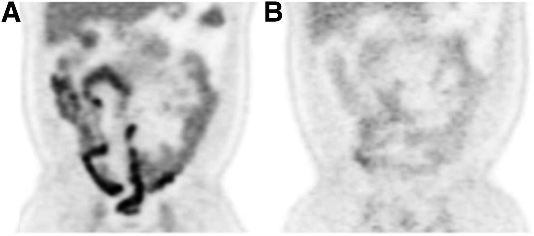
Prominent 18F-FDG bowel uptake, which can compromise image quality, has been identified by multiple investigators with use of metformin (Fig. 2). Gontier et al. conducted a prospective study to determine the impact of antidiabetic medications on 18F-FDG bowel uptake in type 2 diabetic patients (33). They compared the 18F-FDG bowel uptake in 3 groups: type 2 diabetic patients on metformin, patients on oral antidiabetic treatments other than metformin, and a nondiabetic control group. They found that 18F-FDG bowel uptake was significantly higher in patients treated with metformin than in the control group (P < 0.0001), although there was not a significant difference between patients treated with antidiabetic medications other than metformin and the control group. They concluded that metformin significantly increases 18F-FDG uptake in the colon and to a lesser extent in the small intestine.

Effect of metformin on intestinal 18F-FDG uptake. (A) Coronal 18F-FDG PET image of diabetic patient while on metformin demonstrates prominent bowel activity. (B) Prior 18F-FDG PET study of same patient while not on metformin shows only mild tracer activity in bowel.
Several published studies have evaluated the effect of stopping metformin before the PET study and its impact on 18F-FDG uptake. Ozulker et al. studied type 2 diabetic patients who underwent 2 PET studies, one while on metformin and the other after replacing metformin with another oral antidiabetic medication for 3 d before the second PET study (98 ± 7 d after the first PET study) (34). They found that the increased 18F-FDG uptake in the bowel on the first PET study was significantly less on the second study after metformin had been stopped.
Additionally, Oh et al. studied 4 groups of patients: diabetic patients who continued metformin (group A1), diabetic patients who stopped metformin 2 d before the study (group A2), diabetic patients on a regimen that did not include metformin (group B), and nondiabetic individuals, who served as controls (group C) (35). In addition, 10 diabetic patients underwent 2 consecutive PET/CT scans before and after the discontinuation of metformin and the mean intestinal 18F-FDG uptake decreased by 64% without significant changes in the blood glucose level. The high intestinal 18F-FDG uptake in group A1 was significantly reduced in group A2 (P < 0.001). There were no significant differences in intestinal uptake among groups A2, B, and C. No statistically significant differences were noted in the blood glucose levels among the 3 groups of diabetic patients (P > 0.9).
In a written communication (Gholam Reza Berenji, 2013), the author of an abstract presented at the SNMMI 2013 midwinter meeting (36) looked into the effects of peroxisome proliferator-activated receptor–γ agonists (pioglitazone HCl, 30 mg, or rosiglitazone maleate, 8 mg tablets) on 18F-FDG uptake and concluded that there was no significant difference in liver uptake, blood pool uptake, and liver–to–blood pool ratio between type 2 diabetic patients on the agonists and those not on them. The author concluded that peroxisome proliferator-activated receptor–γ agonists do not interfere with 18F-FDG uptake and need not be withheld before an 18F-FDG PET/CT study.
ENVIRONMENTAL CONDITIONS AND PREMEDICATIONS
Prominent 18F-FDG uptake within brown adipose tissue (BAT) (Fig. 3), which normally has a role in nonshivering thermogenesis, can potentially mask or mimic malignant lesions. BAT is also postulated to be protective against diabetes and obesity (37). Other known predictors of the presence of BAT uptake include age (younger), sex (female), body mass index (lower), and maximum outdoor temperature (lower) (38). Cold exposure is known to stimulate BAT via an adrenergic mechanism (18), which is more pronounced during fasting (39). Therefore, to minimize activation of BAT, patients should avoid cold exposure for 2 d before the study and avoid air conditioning on the day of the study. During travel to the PET/CT clinic, they should keep the car windows rolled up and, if necessary, use the car heater on cool days. Patients also should wear warm clothing including long pants or slacks, long sleeves in summer (no shorts or tank tops), and, on cold or even slightly cool days, a sweater, jacket, and cap (40,41). We also maintain a warm room temperature (minimum, 75°F) and provide warm blankets to patients during the uptake period. The SNMMI and others recommend that patients be kept in a quiet, dimly lit, warm room 30–60 min before the injection of 18F-FDG (2,18). Patients should remain as calm as possible, without moving excessively or talking during the uptake phase, to minimize muscle uptake. Other agents and medications such as nicotine and sympathomimetics (ephedrine) are also known to activate BAT and should be withheld before the PET study (42). Williams and Kolodny were the first to demonstrate a decrease in the frequency of 18F-FDG uptake in BAT by incorporating a high-fat, very low carbohydrate, protein-permitted diet 3–5 h before 18F-FDG injection (43).
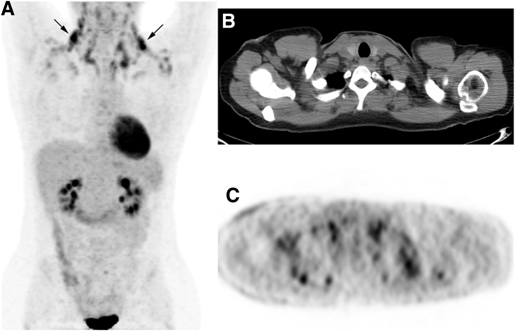
(A) Maximum-intensity-projection image shows intense bilateral radiotracer activity (arrows) in cervical and supraclavicular regions secondary to activation of BAT. (B and C) Axial non–contrast-enhanced CT (B) and PET (C) images localize radiotracer activity to cervical adipose tissue.
Benzodiazepines have been successfully used before PET imaging to relieve anxiety in claustrophobic patients and to relax skeletal muscles. However, the efficacy of benzodiazepines in reducing BAT uptake is questionable (44,45). A randomized controlled trial evaluating the effects of oral diazepam on the neck and upper chest muscles and on BAT uptake found no significant difference between a group of patients receiving 5 mg of oral diazepam and a group receiving a placebo (46). Gelfand et al. have also shown a reduction of interfering 18F-FDG uptake in BAT with use of intravenous fentanyl premedication (44). Other investigators have demonstrated successful reduction of BAT uptake with β-blockers. Soderlund et al., Parysow et al., and Agrawal et al. demonstrated complete or near-complete resolution of BAT uptake with use of 80 mg, 20 mg, and 40 mg of oral propranolol, respectively, 1–2 h before 18F-FDG injection (47–49). We do not routinely administer sedatives or β-blockers in our PET clinic. The SNMMI suggests the administration of lorazepam or diazepam before the injection of 18F-FDG to reduce uptake by BAT or skeletal muscle or the administration of β-blockers to reduce uptake by brown fat (2). The EANM states that there is no reason for routine use of sedatives and suggests that sedatives such as short-acting benzodiazepines be considered in patients with head and neck tumors to reduce muscle uptake or in anxious claustrophobic patients, whereas in children, sedation may be required depending on the age of the child and the type of tumor (3). The NCI, at the discretion of the clinician, recommends the administration of a sedative such as diazepam in patients who are extremely anxious or in whom the area of interest is the head and neck (4). In patients with a history or suspicion of head and neck tumors, the NCI recommends that a benzodiazepine or similar sedative, if not medically contraindicated, be administered orally or intravenously approximately 30 min before injection of the 18F-FDG to ensure a degree of relaxation of the neck muscles. The ACR suggests premedication for anxiety if indicated (5).
TIMING OF PET/CT
The optimal timing of 18F-FDG PET for assessing response after initiating treatment has yet to be clearly determined. Confounding variables related to the timing of the PET study that have a potential impact on image interpretation include inflammation after surgery and radiation, bone marrow effects from use of chemotherapies or colony-stimulating factors, and the effects of chemotherapy on tumor metabolism and macrophage impairment. Acute inflammatory changes with subsequent alterations in 18F-FDG uptake in both tumor and surrounding tissue have been documented after completion of radiation therapy (50). EANM guidelines recommend that the optimum interval between the last chemotherapy cycle and the PET study be at least 10 d, whereas it is best to wait for 3 mo after radiotherapy (3). The NCI working group (4) suggests that posttreatment imaging be performed at least 2 wk after the end of a specific chemotherapy cycle and 6–8 wk or longer after radiation therapy. The Netherlands protocol for standardization and quantification of 18F-FDG whole-body PET studies in multicenter trials suggests that an interval of 4 mo may be required after completion of radiation treatment (6). The timing of 18F-FDG PET/CT after radiofrequency ablation of lung tumors was determined to be at least 3 mo by Higaki et al. (51). The optimal timing of 18F-FDG PET after surgery is controversial as well. In patients with squamous cell carcinoma of the head and neck, Zimmer et al. suggest that 18F-FDG PET imaging be performed no sooner than 2–3 mo after surgery with or without chemoradiation to decrease the number of false-positive findings secondary to inflammation (52). However, other investigators have concluded that despite a high rate of false-positive findings, an earlier postoperative PET/CT examination (median time between surgery and PET/CT, 28 d; range, 13–75 d) significantly changed the adjuvant treatment plan in 15.4% of patients with head and neck cancer (53). In the case of radiation therapy, this delay allows patterns of activity and parenchymal change to stabilize, although increased activity may persist up to 15 mo after the end of radiation therapy (54). To minimize treatment-related false-negative and -positive findings, we suggest that PET studies be done at least 2 wk after the end of the last chemotherapy cycle, 6–8 wk after surgery, and 12 wk after radiation therapy.
CONCLUSION
18F-FDG PET/CT is a frequently used imaging modality in the evaluation of patients with cancer. There is considerable variability in the PET/CT procedure worldwide. Strict adherence to standardized procedures and protocols is an essential requirement toward obtaining good-quality images and ensuring reproducibility across different clinic sites. This review has summarized relevant aspects of patient preparation as outlined by major societies and has proposed a standard patient preparation protocol that can easily be incorporated into daily clinical practice.
Footnotes
↵* NOTE: FOR CE CREDIT, YOU CAN ACCESS THIS ACTIVITY THROUGH THE SNMMI WEB SITE (http://www.snmmi.org/ce_online) THROUGH MARCH 2016.
Published online Feb. 6, 2014.
REFERENCES
- Received for publication September 26, 2013.
- Accepted for publication November 21, 2013.


