


Article Figures & Data
Figures
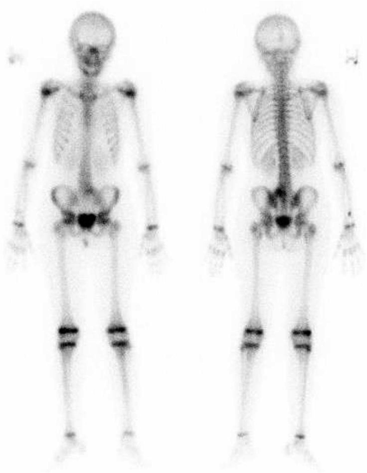
- FIGURE 1.
Whole-body bone scan showing focally increased osteoblastic activity in sacrum.
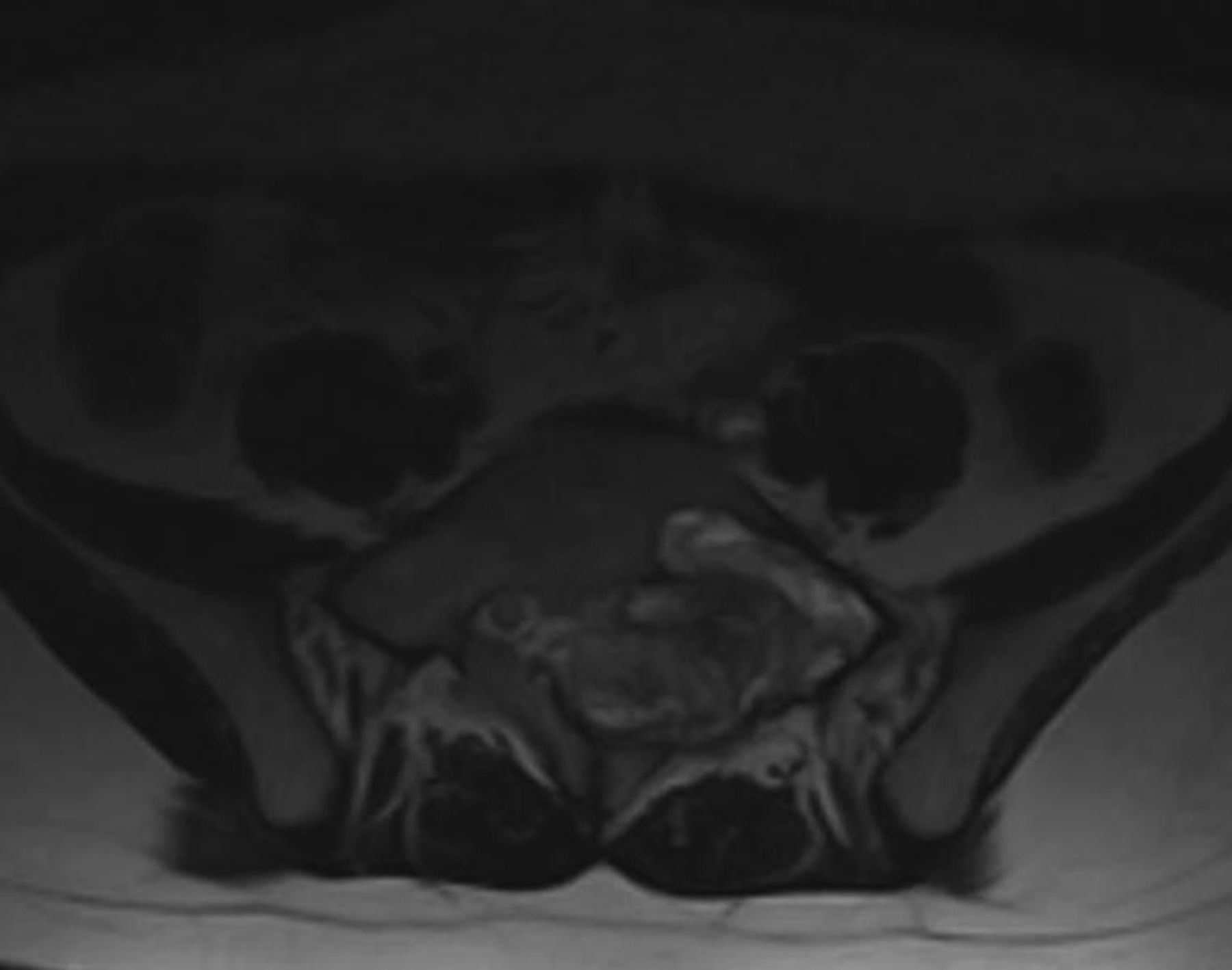
- FIGURE 2.
T2-weighted MR image showing heterogeneous soft-tissue mass in left sacrum encasing sacral nerves.
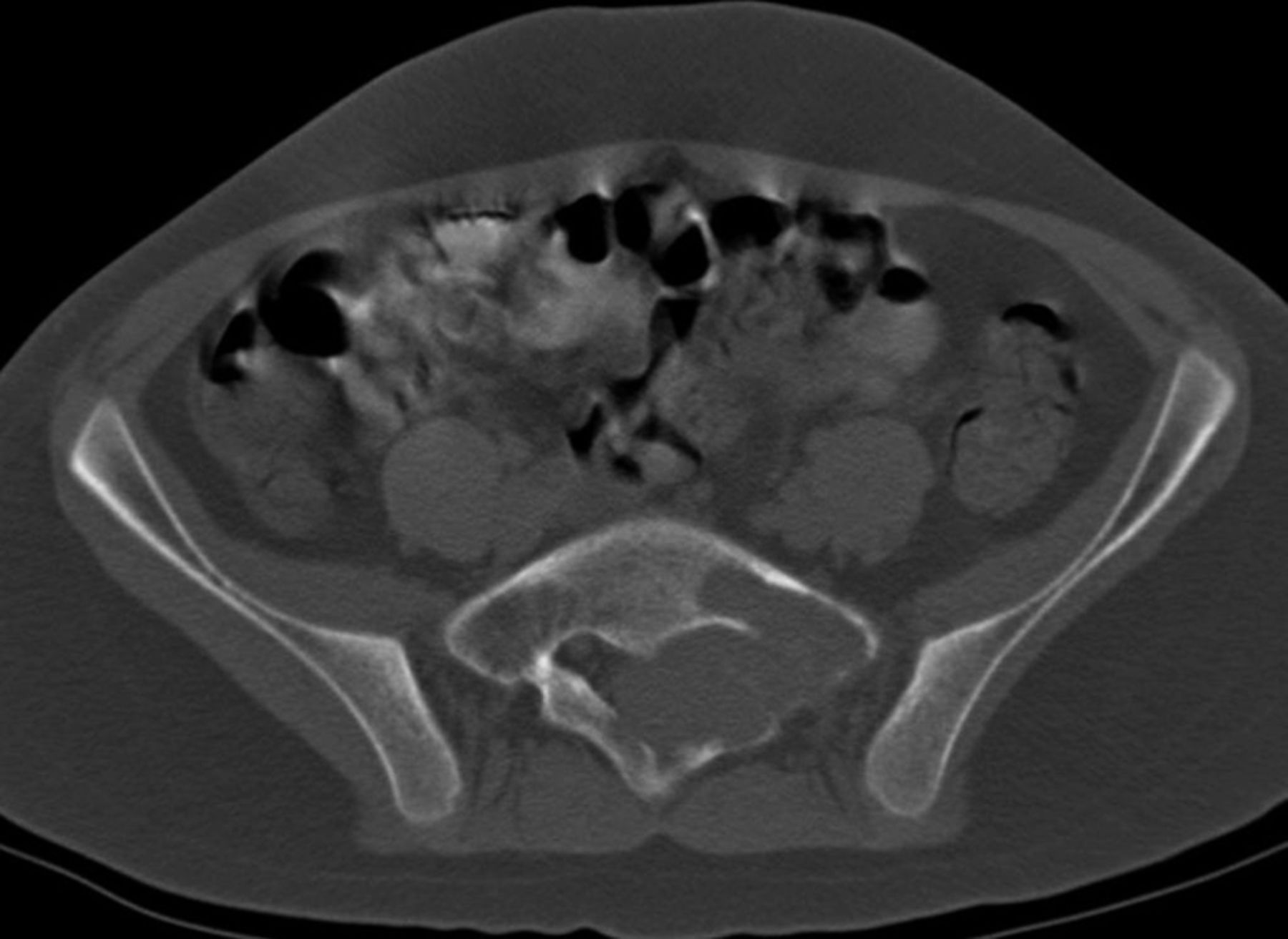
- FIGURE 3.
CT image (bone window) showing well-defined soft-tissue mass in left sacrum with marginal bone sclerosis.
- FIGURE 4.
18F-FDG PET/CT image showing metabolically active left sacral bone tumor.
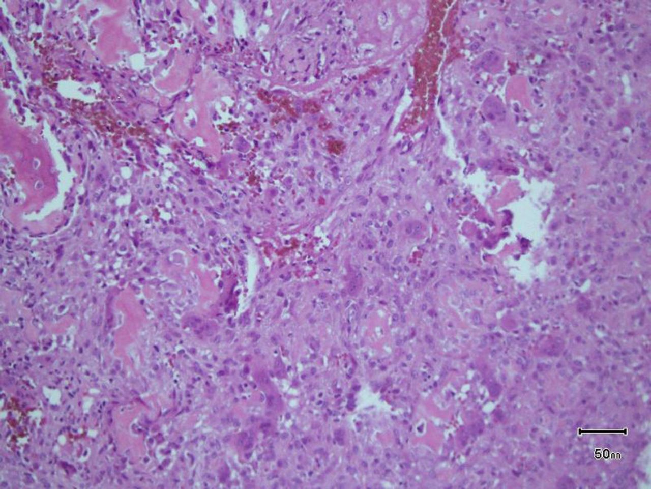
- FIGURE 5.
Histopathologic image showing tumor composed of woven bone spicules and irregular trabeculae lined by benign-looking activated osteoblasts (osteoblastoma).


Jump to section
Related Articles
Cited By...
- No citing articles found.