


Article Figures & Data
Figures
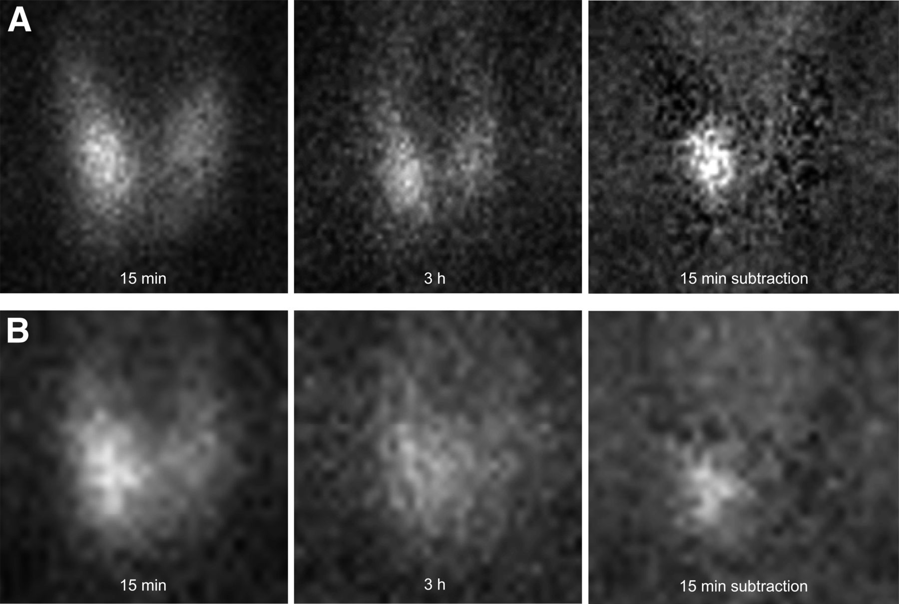
- FIGURE 1.
Pinhole collimator (A) and parallel-hole collimator (B) images of 99mTc-sestamibi at 15 min and 3 h, and image of 99mTc-sestamibi minus 123I at 15 min, in patient with hyperparathyroidism and 380-mg parathyroid adenoma located inferiorly on right. With pinhole collimation, 2 observers correctly localized adenoma without and with subtraction image with degree-of-certainty grades of 2 and 3 for one observer and 3 and 3 for the other. With parallel-hole collimation, observers did not identify adenoma without subtraction image and did localize adenoma with subtraction image with grades of 1 and 1.
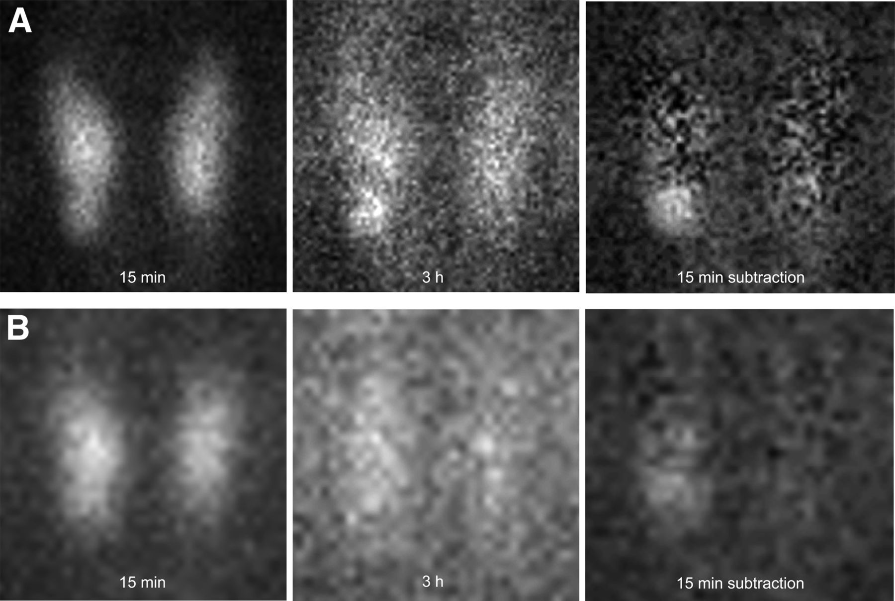
- FIGURE 2.
Pinhole collimator (A) and parallel-hole collimator (B) images of 99mTc-sestamibi at 15 min and 3 h, and image of 99mTc-sestamibi minus 123I at 15 min, in patient with hyperparathyroidism and 1,010-mg parathyroid adenoma located inferiorly on right. With pinhole collimation, 2 observers correctly localized adenoma without and with subtraction image with degree-of-certainty grades of 3 and 2 for one observer and 3 and 3 for the other. With subtraction image, one observer located adenoma at middle level whereas other localizations were all inferior. With parallel-hole collimation, only one observer identified adenoma without subtraction image, grades of 0 and 1, and both observers localized adenoma with subtraction image with grades of 1 and 1.
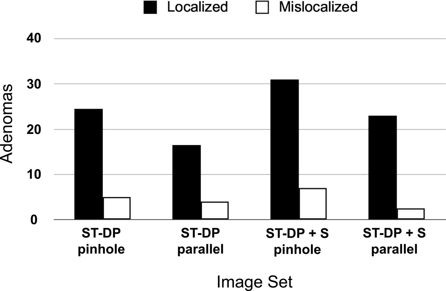
- FIGURE 3.
Performance for each of 4 protocols is shown for correctly localizing position of 37 parathyroid adenomas in 33 patients. Data represent average results for 2 observers.


Tables
Degree of certainty of location† Protocol Observer 0 1 2 3 ST-DP A 19 7 6 5 Pinhole B 6 16 (9) 8 (1) 7 ST-DP A 25 5 (1) 4 3 Parallel B 17 8 (7) 8 4 ST-DP + S A 8 11 (2) 6 (3) 12 (2) Pinhole B 4 13 (7) 10 10 ST-DP + S A 16 7 (1) 7 (1) 7 (1) Parallel B 12 12 (2) 9 4 ↵† 0 = no adenoma seen; 1 = possible adenoma; 2 = probable adenoma; 3 = definite adenoma.
Numbers in parentheses indicate number of localizations that were incorrect.
Localization success Protocol Observer Adenomas Correct 95% CI ST-DP A 18/37 0.49 0.32–0.66 Pinhole B 31/37 0.84 0.68–0.94 ST-DP A 12/37 0.32 0.18–0.50 Parallel B 20/37 0.54 0.37–0.71 ST-DP + S A 29/37 0.78 0.62–0.90 Pinhole B 33/37 0.89 0.75–0.97 ST-DP + S A 21/37 0.57 0.39–0.73 Parallel B 25/37 0.68 0.50–0.82 CI = exact binomial confidence interval.
There were 37 adenomas in 33 patients.


Jump to section
Related Articles
Cited By...
- Primary Hyperparathyroidism: Defining the Appropriate Preoperative Imaging Algorithm
- Parathyroid Imaging with Simultaneous Acquisition of 99mTc-Sestamibi and 123I: The Relative Merits of Pinhole Collimation and SPECT/CT
- The Role of Radionuclide Imaging in the Surgical Management of Primary Hyperparathyroidism
- Preoperative imaging for focused parathyroidectomy: making a good strategy even better