
Main menu

  • Home
  • Content
    • Current
      • JNMT Supplement
    • Ahead of print
    • Past Issues
    • Continuing Education
    • JNMT Podcast
    • SNMMI Annual Meeting Abstracts
  • Subscriptions
    • Subscribers
    • Rates
    • Journal Claims
    • Institutional and Non-member
  • Authors
    • Submit to JNMT
    • Information for Authors
    • Assignment of Copyright
    • AQARA Requirements
  • Info
    • Reviewers
    • Permissions
    • Advertisers
    • Corporate & Special Sales
  • About
    • About Us
    • Editorial Board
    • Contact Information
  • More
    • Alerts
    • Feedback
    • Help
    • SNMMI Journals
  • SNMMI
    • JNMT
    • JNM
    • SNMMI Journals
    • SNMMI

User menu

  • Subscribe
  • My alerts
  • Log in
  • My Cart

Search

  • Advanced search
Journal of Nuclear Medicine Technology
  • SNMMI
    • JNMT
    • JNM
    • SNMMI Journals
    • SNMMI
  • Subscribe
  • My alerts
  • Log in
  • My Cart
Journal of Nuclear Medicine Technology

Advanced Search

  • Home
  • Content
    • Current
    • Ahead of print
    • Past Issues
    • Continuing Education
    • JNMT Podcast
    • SNMMI Annual Meeting Abstracts
  • Subscriptions
    • Subscribers
    • Rates
    • Journal Claims
    • Institutional and Non-member
  • Authors
    • Submit to JNMT
    • Information for Authors
    • Assignment of Copyright
    • AQARA Requirements
  • Info
    • Reviewers
    • Permissions
    • Advertisers
    • Corporate & Special Sales
  • About
    • About Us
    • Editorial Board
    • Contact Information
  • More
    • Alerts
    • Feedback
    • Help
    • SNMMI Journals
  • Watch or Listen to JNMT Podcast
  • Visit SNMMI on Facebook
  • Join SNMMI on LinkedIn
  • Follow SNMMI on Twitter
  • Subscribe to JNMT RSS feeds
Review ArticleTEACHING FILES

67Ga Scintigraphy in the Evaluation of Postsurgical Low Back Pain

Prashant Jolepalem, Raymond Yeow and Paresh Mahajan
Journal of Nuclear Medicine Technology June 2013, 41 (2) 114-116; DOI: https://doi.org/10.2967/jnmt.113.123422
Prashant Jolepalem
1Department of Diagnostic Radiology and Molecular Imaging, Oakland University William Beaumont School of Medicine and William Beaumont Health System, Royal Oak, Michigan; and
  • Find this author on Google Scholar
  • Find this author on PubMed
  • Search for this author on this site
Raymond Yeow
2Oakland University William Beaumont School of Medicine, Rochester, Michigan
  • Find this author on Google Scholar
  • Find this author on PubMed
  • Search for this author on this site
Paresh Mahajan
1Department of Diagnostic Radiology and Molecular Imaging, Oakland University William Beaumont School of Medicine and William Beaumont Health System, Royal Oak, Michigan; and
  • Find this author on Google Scholar
  • Find this author on PubMed
  • Search for this author on this site
  • Article
  • Figures & Data
  • Info & Metrics
  • PDF
Loading

A 62-y-old man presented with chronic, diffuse lower back pain 8 mo after undergoing extensive lumbar spine surgery for osteomyelitis that included an L4 and L5 diskectomy with partial corpectomy, followed by anterior spinal fusion of L4 and L5 with a fibular allograft, and L3 to S1 posterior fusion via spinal instrumentation. Since the surgery, he had multiple admissions due to intractable lower back pain and sepsis. However, multiple bacterial cultures of biopsy samples from the operative site never demonstrated any growth. On the current admission, radiographs (Fig. 1A) and a CT scan of the lumber spine (Fig. 1B) demonstrated erosion of the inferior endplate of L3 and a potential bone fragment anteriorly—findings highly suggestive of osteomyelitis. Bacterial blood cultures were negative. The patient underwent 67Ga scintigraphy, which was compared with an 99mTc-methylene diphosphate (MDP) bone scan performed 2 d prior to the scintigraphy.

FIGURE 1.
  • Download figure
  • Open in new tab
  • Download powerpoint
FIGURE 1.

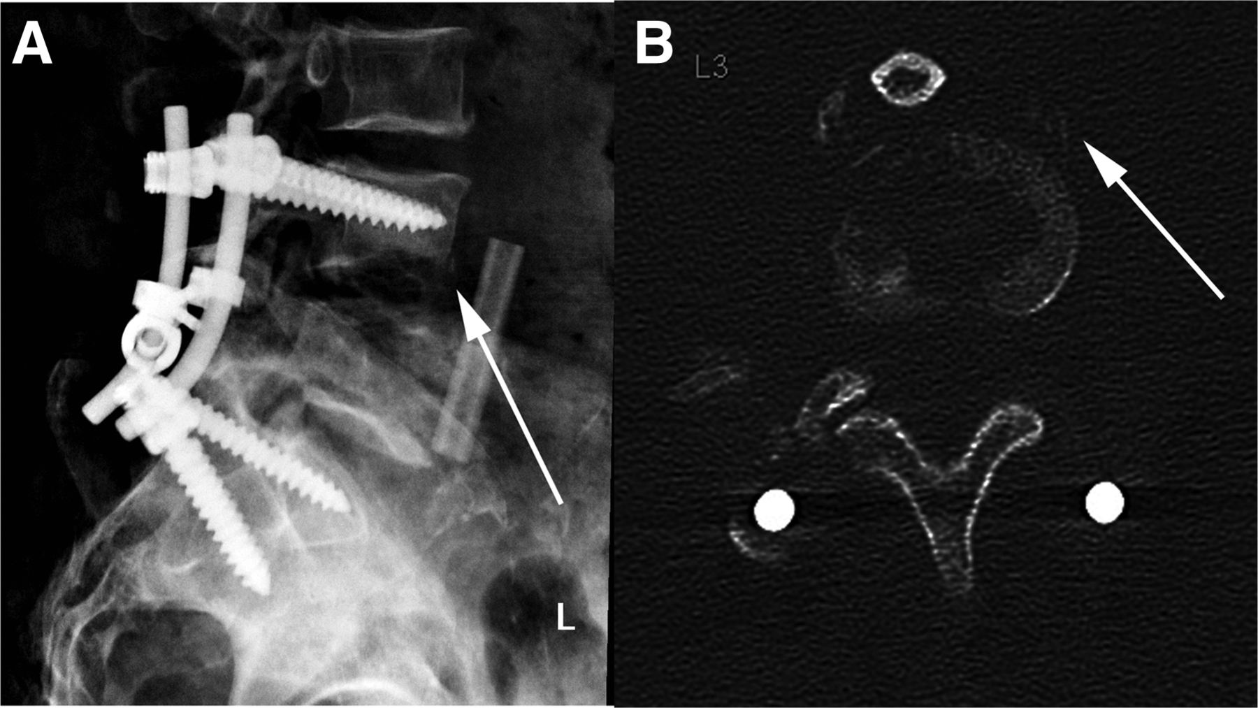
Plain film (A) and axial CT scan of L3 (B) showing inferior endplate erosion and evidence of periostitis (arrows).

The patient was injected with 188.7 MBq (5.1 mCi) of 67Ga-citrate intravenously. A large-field-of-view multipeak camera with a medium-energy parallel-hole collimator was used to obtain scintigrams at 24 and 48 h. Static images of the neck and chest, abdomen and pelvis, and lower extremities were obtained in both the anterior and the posterior projections. SPECT/CT was performed at 48 h for anatomic localization of the abnormal radiotracer uptake and was compared with the previously performed 99mTc-MDP bone scan.

The bone scan demonstrated an area of increased uptake in the body of L3 (Fig. 2A), which was localized to the distal ends of the fixation screws by SPECT/CT (Fig. 3A). Although the location of the activity was suggestive of loosening, the patient had undergone surgery only 8 mo previously; therefore, postsurgical reactive changes could not be excluded. Osteomyelitis involving the hardware remained a possibility, although this location did not correspond to the suggestive findings on correlative imaging. The 67Ga scan was expected to help delineate this differential.

FIGURE 2.
  • Download figure
  • Open in new tab
  • Download powerpoint
FIGURE 2.

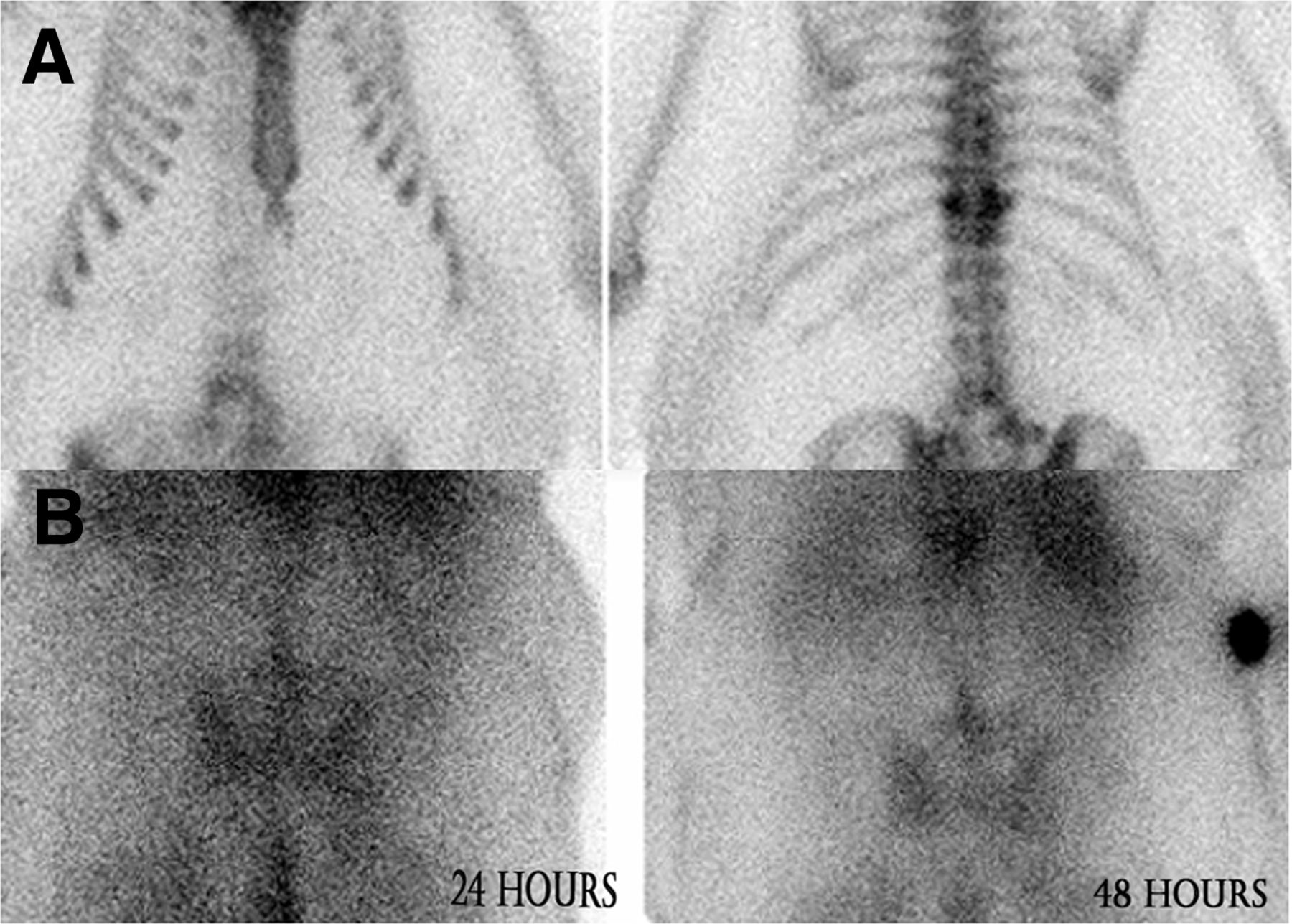
99mTc-MDP bone scan (A) in anterior and posterior projections showing mild activity at L3. More intense uptake is incidentally seen at compression deformity surrounding T11. 67Ga-citrate scan (B) at 24 and 48 h in posterior projection shows progressively increasing uptake in mid lumbar spine.

FIGURE 3.
  • Download figure
  • Open in new tab
  • Download powerpoint
FIGURE 3.

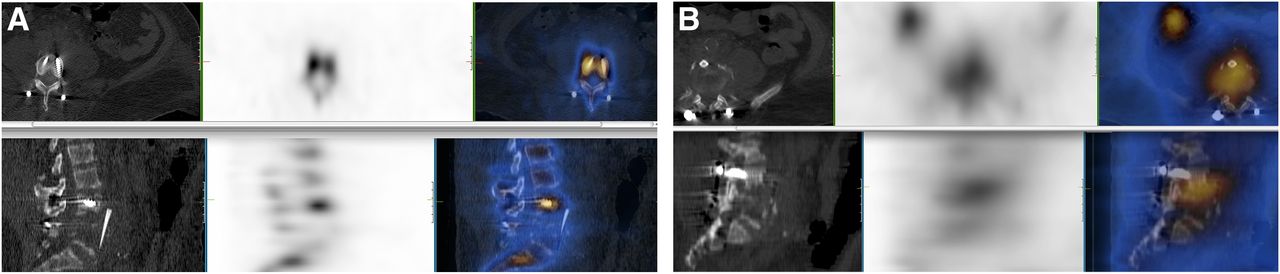
(A) SPECT/CT of 99mTc-MDP bone scan showing that activity is localized to fixation screws and does not involve disk space or bone fragments. (B) SPECT/CT of 67Ga scan showing that spatially discordant uptake involves disk space and inferior endplate of L3 and extends anteriorly to allograft.

On the 67Ga planar images (Fig. 2B), there was progressively increasing uptake in the mid lumbar spine. On SPECT/CT performed 48 h after radiotracer administration (Fig. 3B), there was spatial discordance of the uptake near L3 when compared with the 99mTc-MDP bone scan. The uptake was clearly localized to the inferior endplate of L3 and involved the disk space, allograft site, and anterior bone fragment. These findings were compatible with osteomyelitis or diskitis in this location. The fixation hardware was not involved in the infectious process. Another biopsy sample was taken, guided by the site of activity from the 67Ga scan, and the sample grew Candida.

QUESTION 1

What property of 67Ga makes it a valuable agent in imaging inflammatory processes?

  • A. Localizes to white blood cells in bone marrow.

  • B. Binds to transferrin.

  • C. Localizes to infection sites by binding to leukotaxis.

  • D. Binds to interleukin-8.

QUESTION 2

What is the target organ with 67Ga administration?

  • A. Large bowel.

  • B. Small bowel.

  • C. Liver.

  • D. Salivary glands.

QUESTION 3

Which one of the following is not a common cause of false-negative 67Ga findings?

  • A. Recent gadolinium administration.

  • B. Blood transfusions.

  • C. Inadequate bowel preparation.

  • D. Antibiotic use.

QUESTION 4

In which of the following conditions would 67Ga scintigraphy not be useful?

  • A. Fever of unknown origin.

  • B. Inflammatory bowel disease.

  • C. Sarcoidosis.

  • D. Lymphoma.

CASE DISCUSSION

67Ga is not an ideal imaging agent because of multiple high-energy photopeaks (93, 185, 288, and 394 keV), low abundance, and a high scatter fraction. However, the physiologic properties of 67Ga still make it an extremely useful agent in appropriate clinical scenarios. It can often uncover an underlying cause of fever or infection that clinicians had not previously considered. It is considered superior to radiolabeled leukocytes in cases of pulmonary inflammatory diseases, drug-induced inflammation, leukopenia, diskitis, vertebral osteomyelitis, and chronic-phase infections. 67Ga may also be the better choice in fever of unknown origin depending on the patient history and suspected sources. Additionally, SPECT/CT can help to mitigate some of the shortfalls of its imaging characteristics.

In our case, the patient had signs, symptoms, and imaging findings suggestive of osteomyelitis in his lumbar spine for several months. However, there was no pathologic evidence, and many courses of antibiotics did not relieve his symptoms. After 67Ga localized an infectious process in the inferior endplate of L3 involving the allograft site, a more targeted biopsy was performed with a subsequent fungal culture, which demonstrated growth of Candida. This was an unexpected finding and an extremely rare case of fungal diskitis in an immunocompetent patient. The patient was started on appropriate long-term antifungal therapy, and his symptoms eventually resolved.

Footnotes

  • Published online Apr. 17, 2013.

  • * For the answers, see page 120.

BIBLIOGRAPHY

    1. Ziessman HA,
    2. O’Malley JP,
    3. Thrall JH
    . Nuclear Medicine (The Requisites in Radiology). 3rd ed. Philadelphia, PA: Mosby; 2006.
    1. Palestro CJ,
    2. Love C,
    3. Tronco GC,
    4. Tomas MB
    . Role of radionuclide imaging in the diagnosis of postoperative infection. Radiographics 2000;20:1649–1660.
    OpenUrlPubMed
    1. Love C,
    2. Patel M,
    3. Lonner BS,
    4. et al
    . Diagnosing spinal osteomyelitis: a comparison of bone and gallium scintigraphy and magnetic resonance imaging. Clin Nucl Med 2000;25:963–977.
    OpenUrlCrossRefPubMed
PreviousNext
Back to top

In this issue

Journal of Nuclear Medicine Technology: 41 (2)
Journal of Nuclear Medicine Technology
Vol. 41, Issue 2
June 1, 2013
  • Table of Contents
  • About the Cover
  • Index by author
Print
Download PDF
Article Alerts
Sign In to Email Alerts with your Email Address
Email Article

Thank you for your interest in spreading the word on Journal of Nuclear Medicine Technology.

NOTE: We only request your email address so that the person you are recommending the page to knows that you wanted them to see it, and that it is not junk mail. We do not capture any email address.

Enter multiple addresses on separate lines or separate them with commas.
67Ga Scintigraphy in the Evaluation of Postsurgical Low Back Pain
(Your Name) has sent you a message from Journal of Nuclear Medicine Technology
(Your Name) thought you would like to see the Journal of Nuclear Medicine Technology web site.
Citation Tools
67Ga Scintigraphy in the Evaluation of Postsurgical Low Back Pain
Prashant Jolepalem, Raymond Yeow, Paresh Mahajan
Journal of Nuclear Medicine Technology Jun 2013, 41 (2) 114-116; DOI: 10.2967/jnmt.113.123422

Citation Manager Formats

  • BibTeX
  • Bookends
  • EasyBib
  • EndNote (tagged)
  • EndNote 8 (xml)
  • Medlars
  • Mendeley
  • Papers
  • RefWorks Tagged
  • Ref Manager
  • RIS
  • Zotero
Share
67Ga Scintigraphy in the Evaluation of Postsurgical Low Back Pain
Prashant Jolepalem, Raymond Yeow, Paresh Mahajan
Journal of Nuclear Medicine Technology Jun 2013, 41 (2) 114-116; DOI: 10.2967/jnmt.113.123422
Twitter logo Facebook logo LinkedIn logo Mendeley logo
  • Tweet Widget
  • Facebook Like
  • Google Plus One
Bookmark this article

Jump to section

  • Article
    • QUESTION 1
    • QUESTION 2
    • QUESTION 3
    • QUESTION 4
    • CASE DISCUSSION
    • Footnotes
    • BIBLIOGRAPHY
  • Figures & Data
  • Info & Metrics
  • PDF

Related Articles

  • Nuclear Medicine Instrumentation
  • PubMed
  • Google Scholar

Cited By...

  • No citing articles found.
  • Google Scholar

More in this TOC Section

  • Patient with Acute Shortness of Breath, Elevated D-Dimer, and a Previous History of Cancer
  • Bronchopleural Fistula Mimicking Chronic Obstructive Lung Disease on 133Xe Scan
  • Increased Sacral Uptake on a Bone Scan with SPECT/CT in a Patient with Achondroplasia: Normal or Abnormal?
Show more TEACHING FILES

Similar Articles

SNMMI

© 2025 SNMMI

Powered by HighWire