  • Main menu
  • User menu
  • Search
  • English ▼
    • English
    • Afrikaans
    • Albanian
    • Amharic
    • Arabic
    • Armenian
    • Azerbaijani
    • Basque
    • Belarusian
    • Bengali
    • Bosnian
    • Bulgarian
    • Catalan
    • Cebuano
    • Chichewa
    • Chinese (Simplified)
    • Chinese (Traditional)
    • Corsican
    • Croatian
    • Czech
    • Danish
    • Dutch
    • Esperanto
    • Estonian
    • Filipino
    • Finnish
    • French
    • Frisian
    • Galician
    • Georgian
    • German
    • Greek
    • Gujarati
    • Haitian Creole
    • Hausa
    • Hawaiian
    • Hebrew
    • Hindi
    • Hmong
    • Hungarian
    • Icelandic
    • Igbo
    • Indonesian
    • Irish
    • Italian
    • Japanese
    • Javanese
    • Kannada
    • Kazakh
    • Khmer
    • Korean
    • Kurdish (Kurmanji)
    • Kyrgyz
    • Lao
    • Latin
    • Latvian
    • Lithuanian
    • Luxembourgish
    • Macedonian
    • Malagasy
    • Malay
    • Malayalam
    • Maltese
    • Maori
    • Marathi
    • Mongolian
    • Myanmar (Burmese)
    • Nepali
    • Norwegian
    • Pashto
    • Persian
    • Polish
    • Portuguese
    • Punjabi
    • Romanian
    • Russian
    • Samoan
    • Scottish Gaelic
    • Serbian
    • Sesotho
    • Shona
    • Sindhi
    • Sinhala
    • Slovak
    • Slovenian
    • Somali
    • Spanish
    • Sudanese
    • Swahili
    • Swedish
    • Tajik
    • Tamil
    • Telugu
    • Thai
    • Turkish
    • Ukrainian
    • Urdu
    • Uzbek
    • Vietnamese
    • Welsh
    • Xhosa
    • Yiddish
    • Yoruba
    • Zulu

Main menu

  • Home
  • Content
    • Current
      • JNMT Supplement
    • Ahead of print
    • Past Issues
    • Continuing Education
    • JNMT Podcast
    • SNMMI Annual Meeting Abstracts
  • Subscriptions
    • Subscribers
    • Rates
    • Journal Claims
    • Institutional and Non-member
  • Authors
    • Submit to JNMT
    • Information for Authors
    • Assignment of Copyright
    • AQARA Requirements
  • Info
    • Reviewers
    • Permissions
    • Advertisers
    • Corporate & Special Sales
  • About
    • About Us
    • Editorial Board
    • Contact Information
  • More
    • Alerts
    • Feedback
    • Help
    • SNMMI Journals
  • SNMMI
    • JNMT
    • JNM
    • SNMMI Journals
    • SNMMI

User menu

  • Subscribe
  • My alerts
  • Log in
  • My Cart

Search

  • Advanced search
Journal of Nuclear Medicine Technology
  • SNMMI
    • JNMT
    • JNM
    • SNMMI Journals
    • SNMMI
  • Subscribe
  • My alerts
  • Log in
  • My Cart
Journal of Nuclear Medicine Technology

Advanced Search

English ▼
  • English
  • Afrikaans
  • Albanian
  • Amharic
  • Arabic
  • Armenian
  • Azerbaijani
  • Basque
  • Belarusian
  • Bengali
  • Bosnian
  • Bulgarian
  • Catalan
  • Cebuano
  • Chichewa
  • Chinese (Simplified)
  • Chinese (Traditional)
  • Corsican
  • Croatian
  • Czech
  • Danish
  • Dutch
  • Esperanto
  • Estonian
  • Filipino
  • Finnish
  • French
  • Frisian
  • Galician
  • Georgian
  • German
  • Greek
  • Gujarati
  • Haitian Creole
  • Hausa
  • Hawaiian
  • Hebrew
  • Hindi
  • Hmong
  • Hungarian
  • Icelandic
  • Igbo
  • Indonesian
  • Irish
  • Italian
  • Japanese
  • Javanese
  • Kannada
  • Kazakh
  • Khmer
  • Korean
  • Kurdish (Kurmanji)
  • Kyrgyz
  • Lao
  • Latin
  • Latvian
  • Lithuanian
  • Luxembourgish
  • Macedonian
  • Malagasy
  • Malay
  • Malayalam
  • Maltese
  • Maori
  • Marathi
  • Mongolian
  • Myanmar (Burmese)
  • Nepali
  • Norwegian
  • Pashto
  • Persian
  • Polish
  • Portuguese
  • Punjabi
  • Romanian
  • Russian
  • Samoan
  • Scottish Gaelic
  • Serbian
  • Sesotho
  • Shona
  • Sindhi
  • Sinhala
  • Slovak
  • Slovenian
  • Somali
  • Spanish
  • Sudanese
  • Swahili
  • Swedish
  • Tajik
  • Tamil
  • Telugu
  • Thai
  • Turkish
  • Ukrainian
  • Urdu
  • Uzbek
  • Vietnamese
  • Welsh
  • Xhosa
  • Yiddish
  • Yoruba
  • Zulu
  • Home
  • Content
    • Current
    • Ahead of print
    • Past Issues
    • Continuing Education
    • JNMT Podcast
    • SNMMI Annual Meeting Abstracts
  • Subscriptions
    • Subscribers
    • Rates
    • Journal Claims
    • Institutional and Non-member
  • Authors
    • Submit to JNMT
    • Information for Authors
    • Assignment of Copyright
    • AQARA Requirements
  • Info
    • Reviewers
    • Permissions
    • Advertisers
    • Corporate & Special Sales
  • About
    • About Us
    • Editorial Board
    • Contact Information
  • More
    • Alerts
    • Feedback
    • Help
    • SNMMI Journals
  • Watch or Listen to JNMT Podcast
  • Visit SNMMI on Facebook
  • Join SNMMI on LinkedIn
  • Follow SNMMI on Twitter
  • Subscribe to JNMT RSS feeds
Case ReportCASE REPORTS

Hormone Therapy in Prostate Cancer

Geoffrey M. Currie, Matthew Haase, Rashid Hashmi and Hosen Kiat
Journal of Nuclear Medicine Technology March 2013, 41 (1) 49-51; DOI: https://doi.org/10.2967/jnmt.112.118034
Geoffrey M. Currie
1Faculty of Science, Charles Sturt University, Wagga Wagga, New South Wales, Australia
2Australian School of Advanced Medicine, Macquarie University, Sydney, Australia; and
  • Find this author on Google Scholar
  • Find this author on PubMed
  • Search for this author on this site
Matthew Haase
3RIL/Imed, Wagga Wagga, Australia
  • Find this author on Google Scholar
  • Find this author on PubMed
  • Search for this author on this site
Rashid Hashmi
3RIL/Imed, Wagga Wagga, Australia
  • Find this author on Google Scholar
  • Find this author on PubMed
  • Search for this author on this site
Hosen Kiat
1Faculty of Science, Charles Sturt University, Wagga Wagga, New South Wales, Australia
2Australian School of Advanced Medicine, Macquarie University, Sydney, Australia; and
  • Find this author on Google Scholar
  • Find this author on PubMed
  • Search for this author on this site
  • Article
  • Figures & Data
  • Info & Metrics
  • PDF
Loading

Abstract

The role of nuclear medicine diagnostic bone scanning is well established in prostate cancer. This case provides an insight into the specific role that bone scanning plays in monitoring response to hormone therapy and an example of significant global skeletal response. The case highlights the remarkable efficacy of timely hormone therapy in high-grade prostate cancer with widespread bony metastasis. In addition, the range of hormone therapy currently available for clinical application in the management of metastatic prostate cancer is detailed. Finally, the case represents an incidental diagnosis of prostate cancer after evaluation of nonspecific symptoms.

  • prostate carcinoma
  • hormone ablation therapy
  • GnRH agonist
  • ADT
  • bone scintigraphy

We present a case study that provides an insight into the specific role that bone scanning plays in monitoring response to hormone therapy. The case highlights the remarkable efficacy of timely hormone therapy in high-grade prostate cancer with widespread bony metastasis.

CASE REPORT

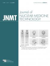
An 83-y-old man presented for evaluation of reported weight loss and constipation. These symptoms were initially assessed by CT of the chest, abdomen, and pelvis. The findings were unremarkable, although a left renal staghorn calculus was noted. Despite no previous history of neoplasia, extensive sclerotic lesions were noted throughout the axial skeleton. This finding, combined with the observation of an irregular prostate contour (without enlargement), raised suspicion of previously undiagnosed widespread metastatic prostate carcinoma. Subsequent follow-up revealed a prostate-specific antigen (PSA) level of 823 μg/L (normal, <4 μg/L) and bone scan findings consistent with widespread skeletal metastases (Fig. 1). Subsequent prostate biopsy confirmed aggressive prostate carcinoma with a Gleason score of 9 (1). The patient commenced hormone therapy using bicalutamide (150 mg; Cosudex [AstraZeneca]) daily (oral tablet) and leuprorelin (30 mg; Lucrin [AbbVie]) depot (intramuscular injection) every 4 mo. The medications were well tolerated. At week 4, goserelin (Zoladex; AstraZeneca), a 10.8-mg subcutaneous implant (1 dose at 3 monthly intervals), was added to the patient’s antitumor therapeutic armamentarium.

FIGURE 1.
  • Download figure
  • Open in new tab
  • Download powerpoint
FIGURE 1.

Baseline nuclear medicine bone scan demonstrating widespread metastatic disease associated with prostate carcinoma.

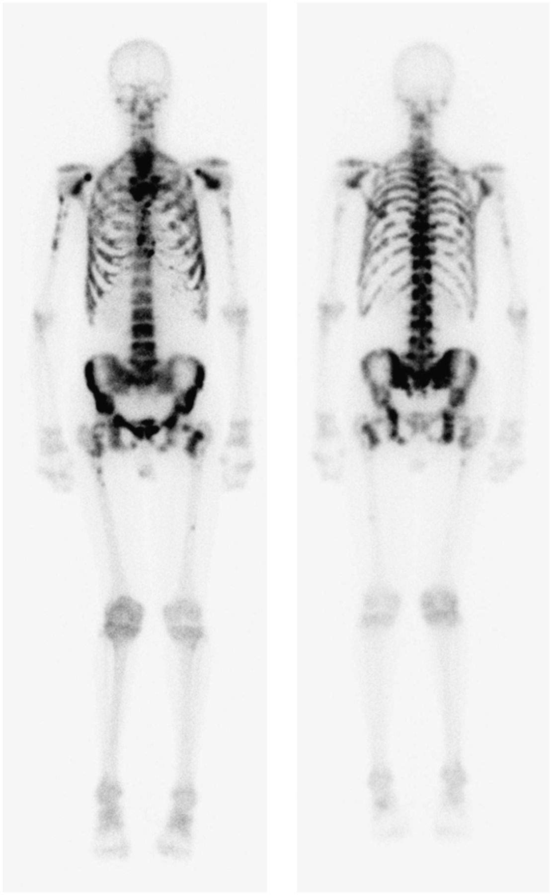
At 5 mo after the commencement of therapy, the PSA level had normalized to 0.39 ug/L. It dropped further to 0.11 μg/L at 10 mo, corresponding to scintigraphic resolution of bony metastatic changes on the follow-up nuclear medicine bone scan (Fig. 2).

FIGURE 2.
  • Download figure
  • Open in new tab
  • Download powerpoint
FIGURE 2.

Follow-up nuclear medicine bone scan demonstrating remission of metastatic disease after hormone ablation therapy.

DISCUSSION

Hormone Therapy

The approach taken in the treatment of prostate cancer will depend on several factors, including the clinical presentation, Gleason score, and PSA (2). For patients with metastatic spread of prostate cancer, such as the patient in this case, the treatment of choice is androgen-deprivation therapy (ADT) (2,3). ADT has been reported to lead to remission in as many as 80% of patients with advanced prostate carcinoma (3,4). ADT is used in advanced prostate cancer to relieve pain, prevent pathologic fractures, and prevent neurologic complications (4). Typically, androgen deprivation is achieved through 3 mechanisms in prostate carcinoma patients, gonadotrophin-releasing hormone (GnRH) agonists, antiandrogens, and GnRH antagonists.

GnRH agonists (goserelin, leuprorelin, and triptorelin) are considered the leading approach to ADT in prostate carcinoma (5). GnRH is released in pulses from the hypothalamus to stimulate pituitary release of luteinizing hormone, which, in turn, stimulates the secretion of testosterone in the testes (5,6). GnRH agonists decrease testosterone levels by overstimulation, downregulation, and eventually receptor desensitization (5–7). There is, however, an initial flare or surge in testosterone production before suppression is effected (5). Unfortunately, testosterone suppression also increases the risk of osteoporosis and fracture (loss of bone mineral density), diabetes (altered glucose tolerance), and cardiovascular disease (7). Some of the more common adverse effects of ADT include weight gain, erectile dysfunction, decreased libido, gynecomastia, insomnia, sweats or chills, and gastrointestinal disturbances (7).

Antiandrogens (bicalutamide, flutamide, and nilutamide) are nonsteroidal inhibitors of androgen receptors (6,7). The advantage of this class of ADT medications is that testosterone levels are not suppressed, and thus the associated risks discussed above are largely avoided (6,7). That is, antiandrogens inhibit conversion of testosterone to dihydrotestosterine (metabolite) and prevent both from binding to the androgen receptor, without suppressing serum testosterone levels. The flare in serum testosterone associated with GnRH agonists means that they should be avoided as monotherapy in patients with severe pain or neurologic problems. Antiandrogens can be used as adjunctive therapy to block the flare (4).

GnRH antagonists (degarelix) have only recently received attention in the literature. GnRH agonists can cause an early increase in testosterone levels which, in turn, can promote tumor spread and worsen symptoms (2). GnRH antagonists can be used before the GnRH agonist therapy to block testosterone increases (2,7). GnRH antagonists can also be used as an alternative approach to androgen deprivation by inhibiting gonadotrophin production, leading to a reduction in the synthesis of androgens in the testes (7).

Bone Scan

ADT has been shown to result in remission in as many as 80% of patients, which is reflected in a median period of 12–33 mo of progression-free survival (4,6). That is, ADT prevents the progression of metastatic spread. Typically the nuclear medicine bone scan will demonstrate partial improvement or stabilization (nonprogression) of metastatic disease in response to ADT, but it is a rare occurrence for the bone scan to revert to normal (4). This observation reflects the fact that ADT rarely provides an overall cure and the likelihood of cure is inversely proportional to the extent of disease at the time of commencement of ADT. Another reason for the infrequency of a completely normal follow-up bone scan is that remodeling of bone in response to healing will mimic metastatic progression. The reversion to normal in this case is remarkable given the advanced stage of disease (Gleason score of 9) and extent of metastatic disease at the commencement of ADT.

After the progression-free survival period, there will be transformation of the disease to an androgen-independent phenotype that will not respond to ADT (6). At this point, alternative therapy (e.g., docetaxel) may be indicated. The mean patient survival (from commencement of ADT) is 23–37 mo (6). Androgen-independent disease is marked by disease progression despite testosterone levels below castrate levels (<50 ng/mL).

CONCLUSION

The bone scan provides a valuable tool in monitoring response to ADT because, independently of PSA levels and anatomic imaging (e.g., radiography and CT), this modality demonstrates metabolic bone changes. This case demonstrates an unusually dramatic improvement in bone scan appearance after hormone therapy in prostate cancer.

DISCLOSURE

No potential conflict of interest relevant to this article was reported.

Acknowledgments

We acknowledge urologist Dr. Steven Sowter for providing information to aid the formulation of the case study.

Footnotes

  • Published online Feb. 12, 2013.

REFERENCES

  1. 1.↵
    1. Thorson P,
    2. Humphrey P
    . Minimal adenocarcinoma in prostate needle biopsy tissue. Am J Clin Pathol. 2000;114:896–909.
    OpenUrlAbstract/FREE Full TextGoogle Scholar
  2. 2.↵
    1. Choi S,
    2. Lee AK
    . Efficacy and safety of gonadotropin-releasing hormone agonists used in the treatment of prostate cancer. Drug Healthc Patient Saf. 2011;3:107–119.
    OpenUrlPubMedGoogle Scholar
  3. 3.↵
    1. Hammerer P,
    2. Madersbacher S
    . Landmarks in hormonal therapy for prostate cancer. BJU Int. 2012;110:23–29.
    OpenUrlCrossRefPubMedGoogle Scholar
  4. 4.↵
    1. Huben RP
    . Hormone therapy of prostatic bone metastases. Adv Exp Med Biol. 1992;324:305–316.
    OpenUrlCrossRefPubMedGoogle Scholar
  5. 5.↵
    1. Lepor H,
    2. Shore ND
    . LHRH agonists for the treatment of prostate cancer: 2012. Rev Urol. 2012;14:1–12.
    OpenUrlPubMedGoogle Scholar
  6. 6.↵
    1. Hellerstedt BA,
    2. Pienta KJ
    . The current state of hormonal therapy for prostate cancer. CA Cancer J Clin. 2002;52:154–179.
    OpenUrlPubMedGoogle Scholar
  7. 7.↵
    1. Rossi S
    , ed. Australian Medicines Handbook 2012. Adelaide, Australia: Australian Medicines Handbook Pty Ltd.; 2012:591–598.
    Google Scholar
  • Received for publication December 2, 2012.
  • Accepted for publication January 28, 2013.
PreviousNext
Back to top

In this issue

Journal of Nuclear Medicine Technology: 41 (1)
Journal of Nuclear Medicine Technology
Vol. 41, Issue 1
March 1, 2013
  • Table of Contents
  • About the Cover
  • Index by author
Print
Download PDF
Article Alerts
Email Article
Citation Tools
Share
Hormone Therapy in Prostate Cancer
Geoffrey M. Currie, Matthew Haase, Rashid Hashmi, Hosen Kiat
Journal of Nuclear Medicine Technology Mar 2013, 41 (1) 49-51; DOI: 10.2967/jnmt.112.118034
Twitter logo Facebook logo LinkedIn logo Mendeley logo
  • Tweet Widget
Bookmark this article

Jump to section

  • Article
    • Abstract
    • CASE REPORT
    • DISCUSSION
    • CONCLUSION
    • DISCLOSURE
    • Acknowledgments
    • Footnotes
    • REFERENCES
  • Figures & Data
  • Info & Metrics
  • PDF

Related Articles

  • No related articles found.
  • PubMed
  • Google Scholar

Cited By...

  • No citing articles found.
  • Google Scholar

More in this TOC Section

  • Post-90Y Radioembolization PET/CT Scan with Respiratory Gating Using Time-of-Flight Reconstruction
  • Primary Hyperparathyroidism–Related Brown Tumors Mimicking Other Giant Cell–Containing Skeletal Tumors: Role of Correlative Imaging in Diagnosis
  • False-Positive Scalp Activity in 131I Imaging Associated with Hair Coloring
Show more Case Reports

Similar Articles

  • Chapter Bios
  • Chapter Bios
  • Chapter Bios
  • SNMMI Clinical Trials Network Research Series for Technologists: An Introduction to Conducting Theranostic Clinical Trials
  • What Is in a Name?
See more

Keywords

  • prostate carcinoma
  • hormone ablation therapy
  • GnRH agonist
  • ADT
  • bone scintigraphy
SNMMI

© 2025 SNMMI

Powered by HighWire
Alerts for this Article
Sign In to Email Alerts with your Email Address
Email this Article

Thank you for your interest in spreading the word on Journal of Nuclear Medicine Technology.

NOTE: We only request your email address so that the person you are recommending the page to knows that you wanted them to see it, and that it is not junk mail. We do not capture any email address.

Enter multiple addresses on separate lines or separate them with commas.
Hormone Therapy in Prostate Cancer
(Your Name) has sent you a message from Journal of Nuclear Medicine Technology
(Your Name) thought you would like to see the Journal of Nuclear Medicine Technology web site.
Citation Tools
Hormone Therapy in Prostate Cancer
Geoffrey M. Currie, Matthew Haase, Rashid Hashmi, Hosen Kiat
Journal of Nuclear Medicine Technology Mar 2013, 41 (1) 49-51; DOI: 10.2967/jnmt.112.118034

Citation Manager Formats

  • BibTeX
  • Bookends
  • EasyBib
  • EndNote (tagged)
  • EndNote 8 (xml)
  • Medlars
  • Mendeley
  • Papers
  • RefWorks Tagged
  • Ref Manager
  • RIS
  • Zotero

We use cookies on this site to enhance your user experience

By clicking any link on this page you are giving your consent for us to set cookies.