
Main menu

  • Home
  • Content
    • Current
      • JNMT Supplement
    • Ahead of print
    • Past Issues
    • Continuing Education
    • JNMT Podcast
    • SNMMI Annual Meeting Abstracts
  • Subscriptions
    • Subscribers
    • Rates
    • Journal Claims
    • Institutional and Non-member
  • Authors
    • Submit to JNMT
    • Information for Authors
    • Assignment of Copyright
    • AQARA Requirements
  • Info
    • Reviewers
    • Permissions
    • Advertisers
    • Corporate & Special Sales
  • About
    • About Us
    • Editorial Board
    • Contact Information
  • More
    • Alerts
    • Feedback
    • Help
    • SNMMI Journals
  • SNMMI
    • JNMT
    • JNM
    • SNMMI Journals
    • SNMMI

User menu

  • Subscribe
  • My alerts
  • Log in
  • My Cart

Search

  • Advanced search
Journal of Nuclear Medicine Technology
  • SNMMI
    • JNMT
    • JNM
    • SNMMI Journals
    • SNMMI
  • Subscribe
  • My alerts
  • Log in
  • My Cart
Journal of Nuclear Medicine Technology

Advanced Search

  • Home
  • Content
    • Current
    • Ahead of print
    • Past Issues
    • Continuing Education
    • JNMT Podcast
    • SNMMI Annual Meeting Abstracts
  • Subscriptions
    • Subscribers
    • Rates
    • Journal Claims
    • Institutional and Non-member
  • Authors
    • Submit to JNMT
    • Information for Authors
    • Assignment of Copyright
    • AQARA Requirements
  • Info
    • Reviewers
    • Permissions
    • Advertisers
    • Corporate & Special Sales
  • About
    • About Us
    • Editorial Board
    • Contact Information
  • More
    • Alerts
    • Feedback
    • Help
    • SNMMI Journals
  • Watch or Listen to JNMT Podcast
  • Visit SNMMI on Facebook
  • Join SNMMI on LinkedIn
  • Follow SNMMI on Twitter
  • Subscribe to JNMT RSS feeds
Case ReportCASE REPORTS

Large Abdominal Photopenic Area on 99mTc-Sestamibi Myocardial Perfusion Imaging

Jennifer Lyon, John Spaulding and Paul M. Zack
Journal of Nuclear Medicine Technology December 2012, 40 (4) 281-282; DOI: https://doi.org/10.2967/jnmt.111.102079
Jennifer Lyon
CNMT
  • Find this author on Google Scholar
  • Find this author on PubMed
  • Search for this author on this site
John Spaulding
CNMT
  • Find this author on Google Scholar
  • Find this author on PubMed
  • Search for this author on this site
Paul M. Zack
MD
  • Find this author on Google Scholar
  • Find this author on PubMed
  • Search for this author on this site
  • Article
  • Figures & Data
  • Info & Metrics
  • PDF
Loading

Abstract

99mTc-sestamibi myocardial perfusion imaging is frequently performed in conjunction with exercise or pharmacologic stress testing for evaluation of coronary heart disease. Interpretation of these studies includes systematic review of unprocessed rotating projectional images for evaluation of cardiac size as well as the presence of motion or attenuation artifacts. Occasionally, incidental noncardiac findings are detected on review of the projectional images. We report a case of a patient with a history of autosomal dominant polycystic kidney disease who was found to have a large abdominal photopenic area on the projectional images. The photopenic area corresponded to the location of large intraabdominal cysts on abdominal CT and was consistent with hepatic cysts associated with the patient’s known polycystic kidney disease. We review the differential diagnosis of large abdominal photopenic regions identified on myocardial projectional images and the importance of routinely analyzing these images for incidental noncardiac findings.

  • sestamibi
  • photopenic
  • polycystic kidney disease
  • renal cyst
  • hepatic cyst

Incidental noncardiac findings occasionally are detected on review of projectional 99mTc-sestamibi myocardial perfusion images (1). Here, we report a case in which a large abdominal photopenic area was found on myocardial perfusion imaging in a patient with a history of autosomal dominant polycystic kidney disease.

CASE REPORT

A 69-y-old man was seen in our cardiology specialty office for evaluation of symptoms of shortness of breath with exertion and abnormalities found on a standard exercise test. He had experienced occasional symptoms of dyspnea with exertion during the past 3 mo. He denied symptoms of exertional or rest chest pain. His cardiac risk factors included a history of borderline hypertension and a family history of cardiac disease. The past medical history was significant for known autosomal dominant polycystic kidney disease. He had been found to have large renal and hepatic cysts on an earlier CT scan and was undergoing nephrology evaluation for moderate renal insufficiency.

On physical examination the blood pressure was 121/82 mm Hg, the heart rate 72 beats per minute, and the respiratory rate 16 breaths per minute. The lungs were clear to auscultation. On cardiac examination, there was a regular rhythm with a soft systolic murmur at the left sternal border. On abdominal examination, there was mild diffuse distention without tenderness to palpation.

The electrocardiogram showed sinus rhythm and nonspecific ST-T changes. A recent standard exercise treadmill stress test had demonstrated abnormal electrocardiographic changes at peak exercise in the inferior and lateral precordial leads.

In light of the patient’s symptoms and electrocardiographic abnormalities observed on standard stress testing, repeated treadmill stress testing was performed in conjunction with same-day stress–rest 99mTc-sestamibi myocardial perfusion imaging. The patient reached an exertional level of 10 metabolic equivalents and 100% of maximum predicted heart rate without symptoms of chest pain. Abnormal electrocardiographic changes were again observed in multiple leads at peak heart rate. Gated SPECT myocardial images showed normal left ventricular wall motion with an ejection fraction of 58%. There was normal myocardial perfusion on both stress and rest images, with no evidence of stress-induced ischemia. Review of the unprocessed rotating projectional images, however, demonstrated a large photopenic area within the left and mid abdominal regions (Fig. 1). This was better visualized in an anterior static image acquired at the abdominal and pelvic regions (Fig. 2). Comparison of these findings to an earlier abdominal CT scan demonstrated that the location of the photopenic area corresponded to large hepatic cysts consistent with the patient’s known polycystic kidney disease (Fig. 3).

FIGURE 1.
  • Download figure
  • Open in new tab
  • Download powerpoint
FIGURE 1.

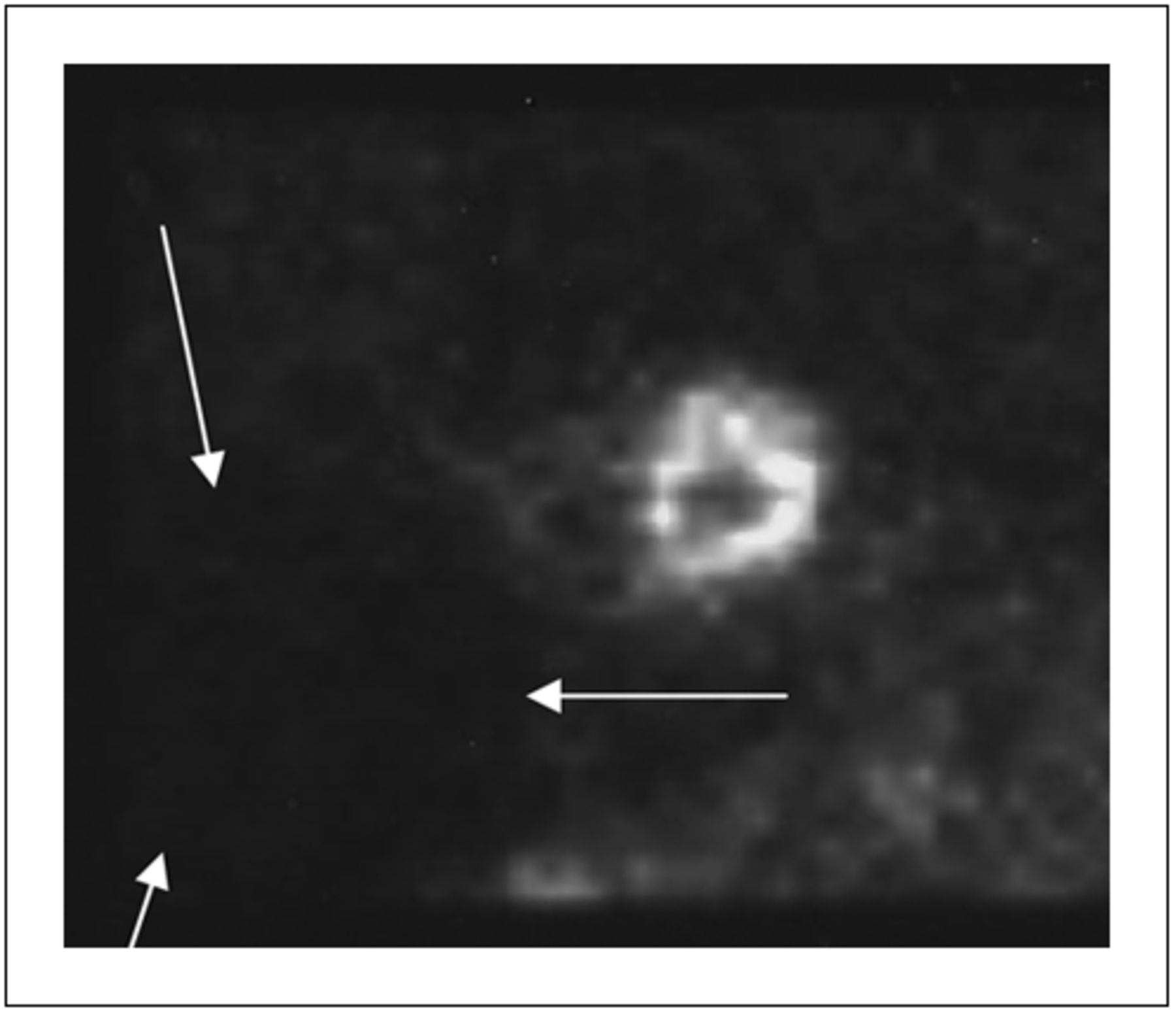
Anterior view of unprocessed projectional images showing large photopenic area within left and mid abdominal regions (arrows).

FIGURE 2.
  • Download figure
  • Open in new tab
  • Download powerpoint
FIGURE 2.

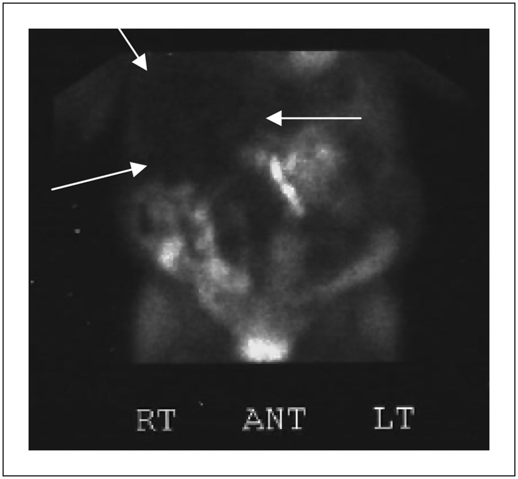
Anterior static image acquired at abdominal and pelvic regions demonstrating large photopenic area (arrows).

FIGURE 3.
  • Download figure
  • Open in new tab
  • Download powerpoint
FIGURE 3.

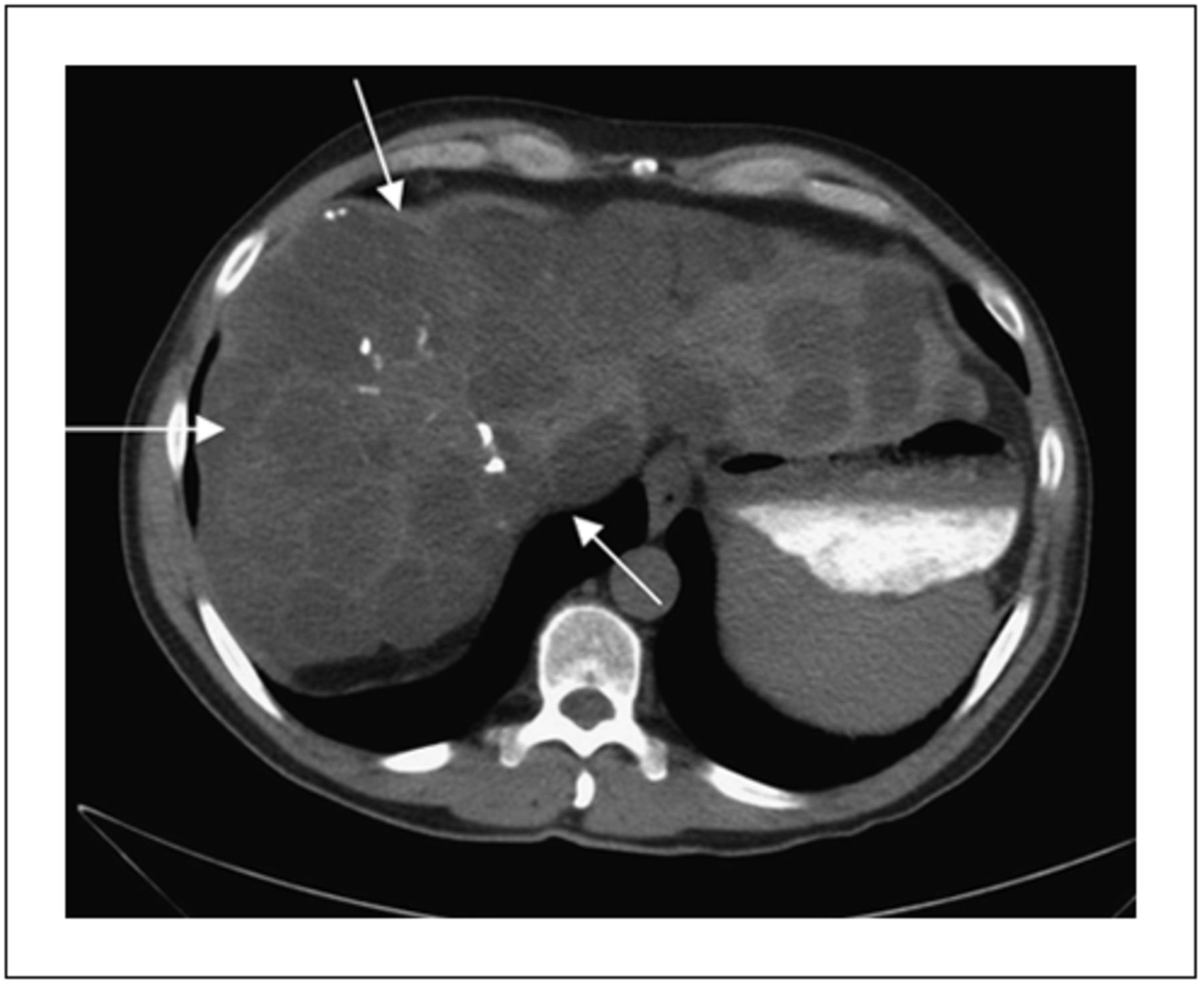
Anterior view of abdominal CT scan demonstrating large hepatic cysts (arrows).

DISCUSSION

Our patient was known to have autosomal dominant polycystic kidney disease, which commonly results in large and multiple renal and hepatic cysts (2). As in our case, these cysts can appear as abdominal photopenic areas on 99mTc-sestamibi imaging (3). Large abdominal photopenic areas are also seen with other types of renal cysts, including acquired cystic renal disease found in patients with end-stage renal disease receiving dialysis and with simple renal cysts (4,5). Other types of hepatic cysts may also result in large abdominal photopenic areas on myocardial perfusion imaging, including simple hepatic cysts and echinococcal cysts (6). Cholecystitis has also been reported to result in an abdominal photopenic area on myocardial perfusion scans (7). When further diagnostic imaging reveals a large solitary cyst, additional investigation may be required to exclude abscess, hematoma, or malignancy (6).

CONCLUSION

This case demonstrates the importance of careful analysis of the rotating unprocessed projectional images in the interpretation of myocardial perfusion imaging. Although the diagnosis of polycystic kidney disease was previously established in our patient, the incidental finding of an extracardiac photopenic area may constitute the first indication of a clinically significant abdominal abnormality.

Acknowledgments

No potential conflict of interest relevant to this article was reported.

Footnotes

  • Published online Sep. 20, 2012.

REFERENCES

  1. 1.↵
    1. Jones SW,
    2. Aziz K,
    3. Yasuda T,
    4. Gewirtz H,
    5. Scott J
    . Importance of systematic review of rotating projection images from Tc99m-sestamibi cardiac perfusion imaging for noncardiac findings. Nucl Med Commun. 2008;29:607–613.
    OpenUrlCrossRefPubMed
  2. 2.↵
    1. Nahm AM,
    2. Henriquez DE,
    3. Ritz E
    . Renal cystic disease (ADPKD and ARPKD). Nephrol Dial Transplant. 2002;17:311–314.
    OpenUrlFREE Full Text
  3. 3.↵
    1. Chang CY,
    2. Lin KH,
    3. Chu YK,
    4. Chang CP,
    5. Liu RS,
    6. Wang SJ
    . Incidental finding of autosomal dominant polycystic kidney disease with liver involvement on Tc-99m sestamibi scintigraphy. Clin Nucl Med. 2011;36:e69–e70.
    OpenUrlCrossRefPubMed
  4. 4.↵
    1. Gowda A,
    2. Chhabra A,
    3. Gavriluke A,
    4. Peddinghaus L,
    5. Jain D
    . Unusual photopenic area in the abdomen on myocardial perfusion imaging. J Nucl Cardiol. 2006;13:e6–e8.
    OpenUrlCrossRefPubMed
  5. 5.↵
    1. Raza M,
    2. Meesala M,
    3. Panjrath G,
    4. Ghanbarinia A,
    5. Jain D
    . Abnormal photopenic area on nuclear perfusion imaging. J Nucl Cardiol. 2005;12:607–609.
    OpenUrlCrossRefPubMed
  6. 6.↵
    1. Chatziioannou SN,
    2. Alfaro-Franco C,
    3. Moore WH,
    4. Alanis-Williams L,
    5. Dhekne RD,
    6. Ford PV
    . The significance of incidental noncardiac findings in TC-99m sestamibi myocardial perfusion imaging: illustrated by a case. Tex Heart Inst J. 1999;26:229–231.
    OpenUrlPubMed
  7. 7.↵
    1. Lamont AE,
    2. Joyce JM,
    3. Grossman SJ
    . Acute cholecystitis detected on a TC-99m sestamibi myocardial imaging. Clin Nucl Med. 1996;21:879.
    OpenUrlCrossRefPubMed
  • Received for publication December 18, 2011.
  • Accepted for publication April 2, 2012.
PreviousNext
Back to top

In this issue

Journal of Nuclear Medicine Technology: 40 (4)
Journal of Nuclear Medicine Technology
Vol. 40, Issue 4
December 1, 2012
  • Table of Contents
  • About the Cover
  • Index by author
Print
Download PDF
Article Alerts
Sign In to Email Alerts with your Email Address
Email Article

Thank you for your interest in spreading the word on Journal of Nuclear Medicine Technology.

NOTE: We only request your email address so that the person you are recommending the page to knows that you wanted them to see it, and that it is not junk mail. We do not capture any email address.

Enter multiple addresses on separate lines or separate them with commas.
Large Abdominal Photopenic Area on 99mTc-Sestamibi Myocardial Perfusion Imaging
(Your Name) has sent you a message from Journal of Nuclear Medicine Technology
(Your Name) thought you would like to see the Journal of Nuclear Medicine Technology web site.
Citation Tools
Large Abdominal Photopenic Area on 99mTc-Sestamibi Myocardial Perfusion Imaging
Jennifer Lyon, John Spaulding, Paul M. Zack
Journal of Nuclear Medicine Technology Dec 2012, 40 (4) 281-282; DOI: 10.2967/jnmt.111.102079

Citation Manager Formats

  • BibTeX
  • Bookends
  • EasyBib
  • EndNote (tagged)
  • EndNote 8 (xml)
  • Medlars
  • Mendeley
  • Papers
  • RefWorks Tagged
  • Ref Manager
  • RIS
  • Zotero
Share
Large Abdominal Photopenic Area on 99mTc-Sestamibi Myocardial Perfusion Imaging
Jennifer Lyon, John Spaulding, Paul M. Zack
Journal of Nuclear Medicine Technology Dec 2012, 40 (4) 281-282; DOI: 10.2967/jnmt.111.102079
Twitter logo Facebook logo LinkedIn logo Mendeley logo
  • Tweet Widget
  • Facebook Like
  • Google Plus One
Bookmark this article

Jump to section

  • Article
    • Abstract
    • CASE REPORT
    • DISCUSSION
    • CONCLUSION
    • Acknowledgments
    • Footnotes
    • REFERENCES
  • Figures & Data
  • Info & Metrics
  • PDF

Related Articles

  • No related articles found.
  • PubMed
  • Google Scholar

Cited By...

  • Incidental Findings on Myocardial Perfusion SPECT Images
  • Google Scholar

More in this TOC Section

  • Post-90Y Radioembolization PET/CT Scan with Respiratory Gating Using Time-of-Flight Reconstruction
  • Primary Hyperparathyroidism–Related Brown Tumors Mimicking Other Giant Cell–Containing Skeletal Tumors: Role of Correlative Imaging in Diagnosis
  • False-Positive Scalp Activity in 131I Imaging Associated with Hair Coloring
Show more Case Reports

Similar Articles

Keywords

  • sestamibi
  • photopenic
  • polycystic kidney disease
  • renal cyst
  • hepatic cyst
SNMMI

© 2025 SNMMI

Powered by HighWire