
Main menu

  • Home
  • Content
    • Current
      • JNMT Supplement
    • Ahead of print
    • Past Issues
    • Continuing Education
    • JNMT Podcast
    • SNMMI Annual Meeting Abstracts
  • Subscriptions
    • Subscribers
    • Rates
    • Journal Claims
    • Institutional and Non-member
  • Authors
    • Submit to JNMT
    • Information for Authors
    • Assignment of Copyright
    • AQARA Requirements
  • Info
    • Reviewers
    • Permissions
    • Advertisers
    • Corporate & Special Sales
  • About
    • About Us
    • Editorial Board
    • Contact Information
  • More
    • Alerts
    • Feedback
    • Help
    • SNMMI Journals
  • SNMMI
    • JNMT
    • JNM
    • SNMMI Journals
    • SNMMI

User menu

  • Subscribe
  • My alerts
  • Log in
  • My Cart

Search

  • Advanced search
Journal of Nuclear Medicine Technology
  • SNMMI
    • JNMT
    • JNM
    • SNMMI Journals
    • SNMMI
  • Subscribe
  • My alerts
  • Log in
  • My Cart
Journal of Nuclear Medicine Technology

Advanced Search

  • Home
  • Content
    • Current
    • Ahead of print
    • Past Issues
    • Continuing Education
    • JNMT Podcast
    • SNMMI Annual Meeting Abstracts
  • Subscriptions
    • Subscribers
    • Rates
    • Journal Claims
    • Institutional and Non-member
  • Authors
    • Submit to JNMT
    • Information for Authors
    • Assignment of Copyright
    • AQARA Requirements
  • Info
    • Reviewers
    • Permissions
    • Advertisers
    • Corporate & Special Sales
  • About
    • About Us
    • Editorial Board
    • Contact Information
  • More
    • Alerts
    • Feedback
    • Help
    • SNMMI Journals
  • Watch or Listen to JNMT Podcast
  • Visit SNMMI on Facebook
  • Join SNMMI on LinkedIn
  • Follow SNMMI on Twitter
  • Subscribe to JNMT RSS feeds
Research ArticleIMAGING

Development of a Relational Database to Capture and Merge Clinical History with the Quantitative Results of Radionuclide Renography

Russell D. Folks, Bital Savir-Baruch, Ernest V. Garcia, Liudmila Verdes and Andrew T. Taylor
Journal of Nuclear Medicine Technology December 2012, 40 (4) 236-243; DOI: https://doi.org/10.2967/jnmt.111.101477
Russell D. Folks
Department of Radiology and Imaging Sciences, Emory University School of Medicine, Atlanta, Georgia
  • Find this author on Google Scholar
  • Find this author on PubMed
  • Search for this author on this site
Bital Savir-Baruch
Department of Radiology and Imaging Sciences, Emory University School of Medicine, Atlanta, Georgia
  • Find this author on Google Scholar
  • Find this author on PubMed
  • Search for this author on this site
Ernest V. Garcia
Department of Radiology and Imaging Sciences, Emory University School of Medicine, Atlanta, Georgia
  • Find this author on Google Scholar
  • Find this author on PubMed
  • Search for this author on this site
Liudmila Verdes
Department of Radiology and Imaging Sciences, Emory University School of Medicine, Atlanta, Georgia
  • Find this author on Google Scholar
  • Find this author on PubMed
  • Search for this author on this site
Andrew T. Taylor
Department of Radiology and Imaging Sciences, Emory University School of Medicine, Atlanta, Georgia
  • Find this author on Google Scholar
  • Find this author on PubMed
  • Search for this author on this site
  • Article
  • Figures & Data
  • Info & Metrics
  • PDF
Loading

Article Figures & Data

Figures

  • Tables
  • FIGURE 1.
    • Download figure
    • Open in new tab
    • Download powerpoint
    FIGURE 1.

    Organization of CLINICAL. History table (A) includes fields summarizing patient’s history of conditions affecting urinary system and other systems, interventional procedures that have been performed, and left and right kidney and ureter findings. Imaging results table (B) contains same set of left and right kidney and ureter fields, derived from imaging study reports rather than narrative history. Interventions and other history are not included on this table. Demographics are present on both tables so that patient records can be matched to their imaging studies.
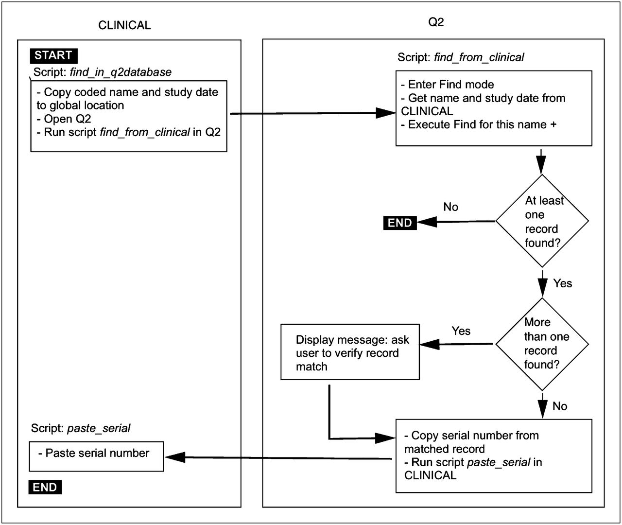
  • FIGURE 2.
    • Download figure
    • Open in new tab
    • Download powerpoint
    FIGURE 2.

    Algorithm for matching patient records across database tables.

  • FIGURE 3.
    • Download figure
    • Open in new tab
    • Download powerpoint
    FIGURE 3.

    Example of how small subset of fields in CLINICAL would be converted to structured text for physician review. (A) Fields as they appear on graphic layout with which user interacts. (B) Database’s internal manipulation of same fields by text functions, performed in calculated field not seen by user. (C) Text file extracted by database script. Fields with value “no data” are not included, greatly simplifying narrative.
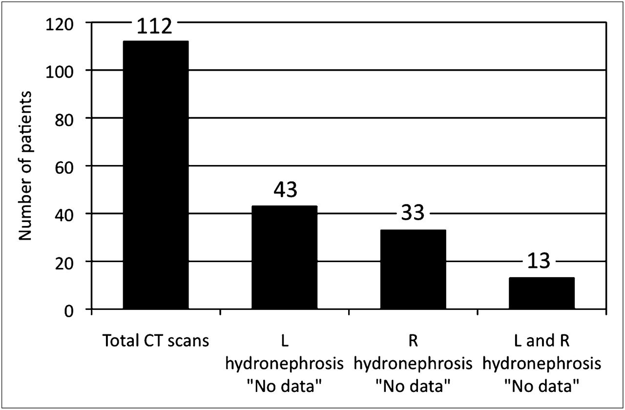
  • FIGURE 4.
    • Download figure
    • Open in new tab
    • Download powerpoint
    FIGURE 4.

    Reporting of hydronephrosis for 112 CT scans performed within 1 y of MAG3. Hydronephrosis was not reported as either present or absent for 38% (43/112) of left kidneys, 29% (33/112) of right kidneys, and 12% (13/112) of both left and right kidneys.

Tables

  • Figures
    • View popup
    TABLE 1

    Profile of Databases

    CLINICAL (2 tables)
    ProfileRENAL (1 table)Q2 (1 table)TotalHistoryImaging
    Fields1428835527580
    Layouts32220155
    Scripts41149**
    Value lists0527**
    • ↵* Same scripts and value lists are used for history and imaging tables.

    • View popup
    TABLE 2

    Database Fields for Clinical History Table

    FieldType of dataValue list contents
    HistoryValue listNormal, absent, no comment
    UrinomaValue listNo data, not present, equivocal, present
    Renal parenchymaValue listNo data, normal, equivocal, atrophied
    Renal scarValue listNo data, not present, equivocal, present
    HydronephrosisValue listNo data, not present, equivocal, present, mild, moderate, severe
    HydroureterValue listNo data, not present, equivocal, present, mild, moderate, severe
    StrictureValue listNo data, not present, equivocal, present
    Renal calculusValue listNo data, not present, equivocal, present
     Largest size (mm)Literal value
    Ureteropelvic junction calculusValue listNo data, not present, equivocal, present
     Largest size (mm)Literal value
    Ureteral calculusValue listNo data, not present, equivocal, present
     Largest size (mm)Literal value
    Calculus, obstructiveValue listNo data, not obstructive, obstructive
    Solid renal massValue listNo data, not present, equivocal, present
     Largest size (cm)Literal value
    Cystic renal massValue listNo data, not present, equivocal, present
     Largest size (cm)Literal value
    Mixed renal massValue listNo data, not present, equivocal, present
     Largest size (cm)Literal value
    Mass, obstructiveValue listNo data, not obstructive, obstructive
    Renal artery stenosisValue listNo data, not present, equivocal, mild, moderate, severe
    SurgeryCheckbox options (1 or more)Prior stent, current stent, prior nephrostomy, current nephrostomy, ureteral reimplantation, pyeloplasty, total nephrectomy, partial nephrectomy, nephrolithotomy
    Stent removal dateLiteral date
    Nephrostomy removal dateLiteral date
    Flank pain on arrivalValue listNo data, not present, equivocal, present
    Flank pain after diureticValue listNo data, not present, equivocal, present
    UreteroceleValue listNo data, not present, equivocal, present
    Duplicated urinary systemValue listNo data, not present, equivocal, present
    • View popup
    TABLE 3

    Joint Judgment of 2 Readers Regarding Presence of Current Ureteral Stent Based on Narrative Summaries of Hospitalizations and Patient Visits

    Reader 2
    Reader 1Stent presentStent absentTotal
    Stent present303
    Stent absent59297
    Total892100
    • View popup
    TABLE 4

    Joint Judgment of 2 Readers Regarding Presence of Obstruction Based on Narrative Summaries of Hospitalizations and Patient Visits

    Reader 2
    Reader 1Hydronephrosis presentHydronephrosis absentTotal
    Hydronephrosis present171936
    Hydronephrosis absent65864
    Total2377100
    • View popup
    TABLE 5

    Joint Judgment of 2 Readers Regarding Presence of Obstruction Based on Imaging Reports

    Reader 2
    Reader 1Hydronephrosis presentHydronephrosis absentTotal
    Hydronephrosis present41849
    Hydronephrosis absent04949
    Total415798
PreviousNext
Back to top

In this issue

Journal of Nuclear Medicine Technology: 40 (4)
Journal of Nuclear Medicine Technology
Vol. 40, Issue 4
December 1, 2012
  • Table of Contents
  • About the Cover
  • Index by author
Print
Download PDF
Article Alerts
Sign In to Email Alerts with your Email Address
Email Article

Thank you for your interest in spreading the word on Journal of Nuclear Medicine Technology.

NOTE: We only request your email address so that the person you are recommending the page to knows that you wanted them to see it, and that it is not junk mail. We do not capture any email address.

Enter multiple addresses on separate lines or separate them with commas.
Development of a Relational Database to Capture and Merge Clinical History with the Quantitative Results of Radionuclide Renography
(Your Name) has sent you a message from Journal of Nuclear Medicine Technology
(Your Name) thought you would like to see the Journal of Nuclear Medicine Technology web site.
Citation Tools
Development of a Relational Database to Capture and Merge Clinical History with the Quantitative Results of Radionuclide Renography
Russell D. Folks, Bital Savir-Baruch, Ernest V. Garcia, Liudmila Verdes, Andrew T. Taylor
Journal of Nuclear Medicine Technology Dec 2012, 40 (4) 236-243; DOI: 10.2967/jnmt.111.101477

Citation Manager Formats

  • BibTeX
  • Bookends
  • EasyBib
  • EndNote (tagged)
  • EndNote 8 (xml)
  • Medlars
  • Mendeley
  • Papers
  • RefWorks Tagged
  • Ref Manager
  • RIS
  • Zotero
Share
Development of a Relational Database to Capture and Merge Clinical History with the Quantitative Results of Radionuclide Renography
Russell D. Folks, Bital Savir-Baruch, Ernest V. Garcia, Liudmila Verdes, Andrew T. Taylor
Journal of Nuclear Medicine Technology Dec 2012, 40 (4) 236-243; DOI: 10.2967/jnmt.111.101477
Twitter logo Facebook logo LinkedIn logo Mendeley logo
  • Tweet Widget
  • Facebook Like
  • Google Plus One
Bookmark this article

Jump to section

  • Article
    • Abstract
    • MATERIALS AND METHODS
    • RESULTS
    • DISCUSSION
    • CONCLUSION
    • Acknowledgments
    • Footnotes
    • REFERENCES
  • Figures & Data
  • Info & Metrics
  • PDF

Related Articles

  • No related articles found.
  • PubMed
  • Google Scholar

Cited By...

  • No citing articles found.
  • Google Scholar

More in this TOC Section

  • Early 10-Minute Postinjection [18F]F-FAPI-42 uEXPLORER Total-Body PET/CT Scanning Protocol for Staging Lung Cancer Using HYPER Iterative Reconstruction
  • Single- Versus Dual-Time-Point Imaging for Transthyretin Cardiac Amyloid Using 99mTc-Pyrophosphate
  • Does Arthrography Improve Accuracy of SPECT/CT for Diagnosis of Aseptic Loosening in Patients with Painful Knee Arthroplasty: A Systematic Review and Metaanalysis
Show more Imaging

Similar Articles

Keywords

  • MAG3 renography
  • databases
  • decision support systems
SNMMI

© 2025 SNMMI

Powered by HighWire