
Main menu

  • Home
  • Content
    • Current
      • JNMT Supplement
    • Ahead of print
    • Past Issues
    • Continuing Education
    • JNMT Podcast
    • SNMMI Annual Meeting Abstracts
  • Subscriptions
    • Subscribers
    • Rates
    • Journal Claims
    • Institutional and Non-member
  • Authors
    • Submit to JNMT
    • Information for Authors
    • Assignment of Copyright
    • AQARA Requirements
  • Info
    • Reviewers
    • Permissions
    • Advertisers
    • Corporate & Special Sales
  • About
    • About Us
    • Editorial Board
    • Contact Information
  • More
    • Alerts
    • Feedback
    • Help
    • SNMMI Journals
  • SNMMI
    • JNMT
    • JNM
    • SNMMI Journals
    • SNMMI

User menu

  • Subscribe
  • My alerts
  • Log in
  • My Cart

Search

  • Advanced search
Journal of Nuclear Medicine Technology
  • SNMMI
    • JNMT
    • JNM
    • SNMMI Journals
    • SNMMI
  • Subscribe
  • My alerts
  • Log in
  • My Cart
Journal of Nuclear Medicine Technology

Advanced Search

  • Home
  • Content
    • Current
    • Ahead of print
    • Past Issues
    • Continuing Education
    • JNMT Podcast
    • SNMMI Annual Meeting Abstracts
  • Subscriptions
    • Subscribers
    • Rates
    • Journal Claims
    • Institutional and Non-member
  • Authors
    • Submit to JNMT
    • Information for Authors
    • Assignment of Copyright
    • AQARA Requirements
  • Info
    • Reviewers
    • Permissions
    • Advertisers
    • Corporate & Special Sales
  • About
    • About Us
    • Editorial Board
    • Contact Information
  • More
    • Alerts
    • Feedback
    • Help
    • SNMMI Journals
  • Watch or Listen to JNMT Podcast
  • Visit SNMMI on Facebook
  • Join SNMMI on LinkedIn
  • Follow SNMMI on Twitter
  • Subscribe to JNMT RSS feeds
Research ArticleIMAGING

Sarcoidlike Reaction of Neoplasia Causing Hypermetabolic Thoracic Adenopathy in Setting of Extrathoracic Malignancy: Report of Two Cases and a Review of the Differential Diagnostic Considerations

Jonathan B. Craun, Kevin P. Banks, Michael N. Clemenshaw and Ronald W. Moren
Journal of Nuclear Medicine Technology December 2012, 40 (4) 231-235; DOI: https://doi.org/10.2967/jnmt.112.102814
Jonathan B. Craun
1Department of Radiology, Brooke Army Medical Center, Fort Sam Houston, Houston, Texas
  • Find this author on Google Scholar
  • Find this author on PubMed
  • Search for this author on this site
Kevin P. Banks
1Department of Radiology, Brooke Army Medical Center, Fort Sam Houston, Houston, Texas
2Department of Radiology, University of Texas Health Science Center at San Antonio, San Antonio, Texas; and
3Rays, Littleton, Colorado
  • Find this author on Google Scholar
  • Find this author on PubMed
  • Search for this author on this site
Michael N. Clemenshaw
1Department of Radiology, Brooke Army Medical Center, Fort Sam Houston, Houston, Texas
2Department of Radiology, University of Texas Health Science Center at San Antonio, San Antonio, Texas; and
  • Find this author on Google Scholar
  • Find this author on PubMed
  • Search for this author on this site
Ronald W. Moren
1Department of Radiology, Brooke Army Medical Center, Fort Sam Houston, Houston, Texas
  • Find this author on Google Scholar
  • Find this author on PubMed
  • Search for this author on this site
  • Article
  • Figures & Data
  • Info & Metrics
  • PDF
Loading

Article Figures & Data

Figures

  • Tables
  • FIGURE 1.
    • Download figure
    • Open in new tab
    • Download powerpoint
    FIGURE 1.

    18F-FDG PET/CT for initial staging of patient A’s laryngeal cancer. (A) Maximum-intensity projection demonstrates single focus of hypermetabolic activity at location of laryngeal cancer with no evidence of locoregional spread of distal metastases. (B) CT shows abnormal soft-tissue mass (arrow) along anterior aspect of left vocal cord. (C and D) Axial attenuation-corrected PET and PET/CT, respectively, demonstrate that lesion is hypermetabolic, consistent with known diagnosis of squamous cell carcinoma.
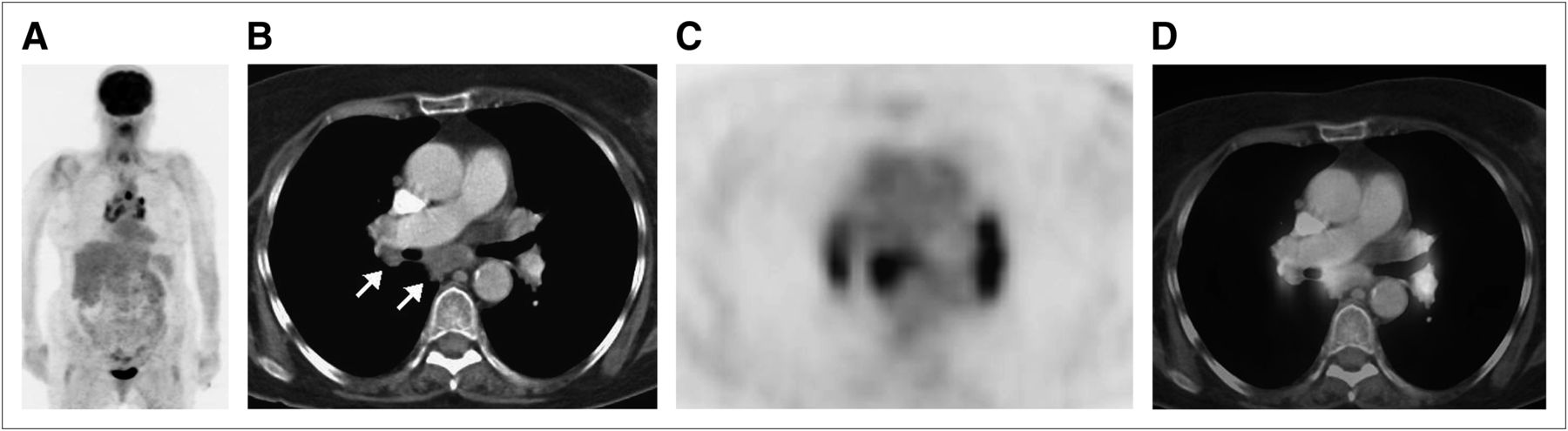
  • FIGURE 2.
    • Download figure
    • Open in new tab
    • Download powerpoint
    FIGURE 2.

    Follow-up 18F-FDG PET/CT for routine surveillance of patient A’s laryngeal cancer. Clinically, patient had no evidence of disease. (A) Maximum-intensity projection demonstrates bilateral abnormal 18F-FDG avidity in hilar regions and mediastinum. (B–D) Axial CT, PET, and PET/CT through hila and mediastinum show these foci to correspond to hypermetabolic lymph nodes (arrows).
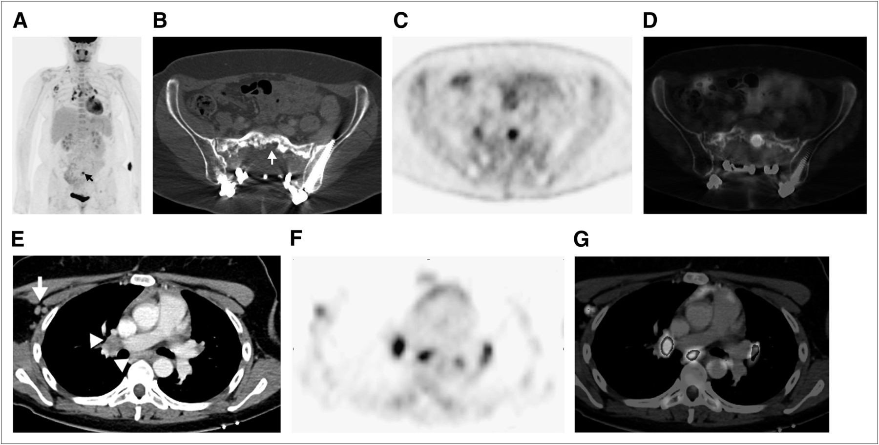
  • FIGURE 3.
    • Download figure
    • Open in new tab
    • Download powerpoint
    FIGURE 3.

    (A) Maximum-intensity projection from follow-up 18F-FDG PET/CT done for routine surveillance of patient B demonstrates abnormal 18F-FDG avidity in sacrum at site of prior disease (arrow). (B, C, and D) Axial CT, PET, and PET/CT, respectively, show that sacral activity corresponds to new hypermetabolic osteolysis consistent with local recurrence. The combination was believed to represent local recurrent disease. Additional findings on maximum-intensity projection included new, bilateral hypermetabolic foci in hilar regions and mediastinum, as well as in right axilla and right neck base. (E, F, and G) Axial CT, PET, and PET/CT, respectively, show these foci to represent hypermetabolic hilar and mediastinal (arrowheads) and right axillary (arrow) lymph nodes. Foci of uptake in right neck base were also shown to be hypermetabolic lymph nodes.

Tables

  • Figures
    • View popup
    TABLE 1

    Features of Hypermetabolic Hilar and Mediastinal Lymph Nodes on 18F-FDG PET

    EntityClinical featuresCT features
    Sarcoidlike reactionHistory of prior malignancy; entity may occur at time of initial cancer diagnosis to years later, often with no clinical evidence of diseaseNo pulmonary changes; patient classically shows bilateral mild to moderately enlarged hilar and mediastinal lymph nodes; enlarged or hypermetabolic lymph nodes may be present elsewhere
    SarcoidBias toward young and middle-aged African–American women; clinical complaints often include erythema nodosum, uveitis, and acute polyarthritis, with nonspecific respiratory symptoms, fatigue, weight loss, and feverPulmonary findings may often be absent though when present show nodular pattern along lymphovascular bundles; patient classically has bilateral lobulated, mild to moderately enlarged hilar and mediastinal lymph nodes; lymph nodes may have peripheral thin egg-shell calcifications
    PneumoconiosisHistory of occupational exposure such as surface mining; patient may be asymptomatic early on and then develop shortness of breath and respiratory symptoms along with pulmonary changesDiffuse reticular or nodular/reticulonodular pattern of pulmonary disease; lung findings can coalesce into massive pulmonary fibrosis; lymph nodes often have egg-shell calcifications
    InfectionNo age or sex predilection; signs and symptoms of respiratory infection are present (i.e., fever, productive cough, leukocytosis, and elevated C-reactive protein)Pulmonary features of infection (bronchopulmonary nodules, infiltrate, or consolidation); lymph nodes ipsilateral to pulmonary abnormality are typically at upper limit of normal size to mildly enlarged
    Radiation-induced inflammationHistory of prior malignancy with treatment by radiotherapy; patient may be asymptomatic or have dyspnea or nonproductive coughSharply marginated, localized fibrotic area of lung; lymph nodes ipsilateral to fibrosis are normal to mildly enlarged
    LymphomaAge predilection based on type of lymphoma; patient may present with weight loss, night sweats, fever, decreased appetiteRarely has pulmonary findings; marked nodal enlargement is often bilateral though may be asymmetric; conglomerate nodal masses may be present
    Bronchogenic carcinomaOlder individuals, often with history of smoking or pulmonary fibrosis, present with weight loss, decreased appetite, hemoptysisLung mass or nodule; nodal enlargement is typically ipsilateral to lesion
    MetastasesHistory of malignancy, possibly with increasing tumor markers suggestive of disease recurrence; is most commonly seen in tumors with predilection for mediastinal or hilar metastases (i.e., esophageal cancer or head and neck cancer)Lymph nodes often asymmetric or unilateral; evidence of metastases may be present in lungs, bones, or elsewhere
PreviousNext
Back to top

In this issue

Journal of Nuclear Medicine Technology: 40 (4)
Journal of Nuclear Medicine Technology
Vol. 40, Issue 4
December 1, 2012
  • Table of Contents
  • About the Cover
  • Index by author
Print
Download PDF
Article Alerts
Sign In to Email Alerts with your Email Address
Email Article

Thank you for your interest in spreading the word on Journal of Nuclear Medicine Technology.

NOTE: We only request your email address so that the person you are recommending the page to knows that you wanted them to see it, and that it is not junk mail. We do not capture any email address.

Enter multiple addresses on separate lines or separate them with commas.
Sarcoidlike Reaction of Neoplasia Causing Hypermetabolic Thoracic Adenopathy in Setting of Extrathoracic Malignancy: Report of Two Cases and a Review of the Differential Diagnostic Considerations
(Your Name) has sent you a message from Journal of Nuclear Medicine Technology
(Your Name) thought you would like to see the Journal of Nuclear Medicine Technology web site.
Citation Tools
Sarcoidlike Reaction of Neoplasia Causing Hypermetabolic Thoracic Adenopathy in Setting of Extrathoracic Malignancy: Report of Two Cases and a Review of the Differential Diagnostic Considerations
Jonathan B. Craun, Kevin P. Banks, Michael N. Clemenshaw, Ronald W. Moren
Journal of Nuclear Medicine Technology Dec 2012, 40 (4) 231-235; DOI: 10.2967/jnmt.112.102814

Citation Manager Formats

  • BibTeX
  • Bookends
  • EasyBib
  • EndNote (tagged)
  • EndNote 8 (xml)
  • Medlars
  • Mendeley
  • Papers
  • RefWorks Tagged
  • Ref Manager
  • RIS
  • Zotero
Share
Sarcoidlike Reaction of Neoplasia Causing Hypermetabolic Thoracic Adenopathy in Setting of Extrathoracic Malignancy: Report of Two Cases and a Review of the Differential Diagnostic Considerations
Jonathan B. Craun, Kevin P. Banks, Michael N. Clemenshaw, Ronald W. Moren
Journal of Nuclear Medicine Technology Dec 2012, 40 (4) 231-235; DOI: 10.2967/jnmt.112.102814
Twitter logo Facebook logo LinkedIn logo Mendeley logo
  • Tweet Widget
  • Facebook Like
  • Google Plus One
Bookmark this article

Jump to section

  • Article
    • Abstract
    • CASE REPORT
    • DISCUSSION
    • CONCLUSION
    • Acknowledgments
    • Footnotes
    • REFERENCES
  • Figures & Data
  • Info & Metrics
  • PDF

Related Articles

  • No related articles found.
  • PubMed
  • Google Scholar

Cited By...

  • Resolution of multiorgan sarcoid-like reaction after treatment of renal cell carcinoma
  • Google Scholar

More in this TOC Section

  • Early 10-Minute Postinjection [18F]F-FAPI-42 uEXPLORER Total-Body PET/CT Scanning Protocol for Staging Lung Cancer Using HYPER Iterative Reconstruction
  • Single- Versus Dual-Time-Point Imaging for Transthyretin Cardiac Amyloid Using 99mTc-Pyrophosphate
  • Robustness of 18F-FDG PET Radiomic Features in Lung Cancer: Impact of Advanced Reconstruction Algorithm
Show more Imaging

Similar Articles

Keywords

  • sarcoidlike reaction
  • 18F-FDG PET/CT
  • hypermetabolic lymph nodes
  • lymph node metastases
SNMMI

© 2025 SNMMI

Powered by HighWire