


Article Figures & Data
Figures
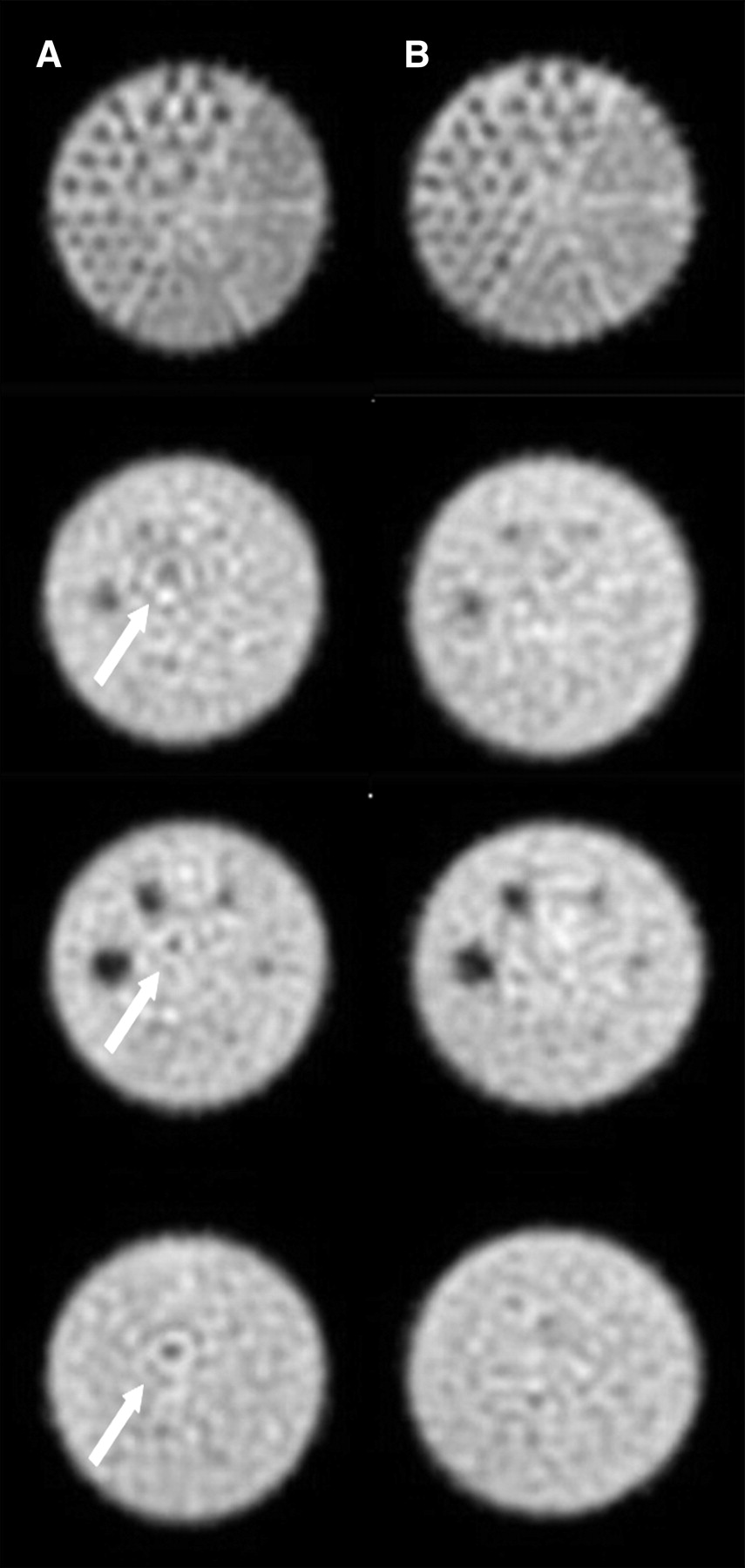
- FIGURE 1.
Phantom images from Vertex: intrinsic uniformity corrected images (A) and extrinsic uniformity corrected images (B). Arrows indicated visualized ring artifacts. A color version of this figure is available as a supplemental file at http://tech.snmjournals.org.
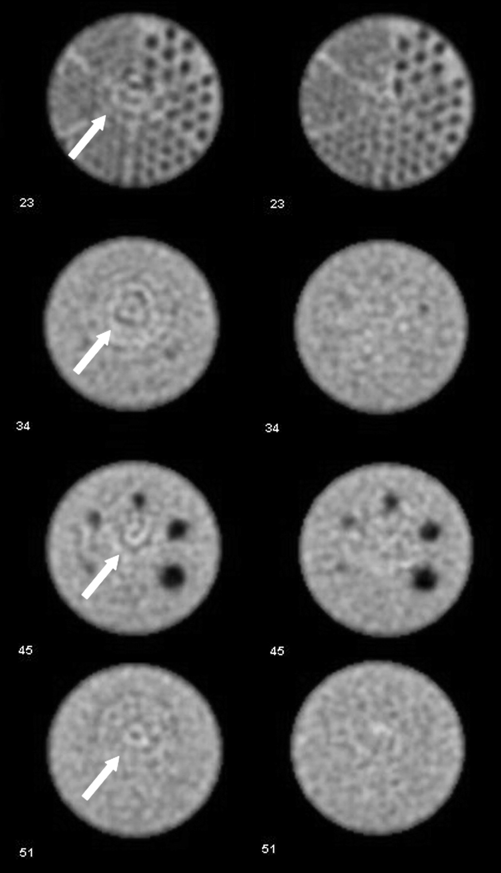
- FIGURE 2.
Phantom images from Infinia: intrinsic uniformity corrected images (A) and extrinsic uniformity corrected images (B). Arrows indicated visualized ring artifacts. A color version of this figure is available as a supplemental file at http://tech.snmjournals.org.
Tables
Parameter Extrinsic Intrinsic Source 57Co sheet source 99mTc point source Activity 740 MBq 37 MBq Counting rate 30,000 counts/s 45,000 counts/s Distance from detector Placed directly on collimator Placed at distance 4–5 times camera diameter Collimator Low energy, high resolution Not applicable Vertex Ultra Infinia Head no. Intrinsic Extrinsic Intrinsic Extrinsic One 1.74% 2.4% 1.1% 1.0% Two 1.61% 2.32% 1.1% 1.1% Parameter Vertex Ultra Infinia No. of stops (azimuths) 128 128 Time per stop (s) (azimuth) 30 30 Degrees of rotation Dual detector, 180°/head Dual detector, 180°/head Matrix 128 × 128 × 16 128 × 128 × 16 Mask size (cm) 38 — Zoom factor 1.46 1.5 Pixel size (mm) 3.17 2.95 Orbit type Noncircular (elliptic) Noncircular (elliptic)
Supplemental Data
Files in this Data Supplement:


Jump to section
Related Articles
Cited By...
- No citing articles found.