


Article Figures & Data
Figures
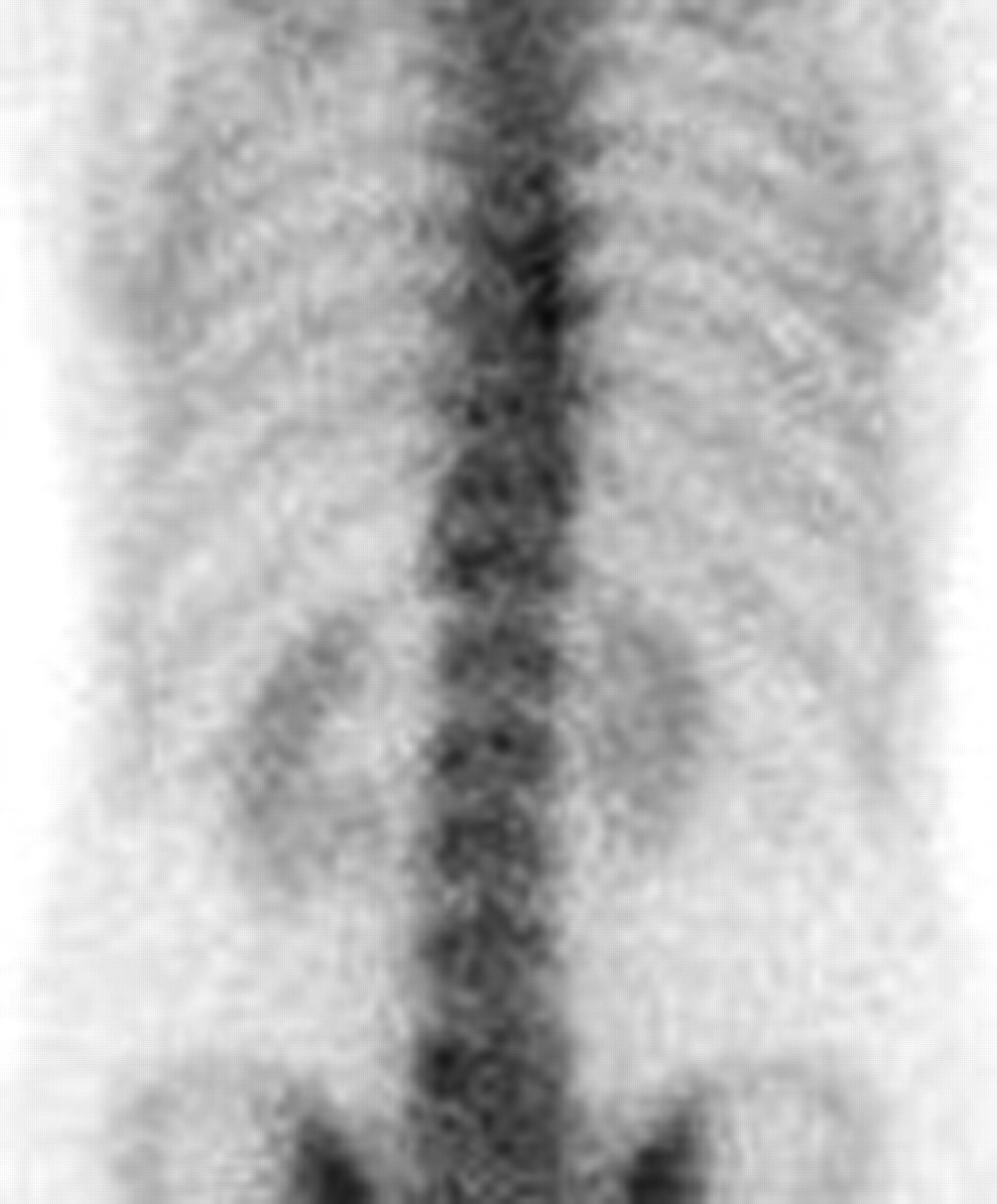
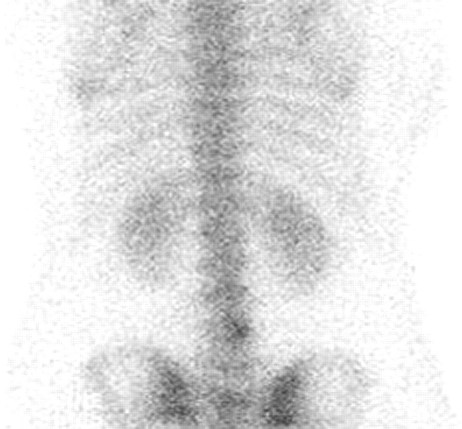
- FIGURE 1.
Baseline planar scan of spine.
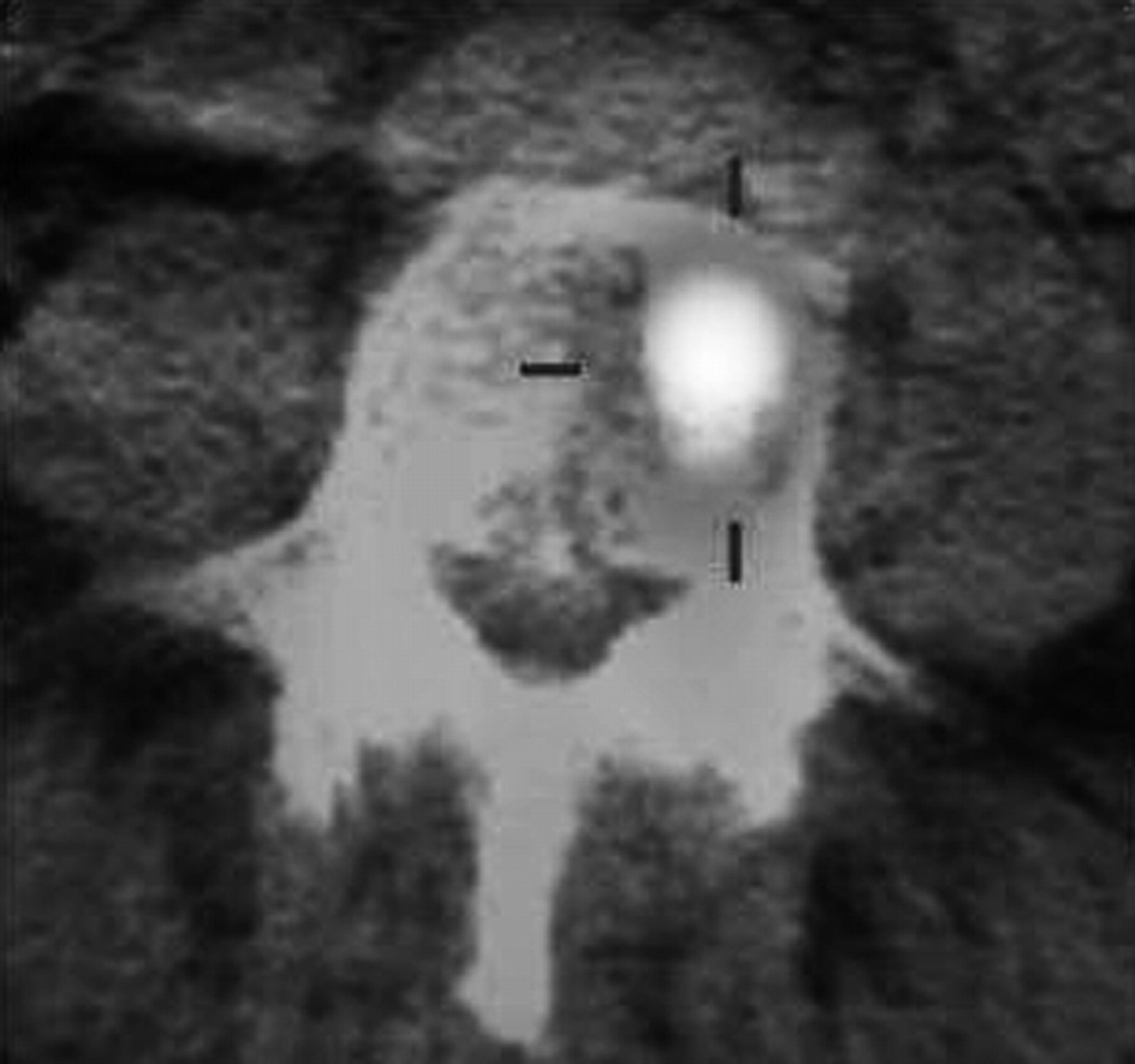
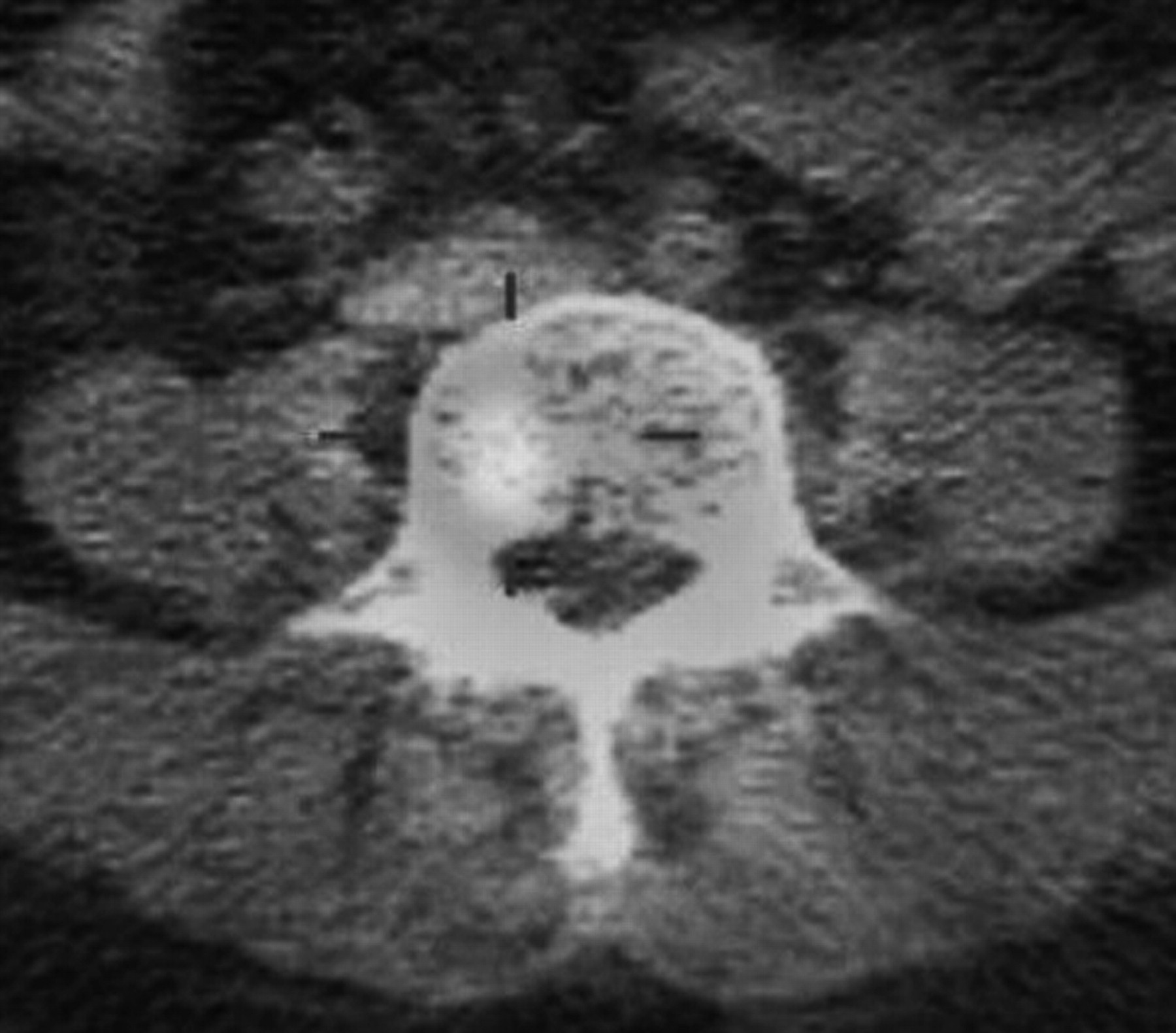
- FIGURE 2.
Baseline SPECT/CT scan. A color version of this figure is available as a supplemental file at http://tech.snmjournals.org.
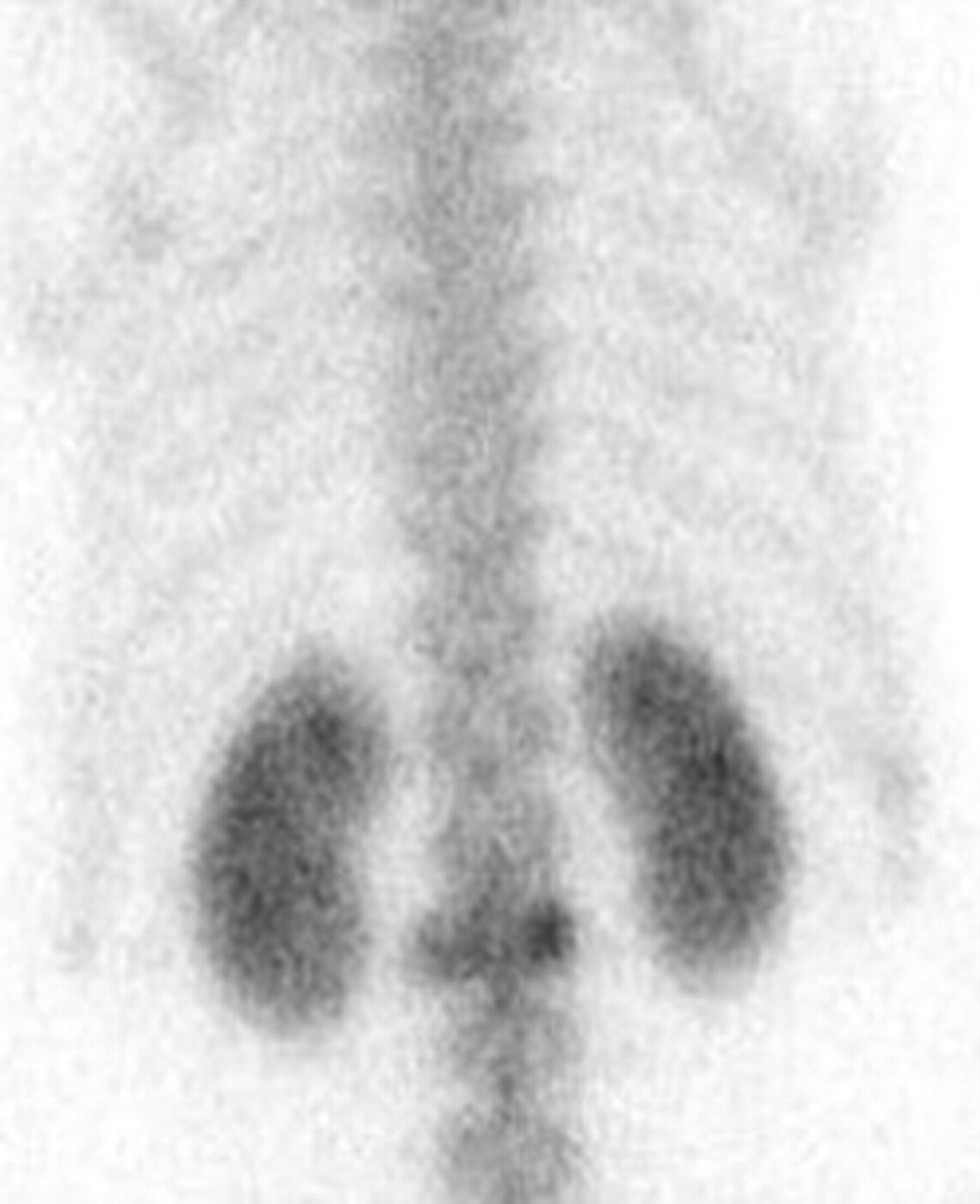
- FIGURE 3.
Bone scan showing L5 degenerative changes.
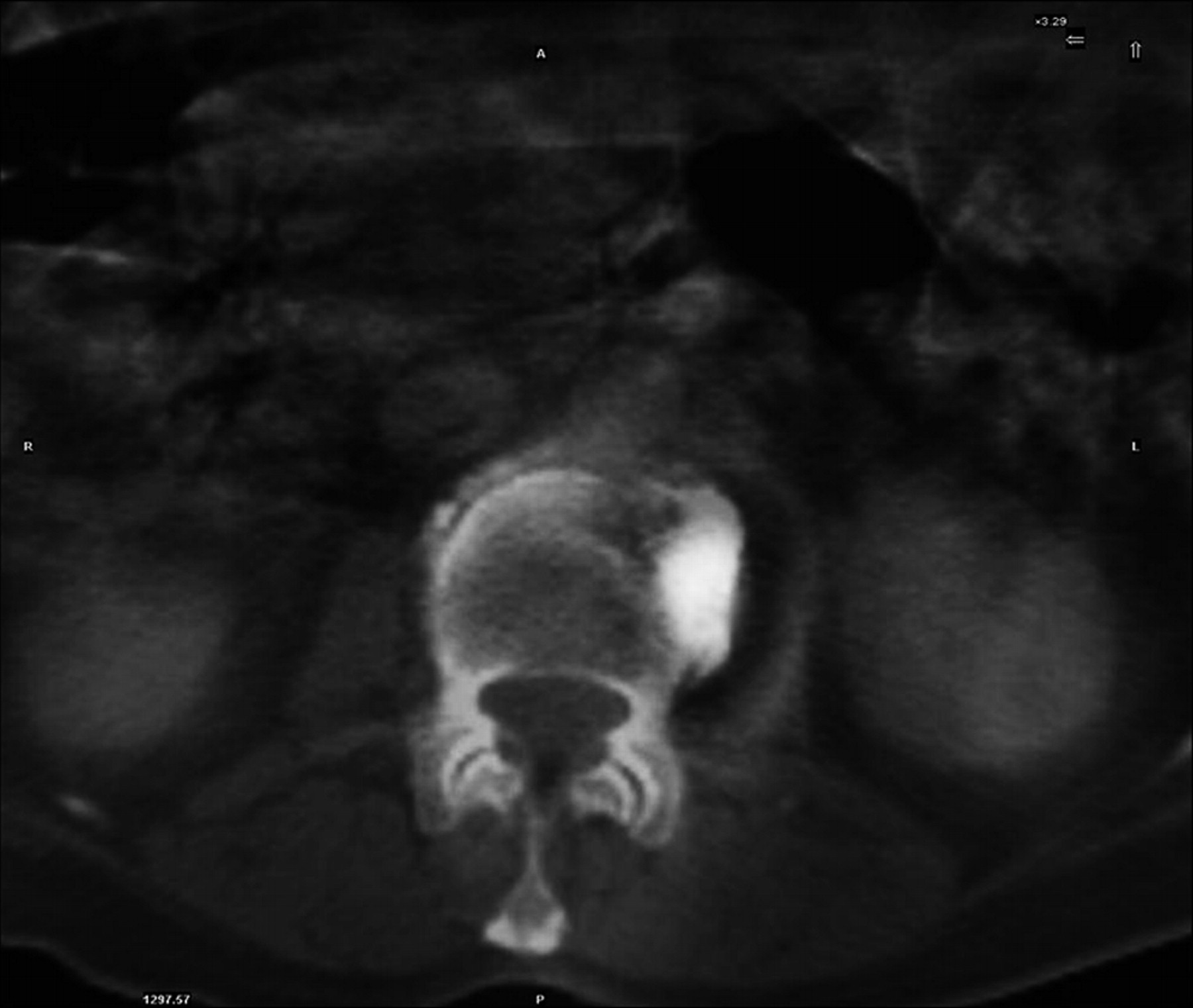
- FIGURE 4.
SPECT/CT scan showing early metastasis. A color version of this figure is available as a supplemental file at http://tech.snmjournals.org.
- FIGURE 5.
Bone scan suggesting posttraumatic or degenerative change.
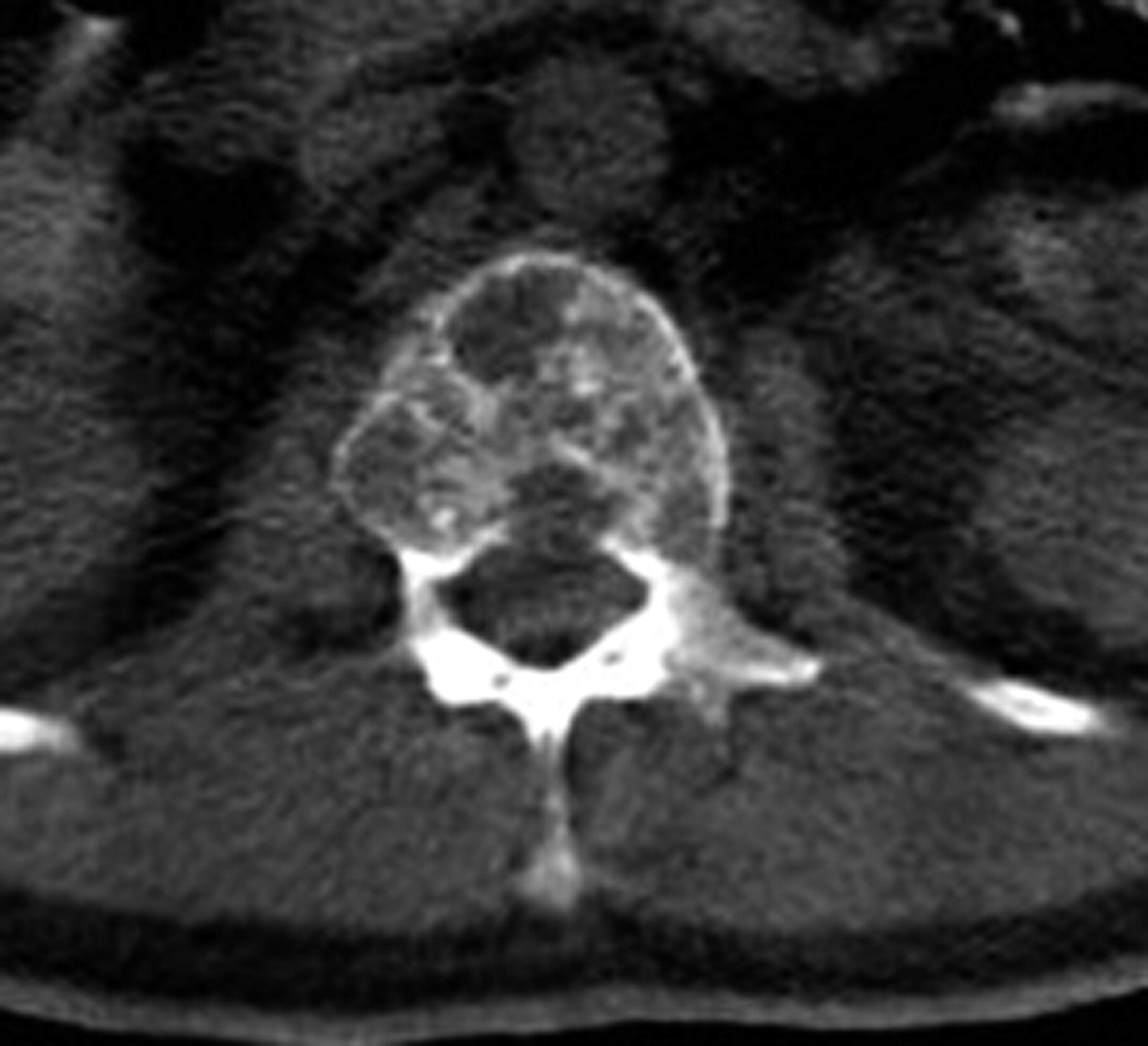
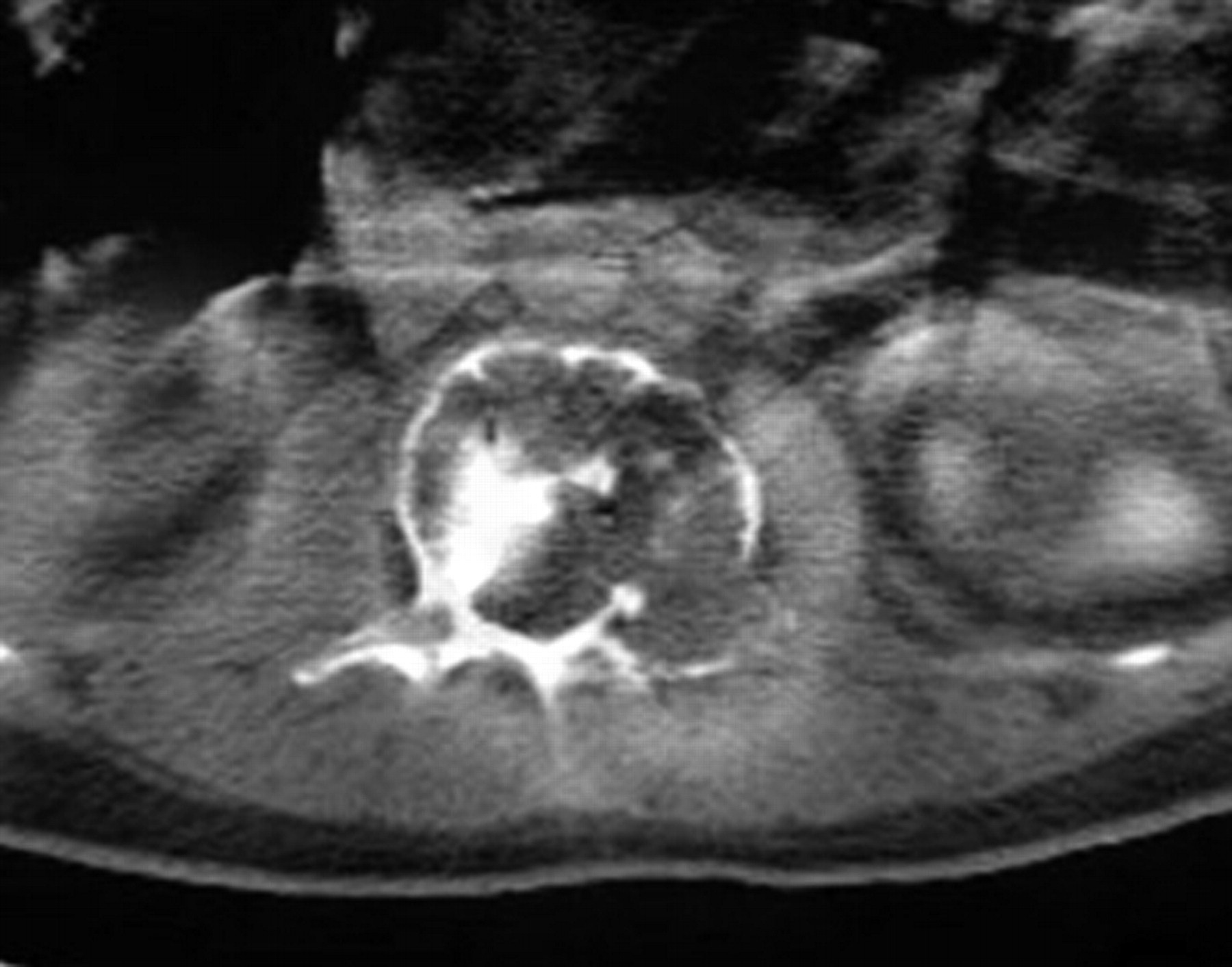
- FIGURE 6.
CT scan showing multiple lytic lesions.
- FIGURE 7.
SPECT/CT scan highlighting metastatic disease. A color version of this figure is available as a supplemental file at http://tech.snmjournals.org.
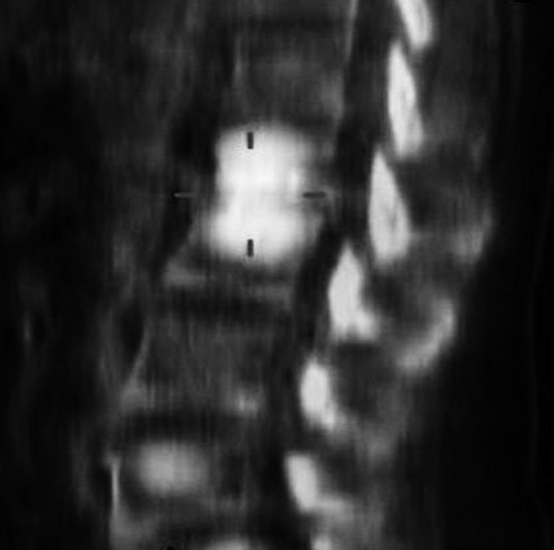
- FIGURE 8.
Bone scan showing L2 lesion.
- FIGURE 9.
SPECT/CT scan showing vertebral body fracture. A color version of this figure is available as a supplemental file at http://tech.snmjournals.org.
- FIGURE 10.
Planar bone scan.
- FIGURE 11.
SPECT/CT scan showing intravertebral disk infection. A color version of this figure is available as a supplemental file at http://tech.snmjournals.org.


Tables
Benign Malignant Facet joint Body and pedicle Costovertebral joint Photon-deficient area (lytic area) Involvement of 2 adjacent vertebrae or endplates of vertebrae Imaging type Indeterminate Benign Malignant Total Planar 51 (63.8%) 27 (33.8%) 2 (2.5%) 80 SPECT/CT 11 (13.8%) 39 (48.8%) 30 (37.5%) 80 - TABLE 3
Outcomes for Baseline and Follow-up Planar Imaging and SPECT/CT in Oncology Cohort
Imaging type Indeterminate Benign Malignant Total Baseline planar 26 (52%) 22 (44%) 2 (4%) 50 Follow-up planar 7 (14%) 20 (40%) 23 (46%) 50 Baseline 5 (10%) 17 (34%) 28 (56%) 50 SPECT/CT 5 (10%) 17 (34%) 28 (56%) 50 Follow-up SPECT/CT 0 (0%) 17 (34%) 33 (66%) 50 Index Baseline planar Baseline SPECT/CT Follow-up planar True-negative 17 15 17 False-negative 31 7 10 True-positive 2 26 23 False-positive 0 2 0 Sensitivity (true-positive rate) 6.1% 78.8% 69.7% Specificity (true-negative rate) 100% 88.2% 100% Prevalence 4% 66% 46% Positive value of positive result 100% 92.9% 100% Positive value of negative result 35.4% 68.2% 63.0% Imaging type Diagnosed Undiagnosed Planar 5 (17%) 25 (83%) SPECT/CT 24 (80%) 6 (20%)
Supplemental Data
Files in this Data Supplement:


